


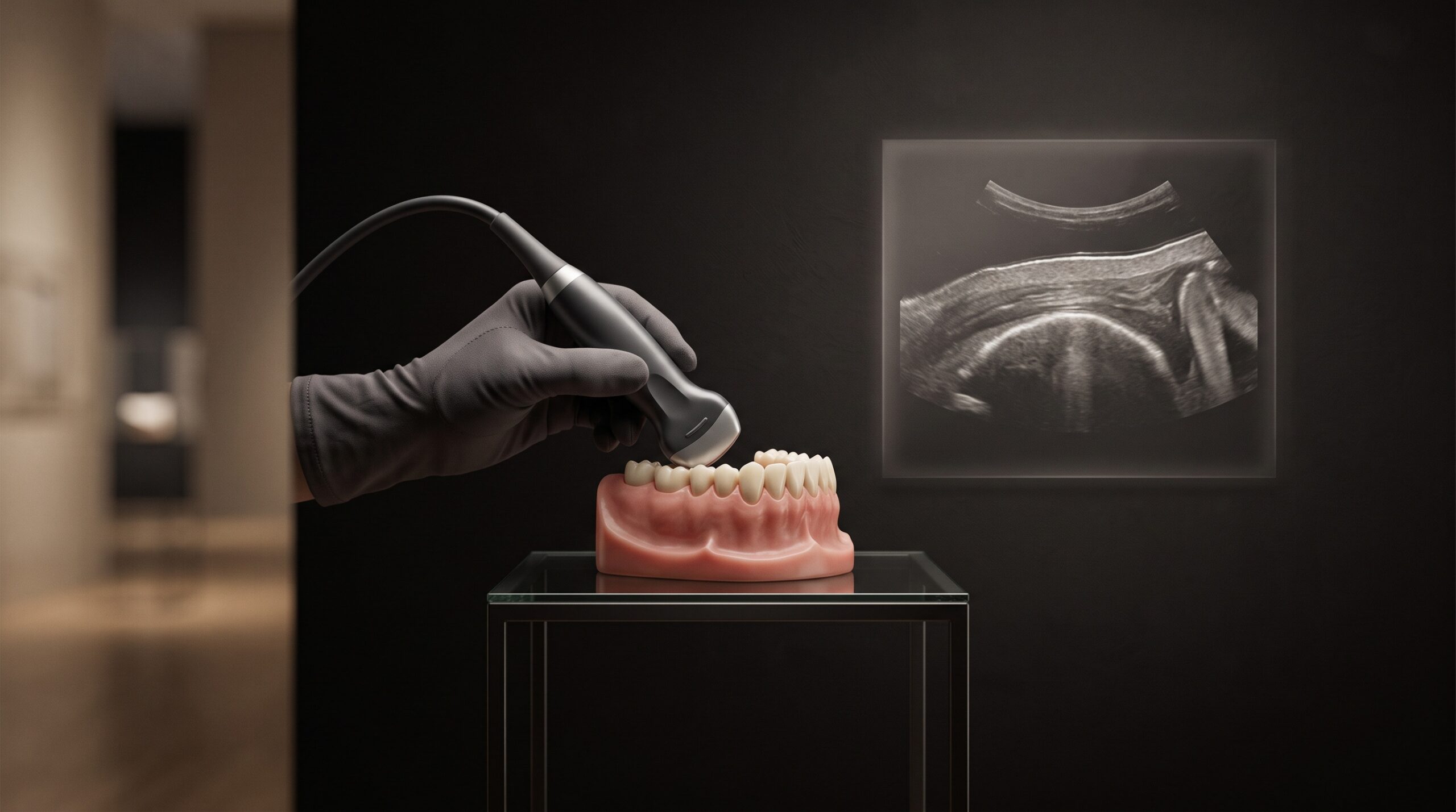
October 19, 2026 Reading the Gum With Sound: High-Frequency Ultrasound and the Measure of Gingival Phenotype
For all the imaging a modern practice can summon, there is a thin layer of the mouth that nearly all of it steps politely around. The radiograph renders bone and enamel in exquisite detail precisely because it is blind to soft tissue; the X-ray beam sails through gum as though it were not there. The intraoral scanner captures the gingiva’s outer surface beautifully but has no way of knowing how thick that surface is, or what lies a fraction of a millimeter beneath it. And yet that layer — the gingiva, and the delicate plate of bone it drapes over — governs some of the most consequential decisions in dentistry: whether a gumline will recede, whether an implant will show a gray shadow through the tissue, whether a graft will take. High-frequency ultrasound is the modality quietly learning to see it, measuring the gum with sound instead of radiation.
The phenotype problem
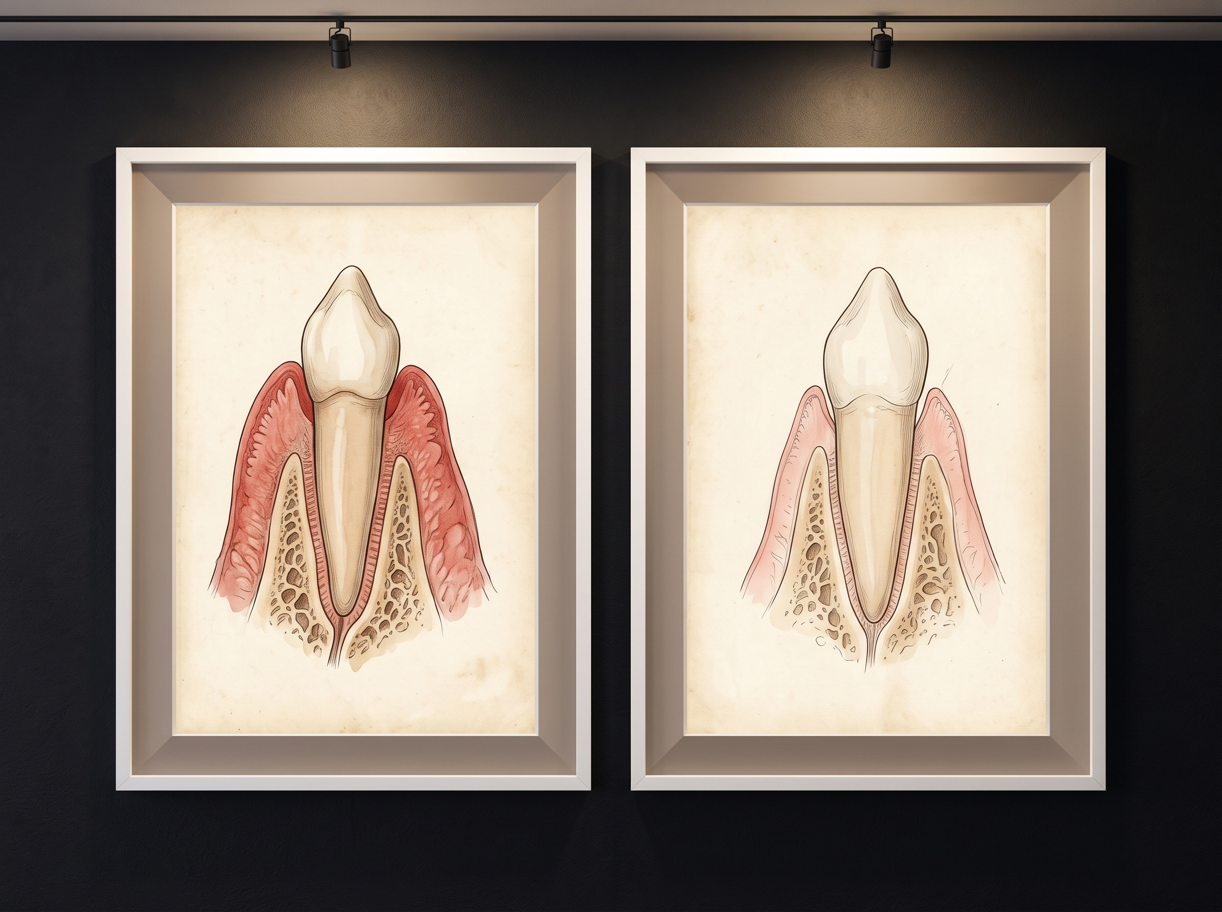
Clinicians have long sorted gingiva into phenotypes — the older word was biotype — along a spectrum from thick and fibrous to thin and scalloped. The distinction is not cosmetic trivia. A thin phenotype recedes more readily, shows the metal or gray of an underlying root or implant more easily, and responds less forgivingly to surgery; a thick phenotype hides margins, resists recession, and heals with more latitude for error. Knowing which one is in front of you is a planning decision, not an afterthought. The trouble has always been measuring it. The quick method is to slide a periodontal probe into the sulcus and judge whether its color shows through the tissue — a binary, subjective call that two clinicians will often make differently. The accurate method, transgingival probing, means anesthetizing the patient and pushing a needle or probe straight through the gum to the bone to read a depth off it. It works, and it hurts, and no one wants to repeat it at every visit to watch a trend. The gum, in other words, has been something we estimate rather than something we image.
How sound reads tissue
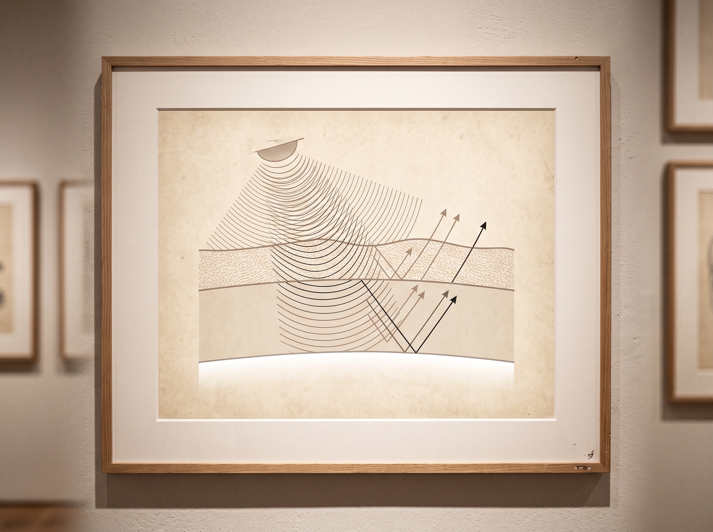
Ultrasound earns its place here by exploiting the one thing soft tissue does readily: it reflects sound at its boundaries. A transducer sends a pulse of high-frequency sound into the tissue and listens for the faint echoes that return wherever the sound crosses from one material into another of different acoustic density — from gum into the fluid of the sulcus, from soft tissue into the hard mineral wall of bone. By timing how long each echo takes to come back, the machine converts time into depth and assembles a cross-sectional image, a slice through the gum as if it had been cut and laid open, without a single cut being made. The governing trade-off is frequency. Diagnostic body ultrasound runs at a few megahertz to see deep organs; dental soft-tissue imaging pushes far higher, into the range of twenty to forty-eight megahertz, because resolution rises with frequency even as penetration falls. For a structure only a millimeter or two thick, shallow is exactly right, and the high frequencies resolve detail a general scanner never could. The result is a map in which the gingival band, the bone crest, and the facial bone plate each declare themselves as distinct bright lines.
What the cross-section finally shows
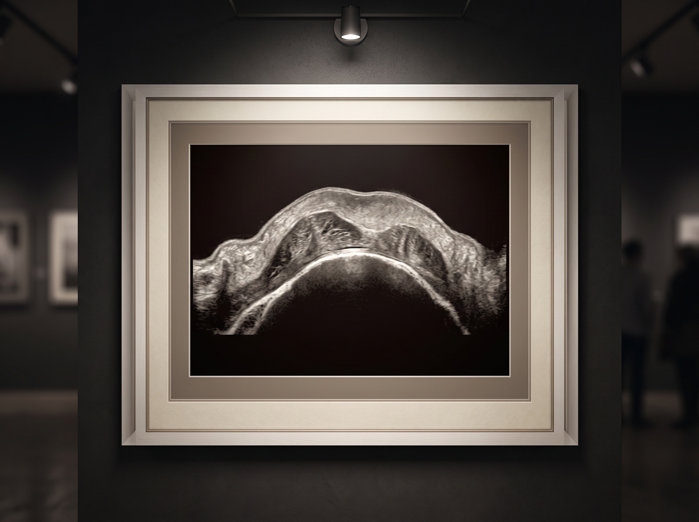
On the screen the phenotype stops being a judgment call. The soft tissue appears as a band of genuine, measurable thickness, read to a tenth of a millimeter at any point the clinician chooses; the crest of bone sits as a sharp reflective line beneath it; the facial plate of bone — whose thickness over an implant site can decide the entire esthetic outcome — can be followed where it is thick enough to return an echo. A single painless scan, repeatable at every recall, replaces both the subjective look and the needle. And because it is radiation-free, it belongs to the same emerging family as the other modalities this gallery has followed for their refusal to irradiate soft tissue to see it — the magnetic-resonance techniques now being adapted to oral tissue and the joint-imaging methods that pair MRI with CBCT. Where those reach for deep structures, ultrasound owns the shallow, the superficial, the unglamorous millimeter of gum that nothing else measures well.
The craft the probe demands
None of this is automatic, and the honest account includes the difficulty. Ultrasound is the most operator-dependent of imaging modalities; the picture lives and dies by how the probe is held. The transducer must couple to the tissue through gel or a soft standoff, must be angled square to the surface it is measuring, and must track a curved, mobile, saliva-slicked anatomy in a space built for fingers, not footprints. Press too hard and the gum compresses, and the thickness you so precisely measured is a thickness you created. The learning curve is real, and the images reward a steady, patient hand the way a difficult photograph does — which is to say this is a craft, not a button. It is also, for now, an adjunct rather than a standard: the equipment is specialized, the intraoral probes are still maturing toward the small footprints the posterior mouth needs, and interpretation asks a clinician to read grayscale the way a radiologist learns to. These are the frictions of a modality on its way in, not reasons to wave it off.
A subject, not a byproduct
There is something fitting, to the sensibility this gallery was built around, in imaging the gum for its own sake. For a century our instruments have treated soft tissue as the thing light and radiation pass through on the way to the structures we cared about — the obstacle, not the subject. Ultrasound inverts that. It makes the gingiva the protagonist of its own picture, a layered, living band with a texture and a thickness and a quiet beauty in cross-section, rendered in the soft grayscale topography that medical ultrasound has always had. To measure the gum is, in a small way, to grant it the dignity of being looked at — the same shift in attention that turns a salivary duct or a worn enamel surface from an incidental finding into a thing worth photographing well.
Future Developments
The near horizon for dental ultrasound is less about the sound and more about what is done with it. Machine-learning segmentation is the obvious next step: software that outlines the gingival band and the bone line automatically, turning a skilled interpretation into a reproducible number and smoothing out the operator dependence that is the method’s chief liability today. Color Doppler, already routine elsewhere, would add blood flow to the picture — a way to see inflammation and healing as function rather than inferring it from appearance. And the most interesting prospect is fusion: a gingival thickness map, captured by sound, registered into the same coordinate space as a CBCT volume and an optical scan to become one more layer of the composite virtual patient, the soft-tissue stratum that model has always been missing. The X-ray gave us the skeleton of the mouth and the scanner gave us its surface. Ultrasound is beginning to give us the part in between — and once an imaging method can measure the gum painlessly and repeatably, the gum stops being something we estimate and becomes, at last, something we can watch.
Sorry, the comment form is closed at this time.