17 Feb Intraoral Photography Best Practices for Treatment Documentation
Intraoral photography has become an indispensable tool in modern dental practice, serving multiple roles from treatment planning and patient education to legal documentation and professional communication. However, achieving consistently high-quality clinical photographs requires more than pointing and shooting – it demands understanding of proper techniques, equipment selection, and systematic approaches to image capture.
Essential Equipment and Setup
Camera Selection and Configuration
Digital SLR cameras remain the gold standard for intraoral photography, offering superior image quality and precise control over exposure settings. Key features to prioritize include:
- High-resolution sensors (minimum 12 megapixels for diagnostic quality)
- Manual exposure controls for consistent lighting
- Live view LCD for accurate composition
- Fast autofocus systems for patient comfort
- Weatherproof construction for infection control protocols
Camera settings should be standardized to ensure consistency across all images. Recommended base settings include manual mode with aperture f/22-f/32 for optimal depth of field, ISO 100-200 for minimal noise, and shutter speeds of 1/60 second or faster to minimize motion blur.
Lens and Macro Photography Considerations
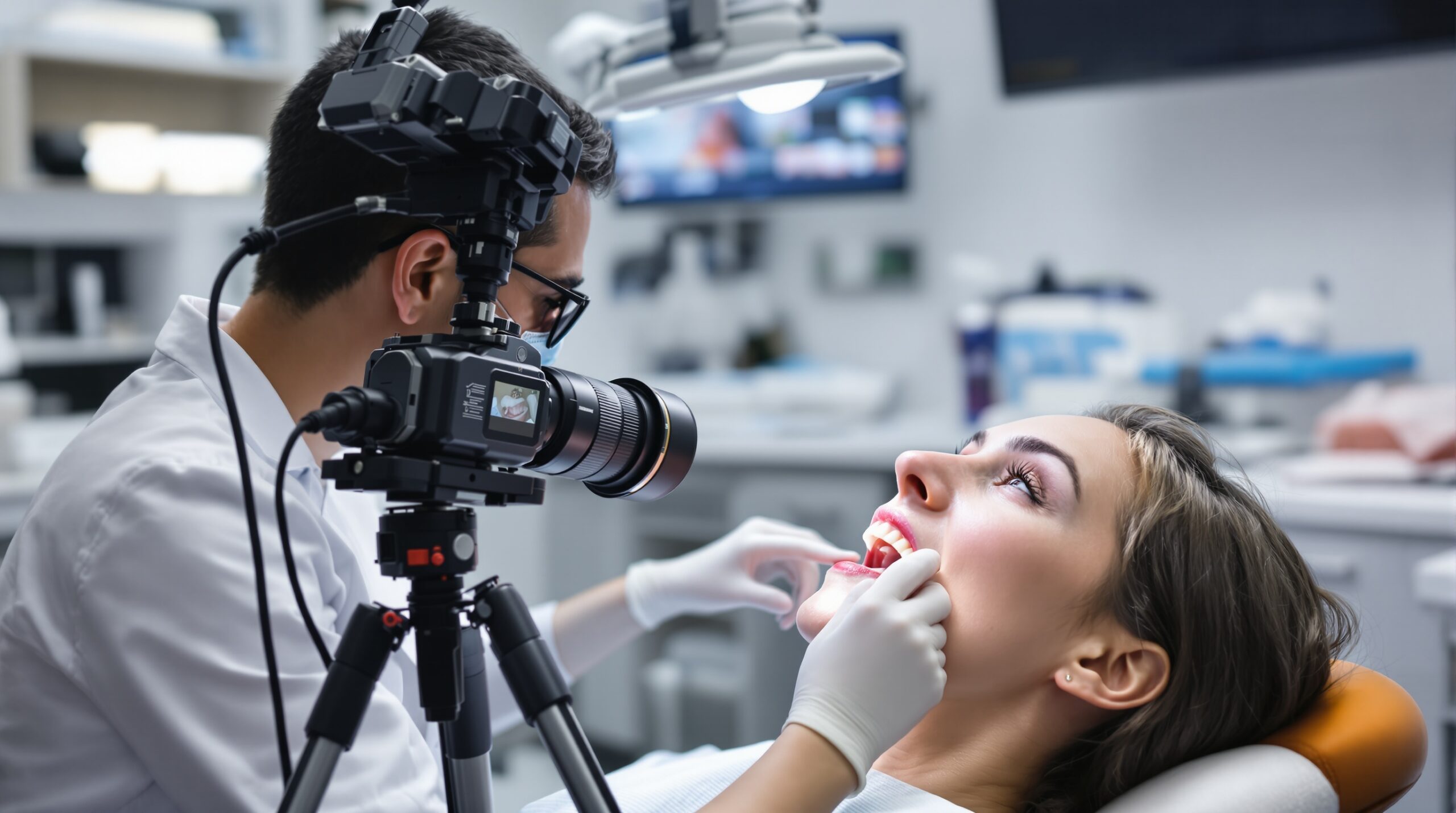
True macro lenses with 1:1 magnification ratios provide the detail resolution necessary for clinical documentation. The 100mm focal length offers ideal working distance, keeping the camera and photographer comfortably away from the patient while achieving proper magnification.
Ring flash systems or twin-point flash units provide even, shadowless illumination essential for diagnostic imaging. These specialized lighting systems eliminate the harsh shadows created by standard camera flashes and ensure consistent color temperature across all images.
Standardized Image Series Development
Comprehensive Documentation Protocol
Effective treatment documentation requires systematic image capture following established protocols. A complete intraoral series typically includes:
- Facial photographs: Frontal smile, profile views, and close-up smile
- Retracted views: Full arch occlusal, buccal corridors, and lateral segments
- Close-up detail shots: Individual teeth, restoration margins, and pathological areas
- Bite registration: Centric occlusion and lateral excursions
- Soft tissue documentation: Gingival health, mucosal conditions, and surgical sites
Each image type serves specific diagnostic and documentation purposes. Standardizing this sequence ensures comprehensive coverage while creating efficient workflows that minimize patient chair time.
Patient Positioning and Comfort
Proper patient positioning is crucial for image quality and patient comfort. The ideal setup positions the patient’s head at photographer eye level, eliminating awkward angles and reducing physical strain. Head stabilization using adjustable headrests prevents movement blur while maintaining patient comfort during extended photography sessions.
Cheek retractors should be inserted gently and positioned to provide adequate visibility without causing discomfort. Different retractor sizes accommodate varying mouth opening capabilities, and proper placement techniques minimize soft tissue distortion that could compromise image accuracy.
Lighting and Exposure Management
Achieving Consistent Illumination
Uniform lighting across the oral cavity presents unique challenges due to the complex three-dimensional anatomy and varying surface reflectance properties of teeth, restorations, and soft tissues. Ring flash systems address these challenges by providing 360-degree illumination that eliminates directional shadows.
Flash power adjustment becomes critical when photographing different oral structures. Darker posterior regions may require increased flash output, while highly reflective anterior restorations might need reduced power to prevent overexposure. Many modern systems offer automatic exposure compensation that adjusts for these variations.
Color Accuracy and White Balance
Accurate color reproduction is essential for treatment planning and shade matching. Custom white balance settings calibrated to the specific flash system ensure consistent color temperature across all images. This becomes particularly important when documenting aesthetic treatments where precise shade communication is critical.
Color calibration targets should be included in the first image of each session, providing reference standards for post-processing and ensuring color accuracy when images are displayed on different devices or printed for patient education materials.
Composition and Framing Techniques
Diagnostic Image Requirements
Effective clinical photography follows specific composition guidelines that maximize diagnostic value. Key principles include:
- Sufficient magnification to show relevant detail without excessive enlargement
- Proper depth of field to maintain focus across the area of interest
- Appropriate framing that includes necessary anatomical landmarks
- Consistent orientation and cropping for easy comparison over time
- Elimination of distracting elements such as fingers or instruments
The rule of thirds applies to intraoral photography, with key structures positioned at intersection points to create visually appealing and diagnostically useful images. However, clinical requirements may override aesthetic considerations when maximum detail visibility is paramount.
Mirror Techniques for Posterior Access
Intraoral mirrors enable visualization of otherwise inaccessible areas, but their use requires specific techniques to achieve high-quality results. Mirror placement should provide direct visualization of the target area while minimizing distortion. Anti-fogging solutions prevent condensation that can obscure detail, while proper mirror angles reduce unwanted reflections.
Larger mirrors provide better coverage but may be uncomfortable for patients with limited mouth opening. Selecting appropriate mirror sizes and shapes for different clinical situations optimizes both image quality and patient comfort.
Digital Workflow Integration
Image Management and Storage
Efficient digital workflows begin with systematic file naming conventions that facilitate quick image retrieval. Recommended naming structures include patient identifier, date, and image type, creating searchable archives that support both clinical and administrative functions.
Cloud-based storage solutions provide secure backup while enabling easy sharing with specialists, laboratories, and patients. These systems typically offer automatic synchronization across multiple devices and locations, ensuring image availability when and where needed.
Integration with Practice Management Systems
Modern practice management software often includes integrated imaging modules that automatically associate photographs with patient records. These systems streamline workflows by eliminating manual file management while maintaining HIPAA compliance through secure, encrypted storage.
Automated backup procedures protect valuable clinical documentation against hardware failures or data corruption. Regular testing of backup systems ensures image accessibility during critical moments.
Quality Control and Standardization
Image Review Protocols
Systematic quality assessment ensures all images meet diagnostic standards before storage. Review criteria should evaluate technical factors such as focus, exposure, and composition, as well as clinical factors including anatomical coverage and diagnostic value.
Immediate image review on the camera’s LCD allows for retakes while the patient is still positioned, preventing the need for additional appointments. However, detailed quality assessment often requires larger computer monitors that reveal subtle technical deficiencies not visible on small camera screens.
Staff Training and Competency
Consistent image quality across all staff members requires comprehensive training programs that address both technical and clinical aspects of intraoral photography. Training should cover equipment operation, patient management, and quality standards specific to the practice’s documentation needs.
Regular competency assessments help maintain standards and identify areas needing additional training. Peer review sessions can provide valuable feedback and promote continuous improvement in imaging techniques.
Patient Communication and Education
Visual Treatment Planning
High-quality intraoral photographs serve as powerful patient education tools, allowing practitioners to show existing conditions and explain proposed treatments in ways that words alone cannot accomplish. Before-and-after comparisons demonstrate treatment success and support case presentation efforts.
Annotation software enables practitioners to highlight specific areas and add explanatory text, creating customized educational materials tailored to individual patient needs. These annotated images can be printed for patient reference or stored electronically for future discussions.
Enhancing Treatment Acceptance
Visual documentation of oral conditions significantly improves patient understanding and treatment acceptance rates. Patients who can see their conditions are more likely to understand the necessity of proposed treatments and value the quality of completed work.
Before treatment images serve as baseline references that highlight improvement, supporting both clinical assessment and patient satisfaction. This documentation becomes particularly valuable for extensive treatments spanning multiple appointments.
Legal and Documentation Considerations
Medico-Legal Protection
Comprehensive photographic documentation provides valuable protection in medico-legal situations. Detailed images showing pre-treatment conditions, surgical procedures, and final results create objective records that support clinical decision-making and treatment outcomes.
Standardized documentation protocols ensure consistency in legal contexts, while proper image storage and backup procedures maintain evidence integrity over extended periods. These practices become increasingly important as treatment complexity and patient expectations continue to rise.
Specialist Communication
High-quality clinical photographs facilitate communication with specialists and laboratories, improving treatment coordination and outcomes. Detailed images allow remote consultation and treatment planning, reducing the need for additional patient appointments while ensuring accurate information transfer.
Standardized image series enable specialists to provide more accurate diagnoses and treatment recommendations, supporting collaborative care approaches that benefit both patients and practitioners.
Future Trends and Technology
AI-Assisted Analysis
Artificial intelligence applications for intraoral photography are emerging, offering automated analysis capabilities that can identify pathological conditions, assess treatment progress, and suggest additional documentation needs. These tools promise to enhance diagnostic accuracy while streamlining clinical workflows.
Machine learning algorithms trained on large image datasets can potentially detect subtle changes that might be overlooked by human observation, supporting earlier intervention and improved treatment outcomes.
3D Integration and Advanced Imaging
The integration of traditional photography with 3D scanning and advanced imaging modalities is creating comprehensive documentation systems that provide unprecedented detail about oral conditions and treatment results.
These multi-modal approaches will likely become standard practice as technology costs decrease and clinical benefits become more apparent.
Conclusion
Mastering intraoral photography requires attention to technical details, systematic approaches, and ongoing commitment to quality improvement. Practices that invest in proper equipment, comprehensive training, and standardized protocols will realize significant benefits in treatment planning, patient communication, and clinical documentation.
The future of dental photography continues to evolve with advancing technology, but the fundamental principles of proper technique, consistent quality, and systematic documentation remain constant. Practitioners who embrace these principles while staying current with technological developments will provide superior patient care while protecting their practices through comprehensive documentation.
Sorry, the comment form is closed at this time.