31 Mar Standardized Dental Photography Protocols: Professional Clinical Documentation and Case Presentation Guidelines
Professional dental photography has evolved from optional documentation to an essential component of comprehensive patient care. Modern dental practices require standardized protocols to ensure consistent, high-quality clinical images that support diagnosis, treatment planning, and patient communication.

Essential Equipment and Setup Standards
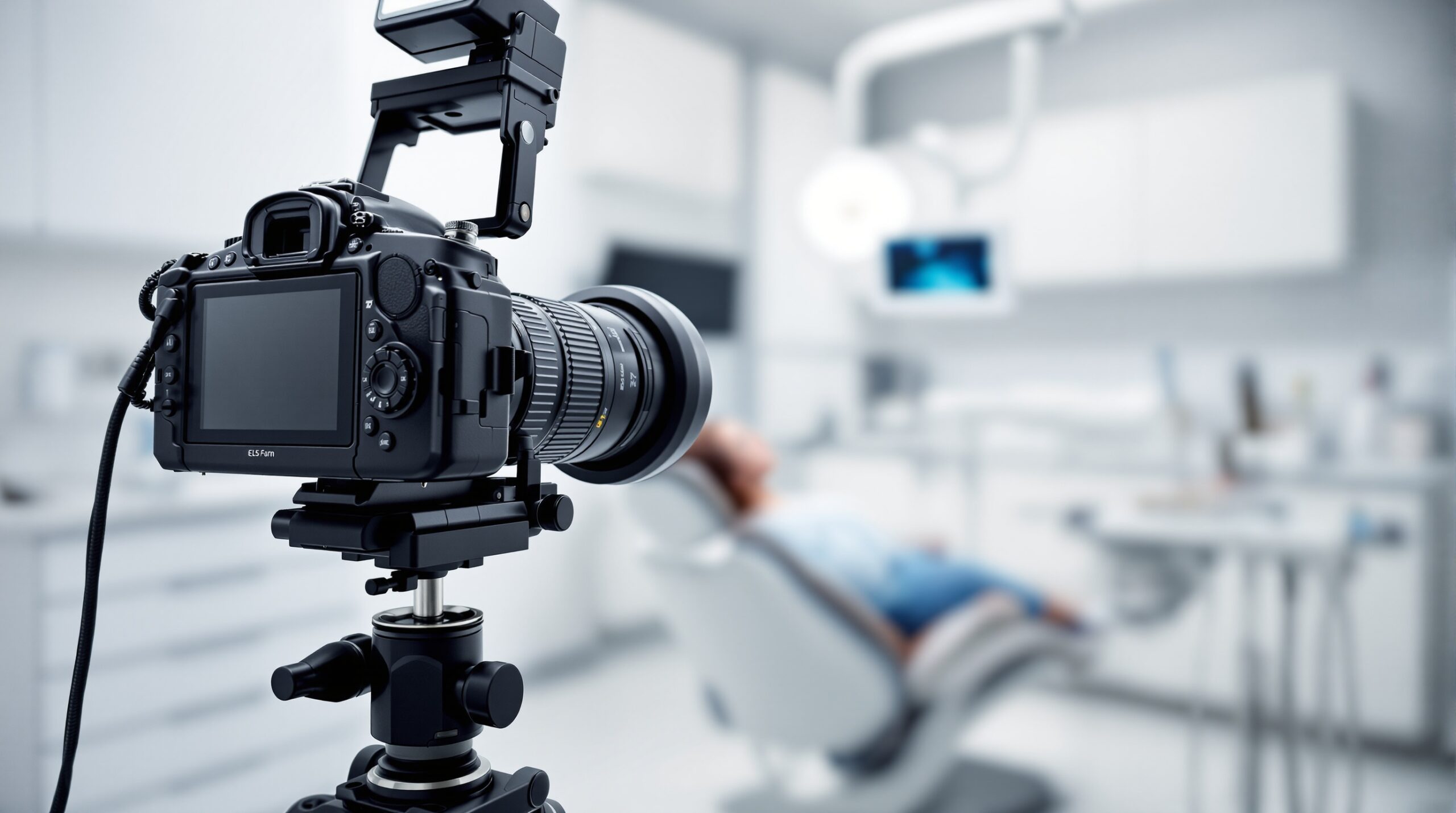
Successful clinical photography begins with proper equipment selection and standardized positioning protocols. A professional DSLR camera equipped with a 100mm macro lens provides optimal magnification and working distance for intraoral documentation. Ring flash systems deliver consistent, shadowless illumination crucial for accurate color reproduction and detail capture.
Camera settings should remain consistent across all documentation sessions. ISO 100-200 minimizes noise while maintaining image quality. Aperture settings between f/16-f/22 ensure adequate depth of field for sharp focus throughout the oral cavity. Manual exposure control prevents inconsistent lighting variations that compromise diagnostic value.
Standardized Patient Positioning and Retraction Techniques
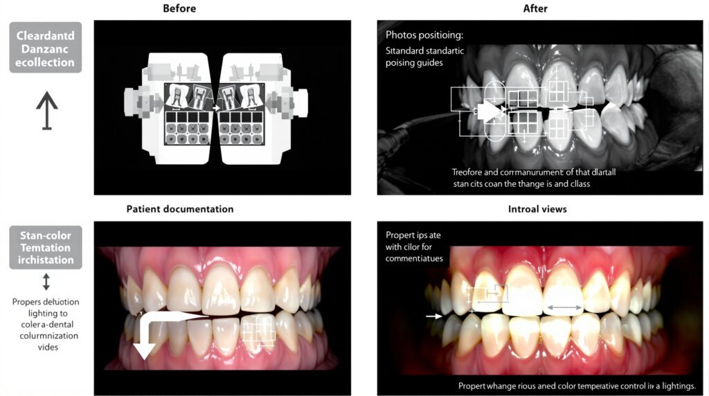
Patient positioning directly impacts image quality and diagnostic accuracy. The patient should be positioned with their head tilted slightly backward, creating optimal access angles for various intraoral views. Proper head support eliminates movement artifacts that degrade image sharpness.
Systematic retraction protocols ensure consistent visualization of target anatomy. Cheek retractors should be positioned to provide maximum tissue displacement without causing patient discomfort. Lip retractors must be carefully placed to avoid obscuring critical anatomical structures while maintaining adequate exposure.
Intraoral Photography Sequence
A comprehensive intraoral series includes eight standardized views: anterior facial, right and left buccal, maxillary occlusal, mandibular occlusal, right and left lateral, and posterior views as needed. Each view serves specific diagnostic purposes and must be captured consistently to enable accurate treatment monitoring over time.
Color Management and Post-Processing Standards
Accurate color reproduction is critical for clinical documentation, particularly when documenting shade matching or pathological conditions. Custom white balance settings should be established for each lighting environment. Color calibration targets can be included in initial setup shots to ensure consistent color accuracy across different viewing devices.
Post-processing should be limited to basic corrections: exposure adjustment, color balance, and cropping. Digital manipulation beyond these parameters compromises the clinical integrity of documentation. All adjustments must be applied consistently across the entire case series to maintain diagnostic validity.
Digital Workflow Integration
Modern dental practices benefit from seamless integration between photography systems and practice management software. Standardized file naming conventions facilitate efficient image retrieval and case presentation. Automated backup systems ensure long-term preservation of clinical documentation while maintaining patient privacy compliance.
Image resolution should be optimized for both clinical review and patient presentation. High-resolution files preserve diagnostic detail for professional consultation, while compressed versions enable efficient patient communication and case presentation workflows.
Quality Assurance Protocols
Regular equipment calibration ensures consistent results over time. Monitor calibration should be performed monthly using professional colorimetry tools. Camera settings and flash output should be verified periodically to maintain standardized exposure parameters across all clinical documentation.
Staff training programs should emphasize both technical proficiency and clinical application. Standardized checklists help maintain consistency when multiple team members participate in clinical photography workflows.
Patient Communication and Case Presentation
Standardized photography protocols enhance patient education and treatment acceptance. Before-and-after documentation provides compelling evidence of treatment success, supporting case presentation and building patient confidence in recommended procedures.
Professional image presentation software enables dynamic case reviews that engage patients in treatment planning discussions. Standardized documentation also supports insurance claim processing and peer consultation when complex cases require additional expertise.
Implementing comprehensive photography protocols transforms clinical documentation from basic record-keeping to powerful diagnostic and communication tools that enhance every aspect of modern dental practice.
Sorry, the comment form is closed at this time.