04 Apr Dental Operating Microscopes: Advancing Clinical Precision Through Enhanced Magnification and Imaging
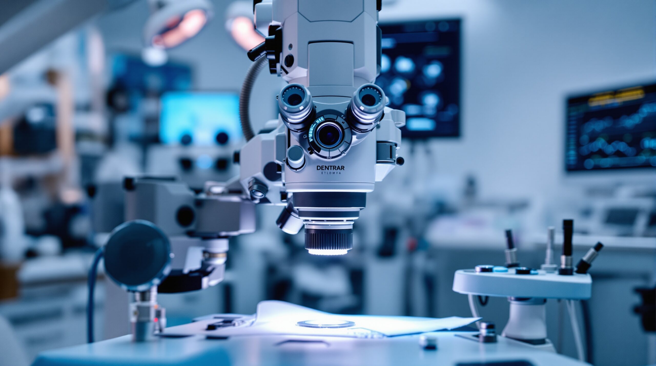
Dental operating microscopes represent a revolutionary advancement in clinical dentistry, providing unprecedented magnification and illumination capabilities that transform diagnostic accuracy and treatment precision. These sophisticated optical instruments enable dental professionals to visualize microscopic structures invisible to the naked eye, fundamentally changing the standard of care across multiple dental specialties.
Enhanced Visualization Through Advanced Optics
Modern dental operating microscopes deliver magnification levels ranging from 6x to 40x, allowing clinicians to identify minute anatomical details, microscopic fractures, and early-stage pathology that would otherwise go undetected. The integration of LED illumination systems provides shadow-free, coaxial lighting that illuminates the treatment area without generating heat, ensuring patient comfort while maintaining optimal visibility.
The optical quality of contemporary microscope systems incorporates apochromatic lenses that eliminate chromatic aberration, delivering true color reproduction essential for accurate tissue assessment and material characterization. This level of optical precision enables dental professionals to distinguish subtle variations in tooth structure, detect hairline fractures, and identify early carious lesions with unprecedented clarity.

Clinical Applications Across Dental Specialties
Endodontics has particularly benefited from microscope technology, with enhanced visualization enabling precise identification of canal orifices, detection of vertical root fractures, and improved success rates in complex root canal procedures. The ability to visualize the pulp chamber floor under high magnification allows for more conservative access preparations and better preservation of tooth structure.
In restorative dentistry, operating microscopes facilitate precision-driven protocols for crown and onlay preparations, ensuring accurate margin placement and optimal tissue preservation. The enhanced visualization capabilities allow clinicians to achieve sub-millimeter accuracy in margin preparation and restoration placement, significantly improving long-term prognosis and patient outcomes.
Periodontal and oral surgical procedures benefit from microscope-enhanced visualization through improved wound closure techniques, precise suturing under magnification, and enhanced ability to detect and remove calculus deposits in deep periodontal pockets. The magnified view enables more conservative surgical approaches and improved healing outcomes.
Integration with Digital Imaging Technology
Contemporary dental operating microscopes seamlessly integrate with digital imaging systems, enabling real-time documentation, patient education, and treatment planning enhancement. Digital capture capabilities allow for high-resolution photography and video recording during procedures, providing valuable documentation for case records and facilitating improved patient communication.

The integration of artificial intelligence-driven diagnostic tools with microscope systems represents the next frontier in dental imaging technology. AI-enhanced image analysis can assist in real-time identification of pathological conditions, automated measurement capabilities, and predictive treatment planning based on microscopic findings.
Future Developments and Emerging Technologies
Emerging technologies in dental microscopy include 3D visualization capabilities, fluorescence imaging integration, and enhanced digital workflow connectivity. These advancements promise to further expand the diagnostic and treatment capabilities of dental operating microscopes while improving clinical efficiency and patient outcomes.
The evolution toward more compact, lightweight microscope designs with improved ergonomics addresses practitioner fatigue concerns while maintaining optical excellence. Advanced autofocus systems and motorized positioning capabilities reduce setup time and enhance workflow efficiency in busy clinical environments.
Training and Adoption Considerations
Successful integration of dental operating microscopes requires comprehensive training and adaptation of clinical workflows. The learning curve associated with microscope-enhanced procedures necessitates dedicated training programs and gradual implementation strategies to maximize the benefits of this advanced technology.
Investment in dental operating microscopes represents a significant commitment to enhanced patient care and clinical excellence. The improved diagnostic capabilities, treatment precision, and patient outcomes achievable through microscope-enhanced dentistry justify the investment for practices committed to delivering the highest standard of care.
As dental education increasingly emphasizes microscope-based techniques, the next generation of dental professionals will be better prepared to leverage these advanced visualization tools, driving broader adoption and standardization across the profession.
Sorry, the comment form is closed at this time.