25 Apr SWIFT MRI Technology in Dentistry: Revolutionary Magnetic Resonance Imaging for Comprehensive Oral Tissue Visualization
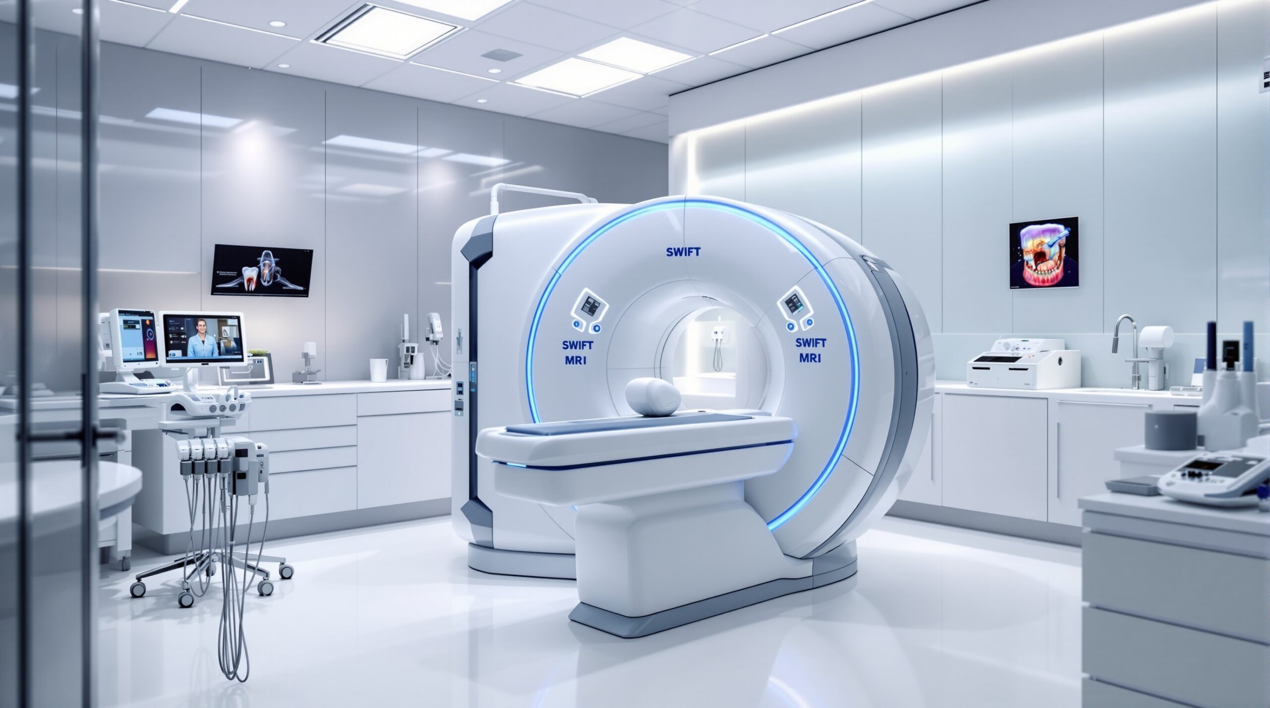
SWeep Imaging with Fourier Transform (SWIFT) represents a groundbreaking advancement in dental magnetic resonance imaging technology, offering unprecedented visualization capabilities for both hard and soft dental tissues. Unlike conventional MRI techniques that struggle with dental applications, SWIFT technology provides clinicians with high-resolution imaging of teeth, supporting structures, and surrounding tissues in remarkably short scanning times.
Understanding SWIFT Technology
SWIFT MRI technology overcomes traditional limitations of magnetic resonance imaging in dentistry by utilizing a unique acquisition method that captures signals from tissues with extremely short T2 relaxation times. This capability is particularly crucial in dental imaging, where enamel and dentin have signal characteristics that are typically difficult to visualize using standard MRI sequences.
The technology employs simultaneous RF excitation and data acquisition, eliminating the dead time present in conventional MRI sequences. This innovation allows SWIFT to capture signals from rapidly decaying tissues, making it exceptionally well-suited for imaging the complete dental structure without the artifacts and signal loss commonly associated with traditional MRI approaches.
Clinical Applications and Advantages
SWIFT MRI technology offers numerous clinical advantages over conventional dental imaging modalities. The technique provides comprehensive visualization of tooth structure, pulp chambers, root canals, and periodontal tissues without ionizing radiation exposure. This radiation-free approach is particularly valuable for pediatric patients, pregnant women, and individuals requiring frequent imaging follow-ups.
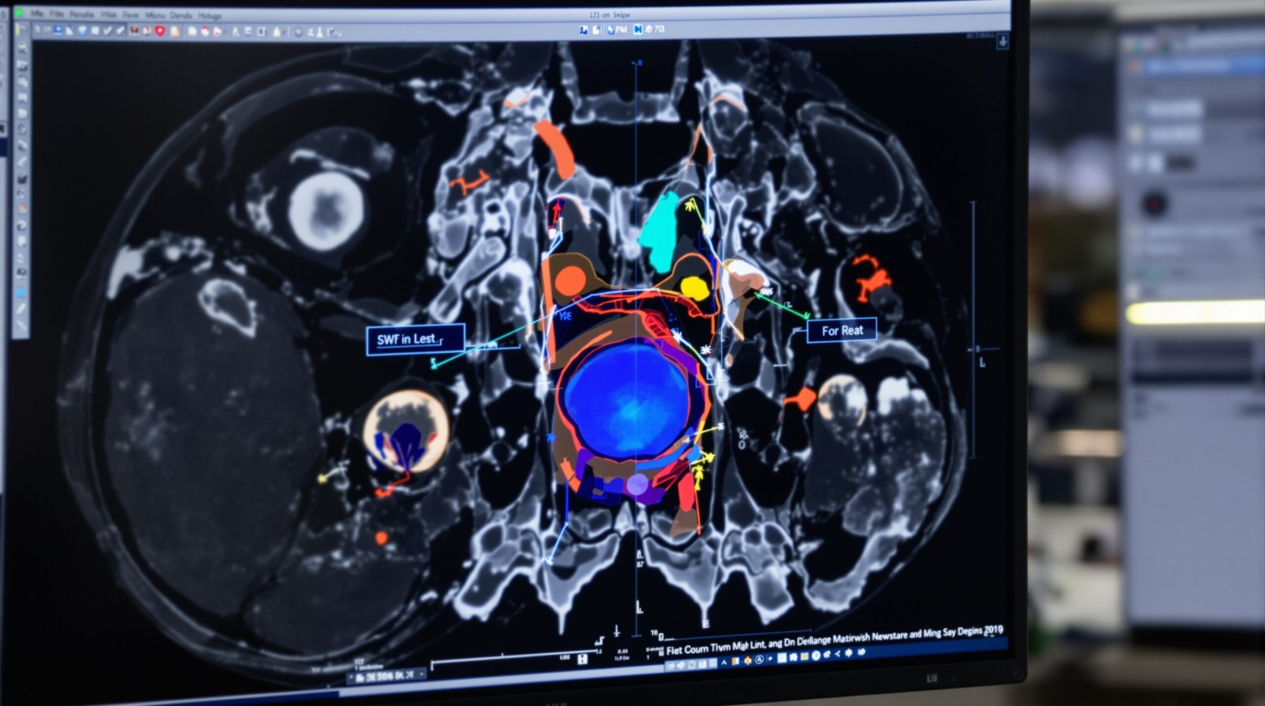
The high sensitivity to pathological changes makes SWIFT MRI exceptional for early detection of dental caries, pulpal inflammation, and periodontal disease. The technology can visualize internal tooth structure with clarity previously unattainable through non-invasive means, enabling more accurate diagnosis and treatment planning.
Comparative Imaging Capabilities
When compared to traditional dental radiography, SWIFT MRI provides superior soft tissue contrast and three-dimensional visualization without geometric distortion. The technology excels in imaging complex anatomical structures such as temporomandibular joints, enabling comprehensive evaluation of both hard and soft tissue components in a single examination.

Technical Specifications and Implementation
SWIFT MRI systems require specialized hardware configurations optimized for dental applications. The technology utilizes ultra-short echo time sequences, typically ranging from microseconds to a few milliseconds, enabling capture of rapidly decaying signals from dental hard tissues.
Implementation considerations include patient positioning protocols, coil selection, and sequence optimization for specific diagnostic requirements. The scanning times for SWIFT dental MRI are significantly reduced compared to conventional MRI, making the technology practical for routine clinical use while maintaining exceptional image quality.
Image Quality and Resolution
SWIFT technology delivers high spatial resolution imaging with excellent contrast differentiation between various dental tissues. The technique provides isotropic voxel sizes enabling multiplanar reconstruction capabilities, allowing clinicians to examine dental structures from any desired orientation without additional scanning.
Future Directions and Research Applications
Ongoing research continues to expand SWIFT MRI applications in dentistry, with emerging studies exploring its potential for monitoring treatment outcomes, evaluating dental implant osseointegration, and assessing orthodontic tooth movement. The technology’s unique capabilities position it as a valuable research tool for investigating dental tissue biology and pathology progression.
Future developments may include further sequence optimization, contrast agent applications, and integration with other advanced imaging modalities to provide comprehensive diagnostic information for complex dental cases.
Conclusion
SWIFT MRI technology represents a significant advancement in dental imaging, offering radiation-free, high-resolution visualization of complete dental structures. Its unique technical capabilities address longstanding limitations in dental MRI applications, providing clinicians with unprecedented diagnostic information for improved patient care. As the technology continues to evolve, SWIFT MRI is positioned to become an integral component of modern dental diagnostic protocols.
Sorry, the comment form is closed at this time.