25 Mar Ultrasound Imaging in Modern Dentistry: Radiation-Free Diagnostic Applications and Clinical Benefits
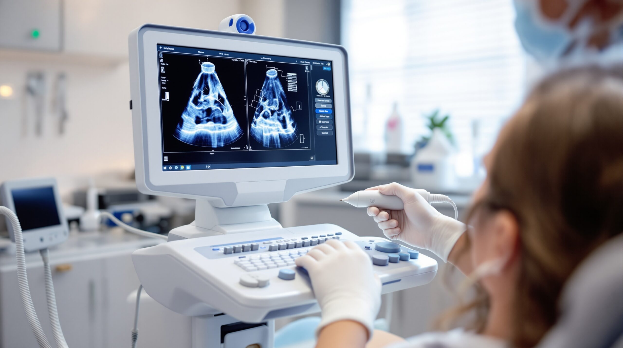
Dental ultrasound imaging represents a revolutionary advancement in diagnostic technology, offering clinicians a radiation-free alternative to traditional radiographic methods. This innovative approach has gained significant momentum in modern dentistry, providing real-time visualization of both hard and soft tissues while eliminating patient exposure to ionizing radiation.
The Science Behind Dental Ultrasound Technology
Ultrasound imaging utilizes high-frequency sound waves to create detailed images of internal structures. Unlike conventional X-rays, which rely on ionizing radiation that can potentially cause cellular damage, ultrasound technology harnesses the power of acoustic waves to generate diagnostic images safely and effectively.
The frequency range used in dental applications typically falls between 10-50 MHz, significantly higher than traditional medical ultrasound devices. This elevated frequency allows for enhanced resolution and improved visualization of the fine anatomical structures within the oral and maxillofacial region.
Clinical Applications in Contemporary Practice
Periodontal Assessment and Monitoring
One of the most promising applications of dental ultrasound lies in periodontal diagnostics. The technology enables precise measurement of periodontal pocket depths, assessment of bone levels, and evaluation of soft tissue health without the need for radiation exposure. Clinical studies have demonstrated that intraoral ultrasonography can reliably assess furcation involvement in mandibular molars with diagnostic accuracy closely matching cone beam computed tomography (CBCT) results.
Peri-implant Tissue Evaluation
Ultrasound imaging has shown exceptional promise in evaluating peri-implant tissues. Research indicates that ultrasound measurements of peri-implant bone defects show no statistically significant differences compared to CBCT imaging, making it an excellent radiation-free alternative for implant monitoring and maintenance protocols.
Soft Tissue Analysis
The non-invasive nature of ultrasound makes it particularly valuable for soft tissue evaluation. Practitioners can assess oral mucosa thickness, especially palatal tissue, which is crucial for periodontal surgery planning. This real-time capability allows for dynamic assessment and intraoperative guidance during various dental procedures.
Comparative Advantages Over Traditional Imaging
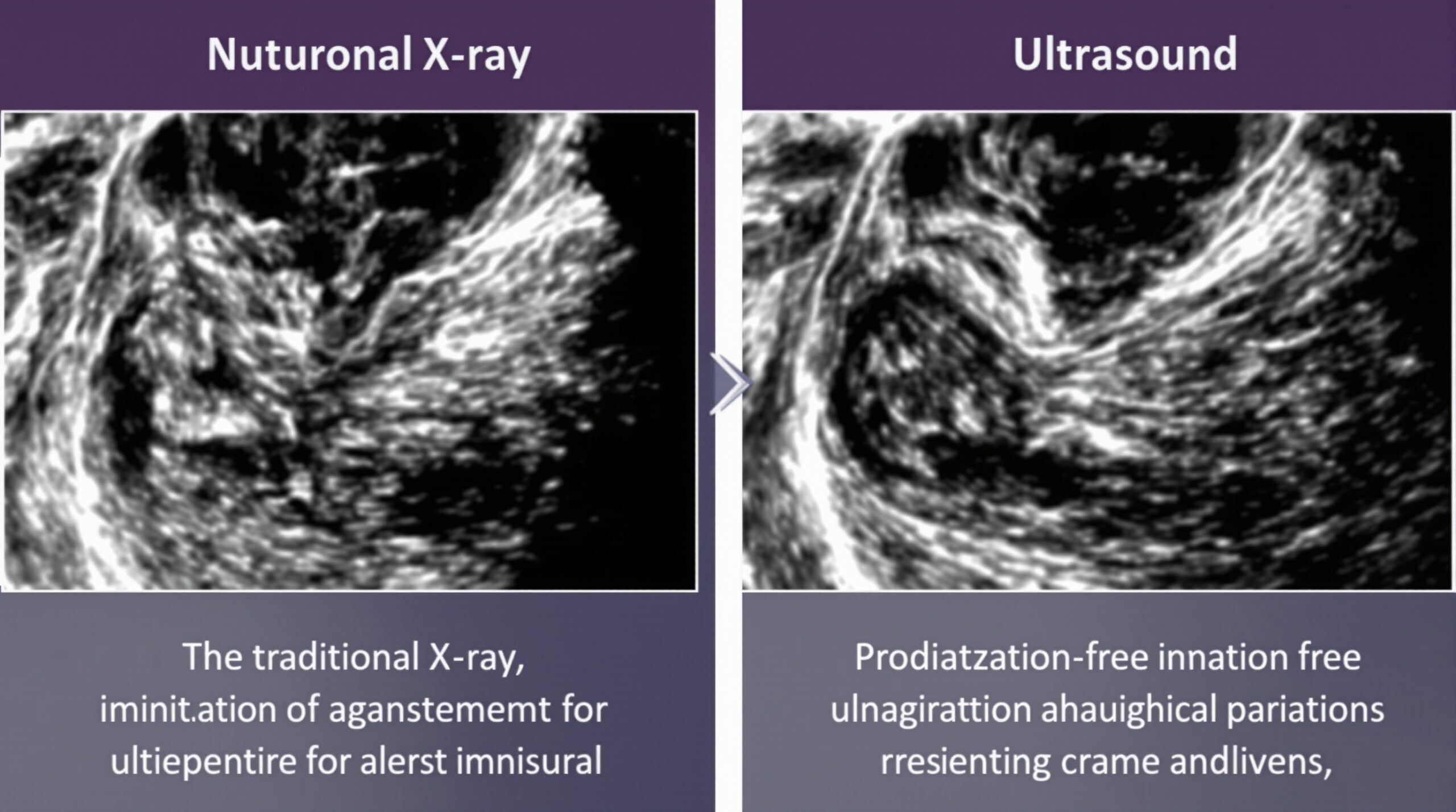
Radiation Safety
The most significant advantage of dental ultrasound is the complete elimination of ionizing radiation exposure. This benefit is particularly important for patients requiring frequent imaging, such as those undergoing orthodontic treatment, implant therapy, or periodontal maintenance. Pregnant patients and children especially benefit from this radiation-free diagnostic approach.
Real-time Imaging Capabilities
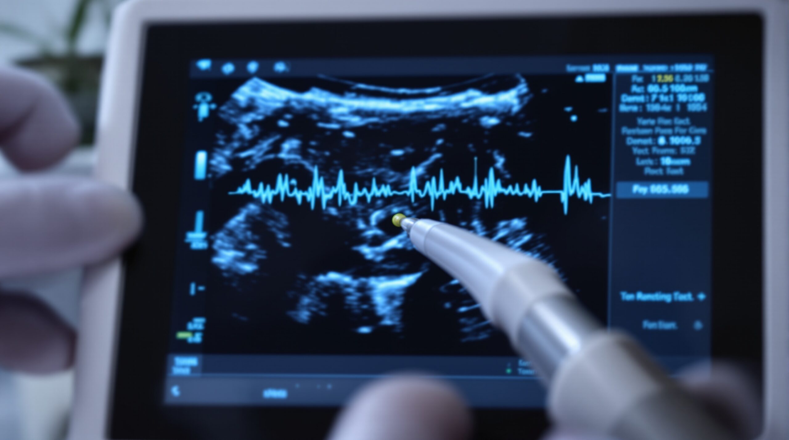
Unlike static radiographic images, ultrasound provides real-time visualization, allowing clinicians to observe tissue movement, blood flow, and dynamic changes during examination. This capability enhances diagnostic accuracy and enables immediate assessment of treatment outcomes.
Cost-effectiveness
Once the initial equipment investment is made, ultrasound imaging eliminates the ongoing costs associated with radiographic film, digital sensors, and radiation safety measures. The technology also reduces the need for retakes due to positioning errors, common with traditional radiography.
Current Limitations and Future Developments
While dental ultrasound technology shows tremendous promise, certain limitations currently exist. Penetration depth through hard tissues remains restricted compared to radiographic methods. Additionally, operator training and technique standardization require further development to ensure consistent results across different practitioners and clinical settings.
Research continues to advance ultrasound applications in dentistry, with ongoing studies exploring enhanced imaging algorithms, improved probe designs, and expanded clinical protocols. Future developments may include integration with artificial intelligence for automated diagnosis and treatment planning assistance.
Integration into Modern Dental Practice
The implementation of ultrasound technology into contemporary dental practice requires careful consideration of training requirements, equipment specifications, and clinical protocols. Successful integration involves understanding the technology’s capabilities and limitations while establishing appropriate patient selection criteria.
Practitioners considering ultrasound adoption should evaluate their specific diagnostic needs, patient demographics, and practice workflow to determine optimal implementation strategies. The technology works best as a complement to existing diagnostic methods rather than a complete replacement for all imaging modalities.
Conclusion
Dental ultrasound imaging represents a paradigm shift toward safer, more versatile diagnostic capabilities in modern dentistry. As technology continues to evolve and clinical protocols become more standardized, ultrasound imaging will likely play an increasingly important role in comprehensive dental care. The radiation-free nature, real-time imaging capabilities, and expanding clinical applications make dental ultrasound a valuable addition to the contemporary diagnostic toolkit, ultimately benefiting both practitioners and patients through enhanced safety and diagnostic precision.
No Comments