Logo Logo Logo Logo Logo
  • Home
  • Download
  • About
  • Blog
  • Home
  • Download
  • About
  • Blog
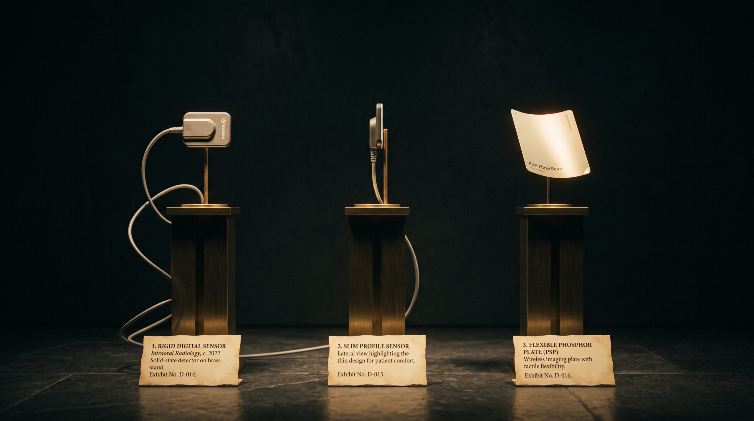
Three dental x-ray detectors displayed as artworks on brass museum plinths: two rigid corded solid-state sensors and one thin flexible phosphor plate.

04 Sep Three Ways to Catch a Photon: CCD, CMOS, and Phosphor Plates Compared

Posted at 10:39h in Dental Imaging by Raymond 0 Comments
0 Likes

Every digital intraoral image begins with a decision made in silicon or phosphor: how to turn an x-ray photon into a number. There are three answers in common use, and each one quietly shapes what the radiograph can and cannot show....

Read More
A single pale cube on a brass museum plinth engraved with a number that appears to shimmer and double, presented as an artwork whose value cannot be trusted.

03 Sep The Number That Lies: Why a CBCT’s Gray Values Are Not True Hounsfield Units

Posted at 10:37h in Dental Imaging by Raymond 0 Comments
0 Likes

On a medical CT, the number in a voxel is a promise: water reads zero, cortical bone reads a thousand, and the scale means the same thing tomorrow as today. On a cone-beam scan, the same-looking number keeps no such promise - and mistaking one...

Read More
A blank digital x-ray sensor panel presented like a primed empty canvas on a brass easel under museum lighting, its surface a faint grain of countless cells.

01 Sep The Sensor’s Fingerprint: Dark Current, Fixed-Pattern Noise, and the Flat-Field Correction That Makes a Detector Honest

Posted at 10:38h in Dental Imaging by Raymond 0 Comments
0 Likes

No two pixels on a digital sensor are born equal. Each carries its own faint bias, its own gain, its own quiet defect - and unless the detector first learns and subtracts that fingerprint, the pattern it prints onto every image is its own, not...

Read More
A gallery wall of small framed dental images arranged in a grid, progressing from faint and grainy to crisp and luminous

30 Aug Two Hundred Patents Deep: What a Two-Decade Portfolio Reveals About How the Dental Image Learned to See

Posted at 10:37h in Dental Imaging by Raymond 0 Comments
0 Likes

A single vendor recently tallied two decades of dental-imaging patents - hundreds of filings, a couple hundred granted. Read as more than a press release, it is a map of how the dental image was engineered, invention by patient invention....

Read More
A framed panoramic dental radiograph on a gallery wall with a faint translucent ghost shadow floating over the opposite side of the jaw

22 Aug The Ghost on the Other Side: How a Panoramic X-Ray Paints a Shadow Where No Bone Exists

Posted at 11:31h in Dental Imaging by Raymond 0 Comments
0 Likes

There is a second image hiding inside every panoramic radiograph: faint, doubled, floating on the wrong side of the jaw. It is not a machine fault. It is geometry - and learning to read it is a quiet mark of the craft....

Read More
Two framed images of the same molar side by side - a color surface scan and a grey radiograph - with light suggesting they are merging

21 Aug Two Ways of Seeing, One Frame: Fusing the Intraoral Scan and the Radiograph Into a Single Image

Posted at 10:38h in Dental Imaging by Raymond 0 Comments
0 Likes

One instrument captures the surface and refuses to look beneath it. The other looks straight through and forgets the surface exists. For decades the clinician fused them in the mind. Now the software does it - and the craft is changing....

Read More
A framed dental radiograph on a gallery wall with a small brass placard beneath it bearing a single glowing number

20 Aug The Dose You Cannot See: Exposure Index, Dose Creep, and the Number a Forgiving Sensor Hides

Posted at 11:39h in Dental Imaging by Raymond 0 Comments
0 Likes

Film punished overexposure by going dark. Digital rewards it with a cleaner picture - which is exactly how dose creeps upward, invisibly. Here is the number that fixes it....

Read More
A framed dental radiograph with a soft translucent grey veil drifting over its lower half, washing out the contrast there

19 Aug The Scattered Photon: How Compton Scatter, Collimation, and the Missing Grid Shape Contrast in Intraoral Radiography

Posted at 10:37h in Dental Imaging by Raymond 0 Comments
0 Likes

A radiograph's contrast is built by the photons that are cleanly absorbed and the ones that pass straight through. The scattered photon belongs to neither, and it is the quiet vandal of the image - a fog with no source that dental imaging fights not...

Read More
A framed dental radiograph on a gallery wall with a small blank museum placard beside it under refined lighting

18 Aug The Placard Beside the Picture: How Structured Reporting Makes a Dental Radiograph Legible

Posted at 10:38h in Dental Imaging by Raymond 0 Comments
0 Likes

In a gallery, the placard beside the artwork decides how much of the work you actually take in. The radiograph has a placard too - the report - and a new comparative study shows that giving it a fixed structure, rather than free-flowing prose, measurably...

Read More
A diagnostic display monitor shown as framed gallery art, its screen holding a dental radiograph in a smooth ramp of grays

17 Aug The Last Instrument: Diagnostic Display Calibration and the DICOM Grayscale Standard for Reading a Radiograph

Posted at 11:38h in Dental Imaging by Raymond 0 Comments
0 Likes

Every radiograph ends its journey on a screen, and the screen is the one instrument in the imaging chain that most practices never calibrate. The DICOM grayscale standard exists because the human eye reads brightness unevenly — and because a monitor that does not account...

Read More
  • 1
  • 2
  • 3
  • 4
  • 5
© Copyright 2020, Raster Builders Inc. All Rights Reserved.