


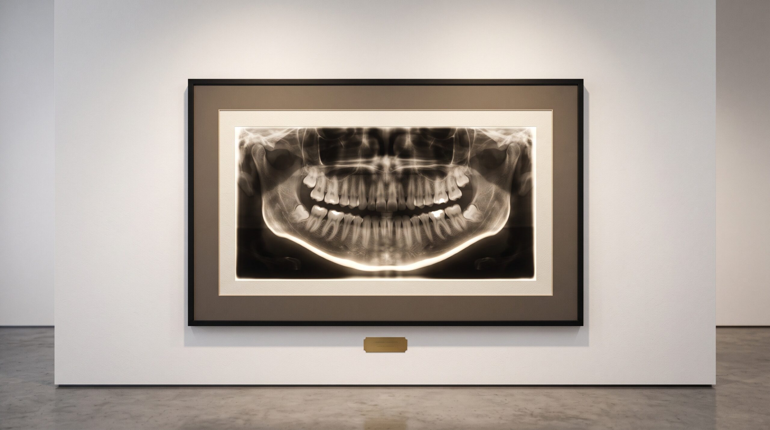
September 16, 2026 The Bone at the Bottom of the Frame: How a Panoramic X-Ray Quietly Screens for Osteoporosis
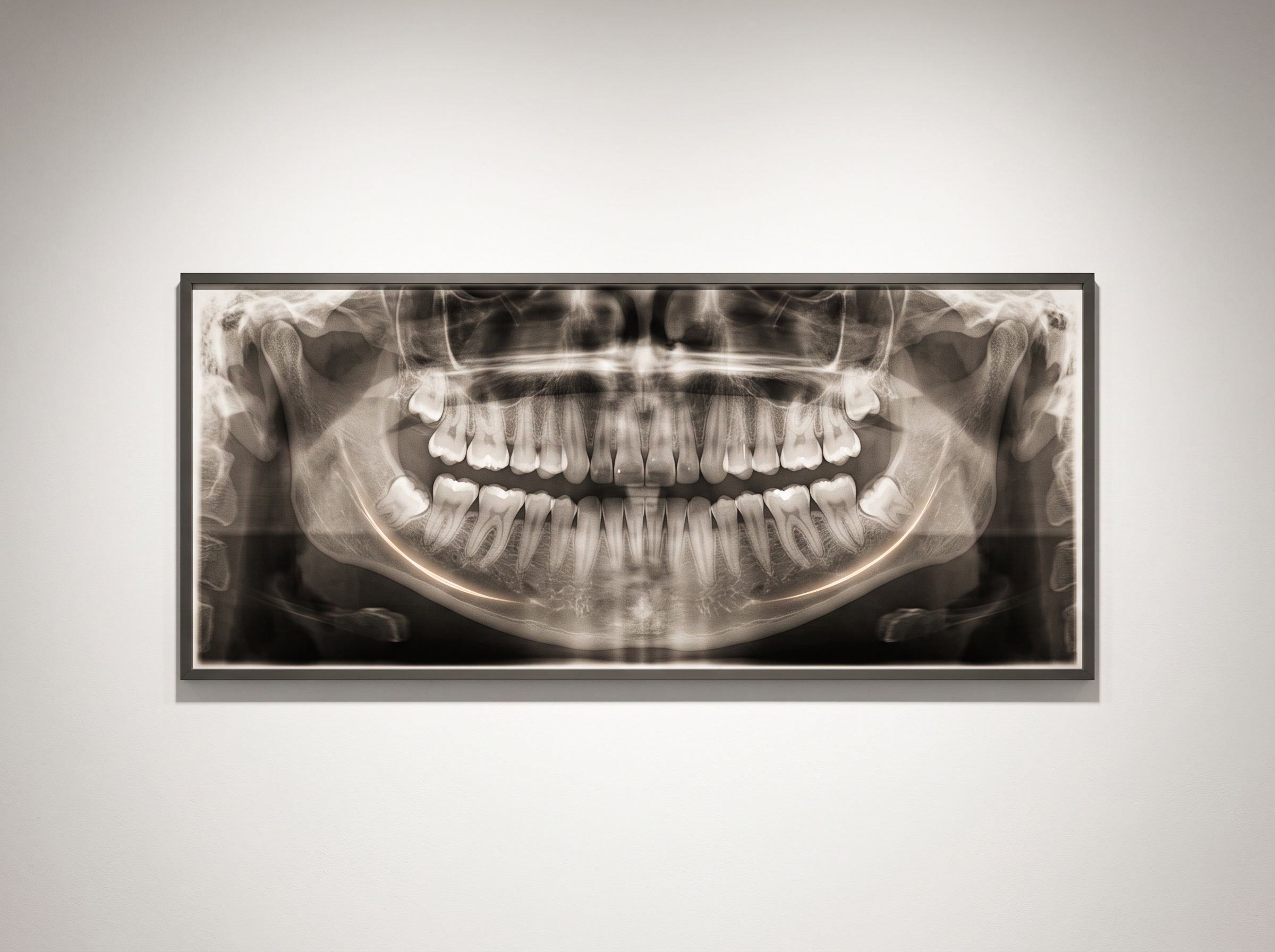
A panoramic radiograph is asked a straightforward question. Survey the arch, count the teeth, check the developing third molars, glance at the sinuses and the condyles – and then it is filed. Yet running along the very bottom of that sweeping horseshoe is a bright, dense ribbon of bone: the inferior cortex of the mandible, the compact outer shell of the lower jaw. No one orders a panoramic to look at it. But that ribbon keeps a quiet second record, because the thickness and the texture of the mandibular cortex track the quality of bone across the whole skeleton. Read with care, the film taken for the teeth can whisper that a patient’s bones are thinning – and that is a message worth hearing, because it usually arrives no other way.
Osteoporosis is a silent disease. It announces itself, most cruelly, with a fracture – a wrist, a hip, a vertebra that gives way under an ordinary load – and by then the loss is long established. The definitive test, dual-energy X-ray absorptiometry, is accurate and widely trusted, but it is a targeted study, not something everyone receives, and plenty of people at risk are never sent for one. Panoramics, by contrast, are taken in enormous numbers for entirely unrelated reasons. That abundance is exactly what makes the mandibular cortex interesting: it is a bone-quality witness already present in millions of images, costing no extra radiation and no extra appointment. This is the same enlargement of vision that lets a dental scan reveal a great deal that lies beyond the teeth themselves – only here the extra reading is not of the airway or the sinuses but of the skeleton’s own condition.
The Cortex at the Bottom of the Frame
Why should the jaw speak for the hip? Because osteoporosis is systemic. As bone mineral density falls, the compact cortical shell everywhere thins and its inner surface, the endosteal margin, loses its clean edge and begins to erode into a porous, moth-eaten border. The lower cortex of the mandible is simply a convenient place to watch that happen: it is thick, consistently positioned, and captured in sharp profile on almost every panoramic. What looks like an incidental strip of white at the edge of the image is, to a trained eye, a legible gauge of how well the body is holding its bone together.
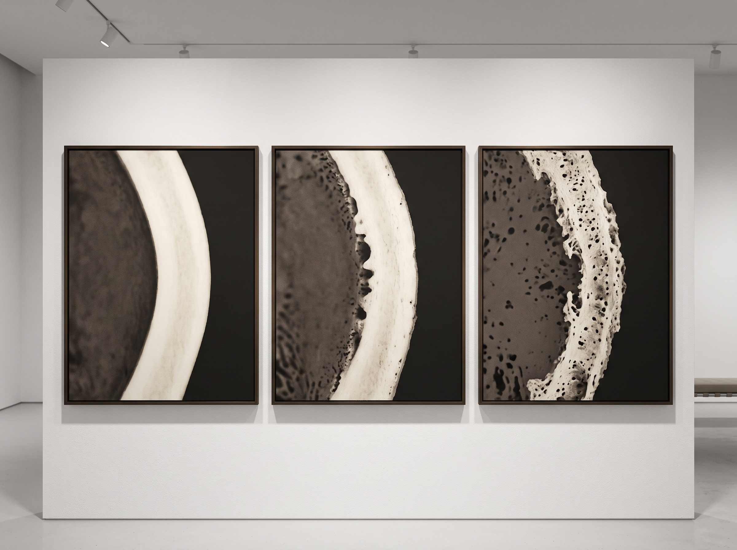
Three Ways to Read a Cortex
Decades of research have distilled that reading into a few reproducible measures – radiomorphometric indices, in the literature’s phrase. The best known is the Mandibular Cortical Index, or Klemetti classification, which grades the inferior cortex into three visual categories: C1, an even cortex with a smooth, sharply defined inner margin; C2, mild to moderate erosion, where the endosteal edge shows semilunar defects and small residues of cortical bone; and C3, a severely eroded cortex that is clearly porous, its margin broken into heavy residues. A C2 or C3 jaw is a candidate for further investigation. The second measure is the Mandibular Cortical Width, sometimes called the mental index – a plain linear measurement of cortical thickness taken at the level of the mental foramen, which thins measurably as density falls. The third, the Panoramic Mandibular Index, refines that into a ratio: cortical thickness divided by the distance from the mental foramen to the lower border, normalising the measurement against the jaw’s own dimensions. Each is a different way of turning a shape into a signal.
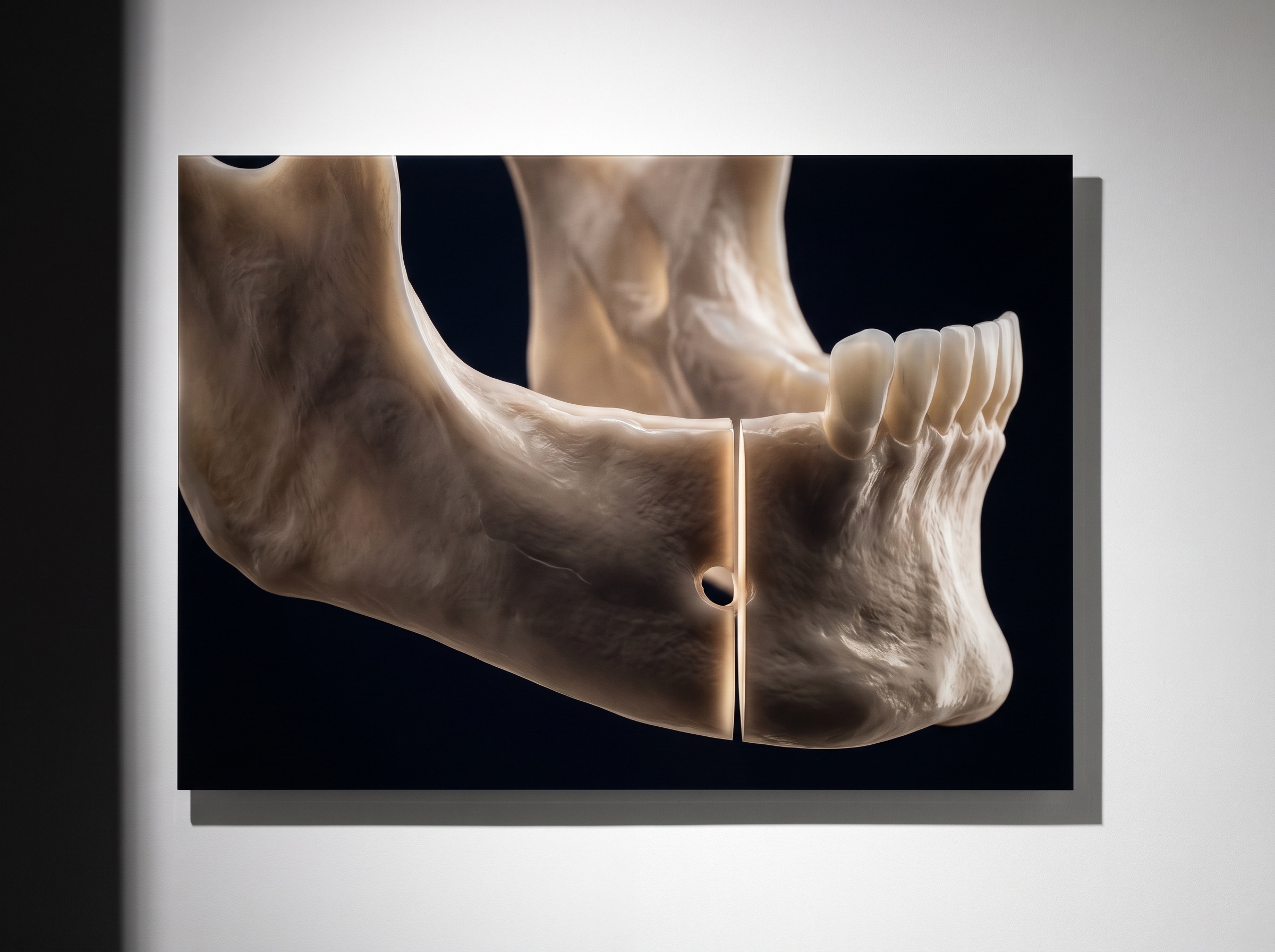
A Triage, Not a Diagnosis
It matters to be precise about what this can and cannot do, because the temptation to overclaim is real. These indices screen; they do not diagnose. A thinned or eroded cortex flags a patient who should be referred for a bone-density scan – it does not, by itself, establish osteoporosis, and absorptiometry remains the standard that confirms or excludes it. The limits run in both directions. Visual grading of the cortex carries genuine inter-observer variability; one clinician’s C2 is another’s C1. Cortical morphology is also shaped by age, by tooth loss, and by the projection geometry and magnification peculiar to panoramic imaging, all of which can nudge a reading. And a normal-looking cortex does not rule the disease out. The honest framing is the useful one: the panoramic identifies people who ought to be tested. It is a triage step performed on an image already justly taken – very much in the spirit of getting more from the studies a patient genuinely needs, rather than exposing them to new ones.
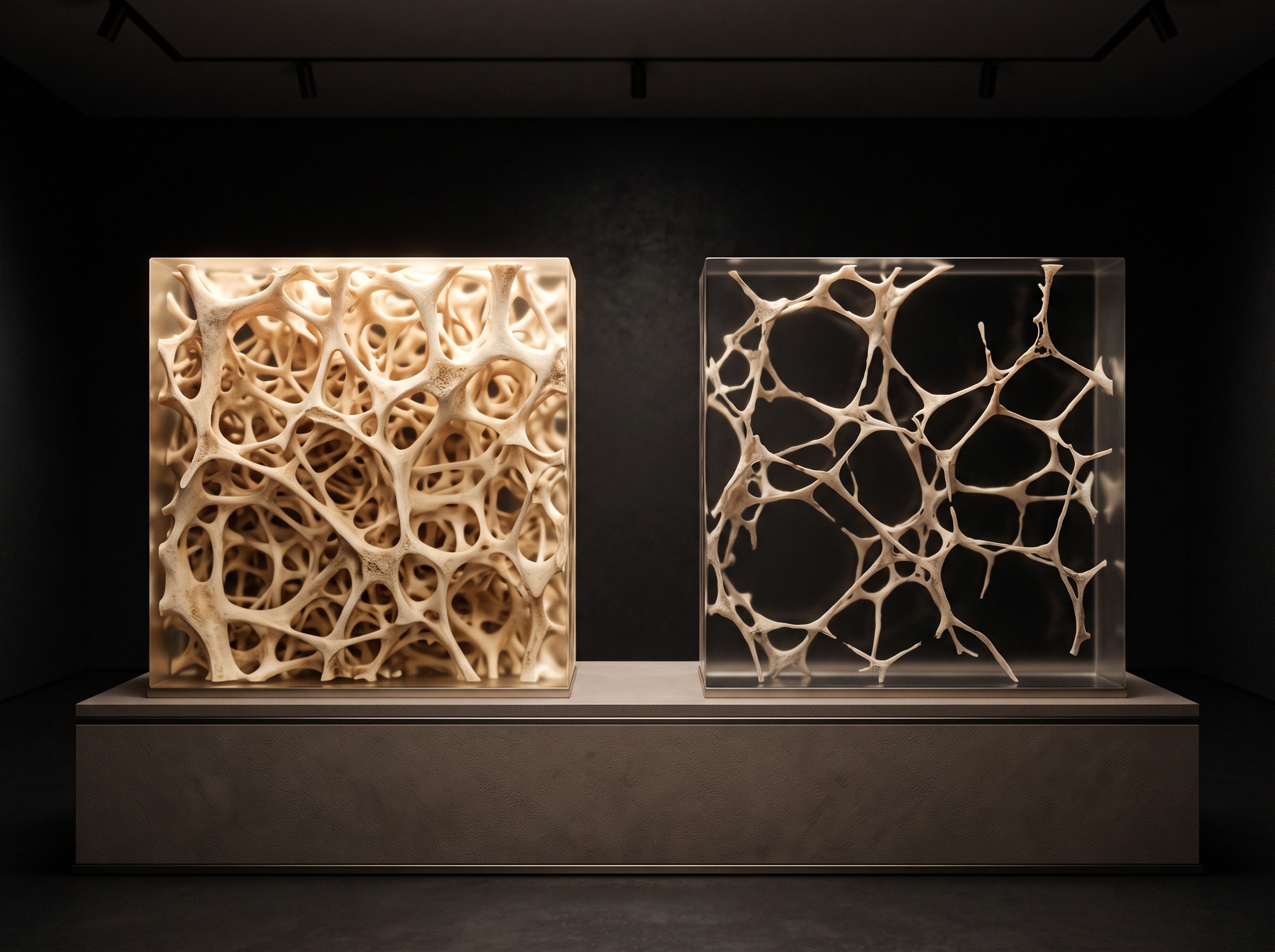
When the Machine Reads the Margin
The weakness of any manual index is that it depends on someone remembering to look, and then looking consistently. This is precisely the kind of task now being handed to software. Convolutional neural networks and radiomic texture analysis can measure cortical width and classify the Mandibular Cortical Index from a panoramic automatically and, crucially, reproducibly – the same image graded the same way every time. The dentomaxillofacial-radiology literature reports deep-learning systems that screen for low bone density directly from the raw panoramic, and radiomics work that quantifies the cortex’s porosity as a continuous number rather than a coarse three-step grade. It is the same competence we have watched a machine bring to other radiographs: the ability to trace precise landmarks on a cephalometric X-ray is not far from the ability to trace the eroding inner edge of a cortex. And because bone loss is a story that unfolds over years, the same tools that let us image change between one radiograph and the next could one day watch a single patient’s cortex thin across a decade of routine panoramics.
Future Developments
The trajectory points toward opportunistic screening that is genuinely automatic – a bone-quality flag computed silently on every panoramic and surfaced only when the cortex crosses a threshold, so the reading no longer depends on anyone remembering to perform it. Tighter integration follows naturally: a flagged jaw linked to a patient’s medical record and to established fracture-risk tools, so that the dental office becomes an unlikely but valuable early sentinel for a disease that dentistry never set out to detect. Further out lies the larger idea this small measurement gestures at – that the dental image is a window onto systemic health, holding traces of far more than the teeth. The panorama, it turns out, has always known more about the patient than we asked it. The craft, as ever at PatientGallery, is learning to read what the image quietly recorded all along.
Sources & further reading:
- Osteoporosis screening support system from panoramic radiographs using deep learning by convolutional neural network (Dentomaxillofacial Radiology, 2022)
- Radiomics-based quantitative assessment of the mandibular cortical index on panoramic radiographs (PMC, 2025)
Sorry, the comment form is closed at this time.