


September 10, 2026 The Art of Seeing Change: Digital Subtraction Radiography and Imaging What the Eye Cannot
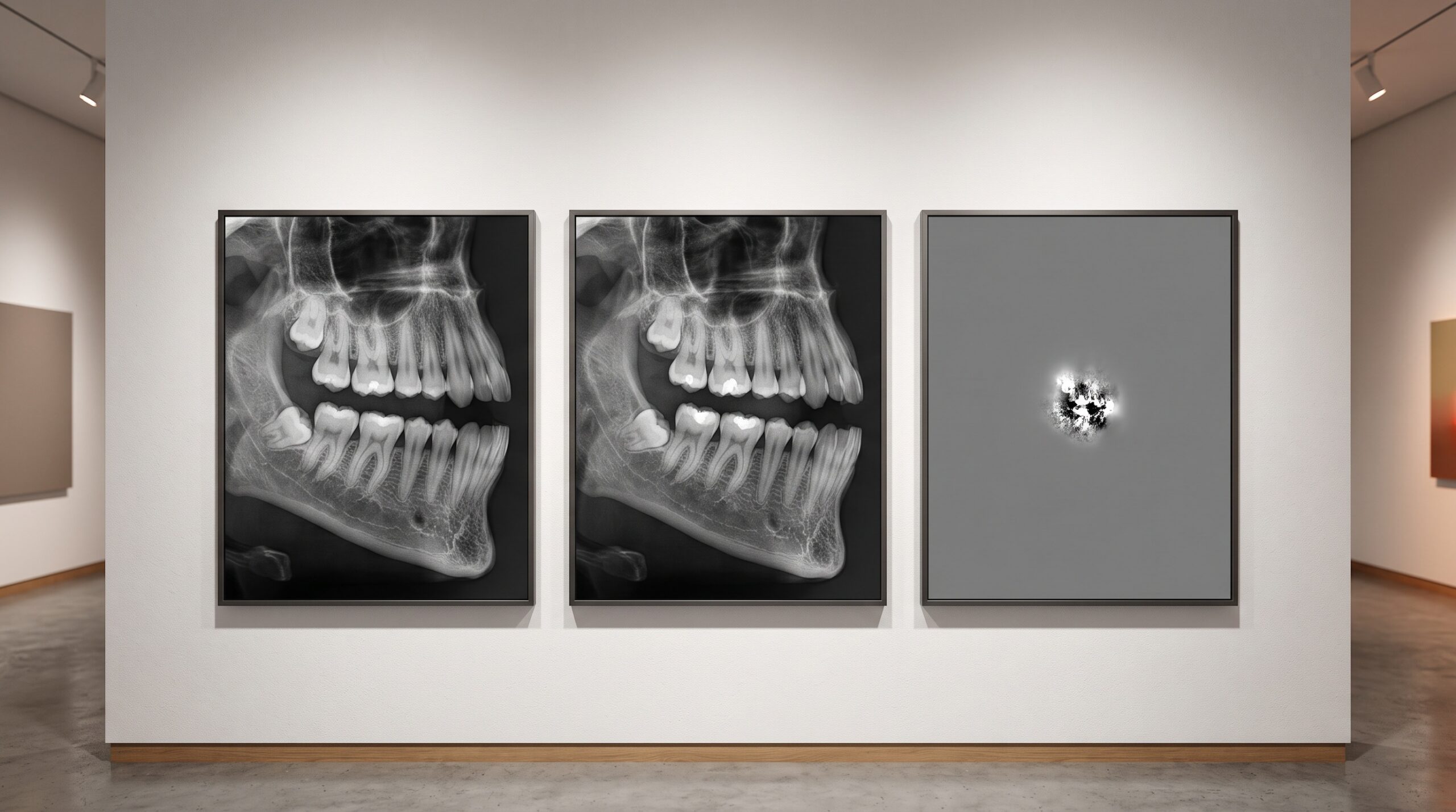
Place two radiographs of the same tooth side by side, taken six months apart, and ask a skilled clinician whether anything has changed. More often than not, the honest answer is that it is impossible to be sure. The eye is a superb detector of edges and patterns, but a poor one for subtle shifts in density spread across a field of bone. By the time alveolar bone loss is unambiguous on a conventional radiograph, a great deal of it – by many estimates a third to a half of the local mineral – is already gone. The disease announces itself only once it has done substantial work. Digital subtraction radiography exists to close that gap, and it does so with an idea of almost austere simplicity: if two images of the same thing are identical except for what changed, then subtract one from the other, and everything that stayed the same will vanish, leaving only the change itself.
When it works, the result is arresting. The unchanged anatomy – the crowns, the roots, the stable bone, the background – cancels to a flat, featureless grey, the visual equivalent of silence. Against that silence, the only thing left with any tone is the region that gained or lost density between the two visits: a bright patch where bone was deposited, a dark one where it was resorbed. A process that was invisible because it was buried in a busy image becomes the only thing in the frame. It is one of the most sensitive non-invasive tools in all of radiographic imaging, capable of revealing change when only a few percent of bone per unit volume has shifted. And its entire difficulty is contained in a single, unforgiving word: alignment.
Why Subtraction Is Really a Problem of Alignment
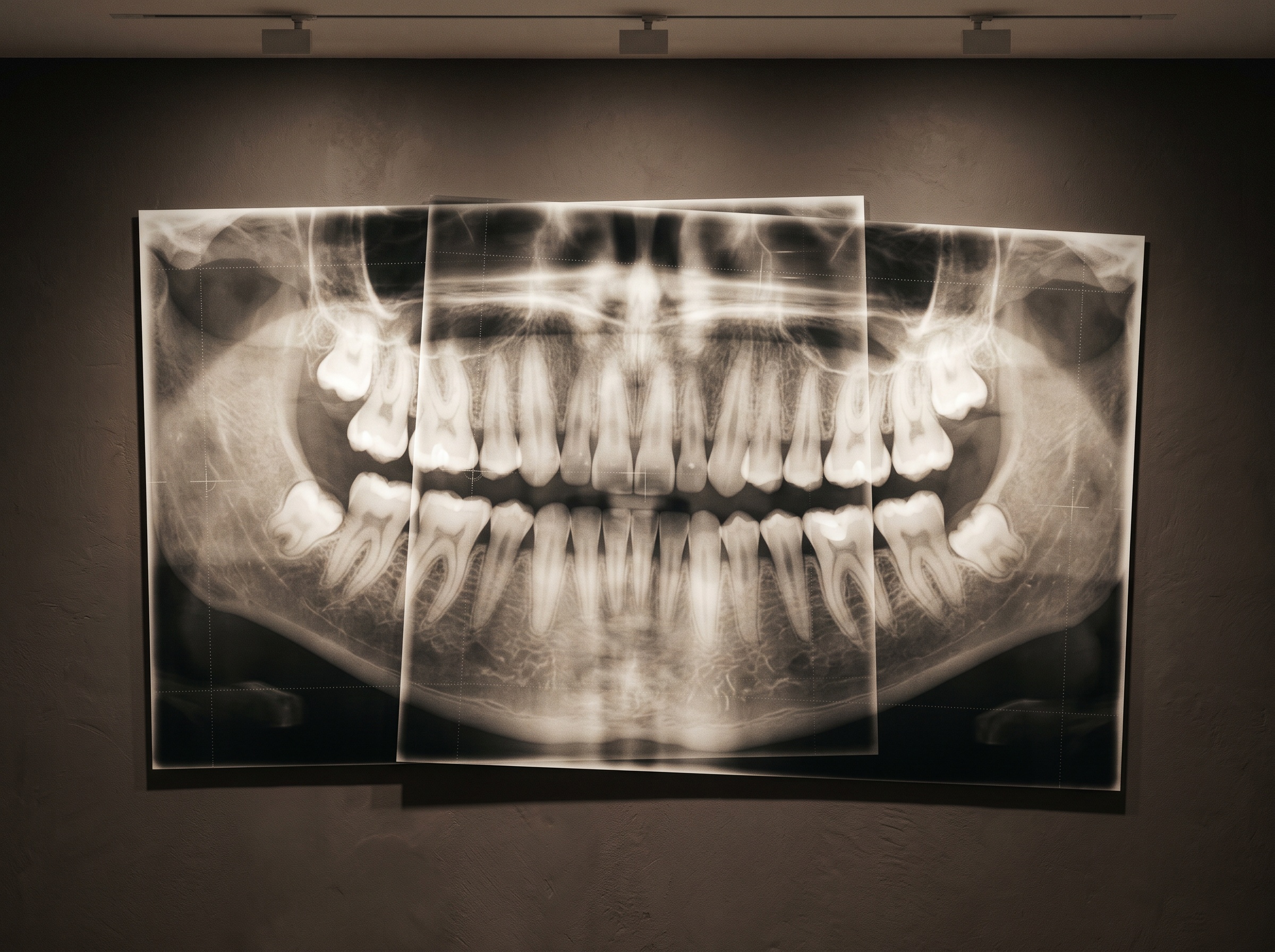
The mathematics of subtraction assumes the two images are perfectly superimposed, pixel for pixel. In practice they never are, not naturally. The patient sat down a second time months later; the sensor went in at a slightly different angle; the tube was a degree off; the head was tilted a fraction. Each of those tiny discrepancies means that a given pixel in the first image and the pixel beneath it in the second are no longer looking at the same piece of anatomy. Subtract misaligned images and the technique turns treacherous: the edges of every root and every restoration fail to cancel, throwing off bright and dark fringes of false ‘change’ that have nothing to do with biology and everything to do with geometry. This misregistration artifact is the central enemy. It can be large enough to bury the real signal entirely, which is why a casually produced subtraction image is worse than useless – it is actively misleading. The whole art, then, is the craft of reproducible geometry, the same honest, repeatable projection geometry that every good radiograph depends on, raised here to an almost absolute standard.
The Historical Answer: Hold the Geometry Still
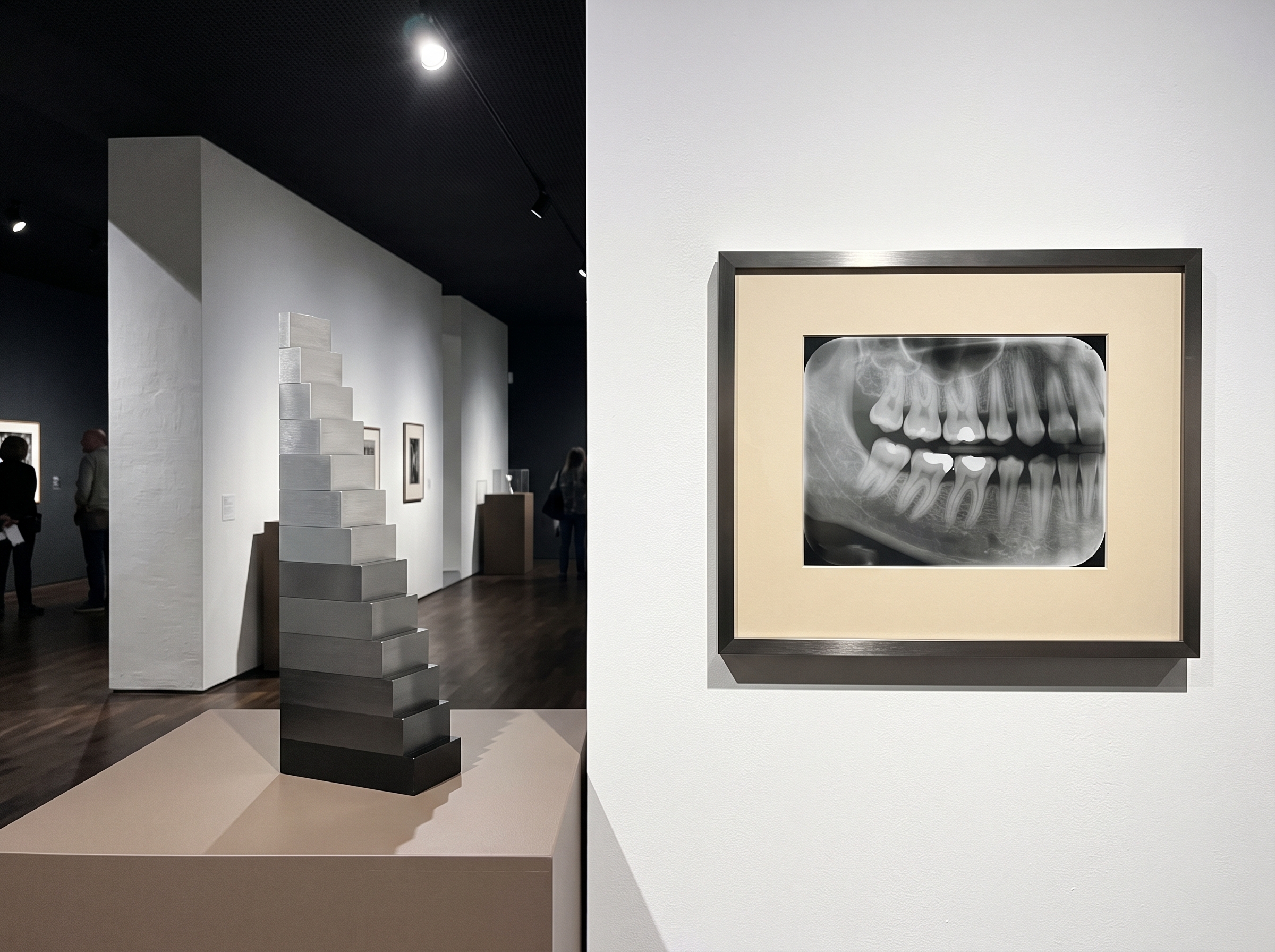
The first solutions were mechanical and uncompromising. The foundational work on DSR for monitoring bone – the longitudinal peri-implant validation carried out by Jeffcoat and Reddy in the early 1990s – leaned on rigid fixation of the projection geometry so that the second image would reproduce the first as faithfully as possible. Bite blocks keyed to individual teeth, customised stents, and in the most exacting cephalostat-stabilised setups, a head-holding frame that returned the patient to within a fraction of a degree of the original position: reported angular disparities on the order of a third of a degree. The more rigidly the geometry was reproduced, the cleaner the cancellation and the more trustworthy the surviving signal; stabilised projections reliably out-performed looser, stent-only approaches at revealing simulated lesions. To read the change quantitatively rather than merely spot it, a calibrated aluminium step-wedge was exposed alongside the tooth – a staircase of known thicknesses giving a reference ladder of grey. It let the software correct for differences in exposure between the two visits and convert a difference in grey level into an estimate of the actual mass of bone gained or lost. The subtraction stopped being a picture and became a measurement.
Sensitive Enough to Matter, Too Demanding to Spread
The payoff was real. For monitoring the bone around an implant – where the earliest marginal remodelling is precisely the signal a clinician most wants and least reliably sees – quantitative subtraction could flag density changes far too subtle for visual reading, offering an early, objective read on integration and peri-implant health. In periodontal research, it tracked the slow advance or arrest of lesions with a precision no naked-eye comparison could match. And yet DSR never became a routine chairside tool. The reason is the same word again: alignment. The geometric discipline it demanded – cephalostats, custom stents, meticulous exposure matching – was feasible in a research clinic and impractical in a busy general practice. The sensitivity was never in doubt; the reproducibility was the barrier. A technique is only as clinically useful as it is easy to do correctly, and for decades DSR was extraordinarily easy to do incorrectly. Its sensitivity also presumes a clean, well-exposed underlying image to begin with; subtracting two noisy or poorly exposed radiographs only subtracts their flaws into a messier result.

The Software Revival: Warping Geometry Back Into Agreement
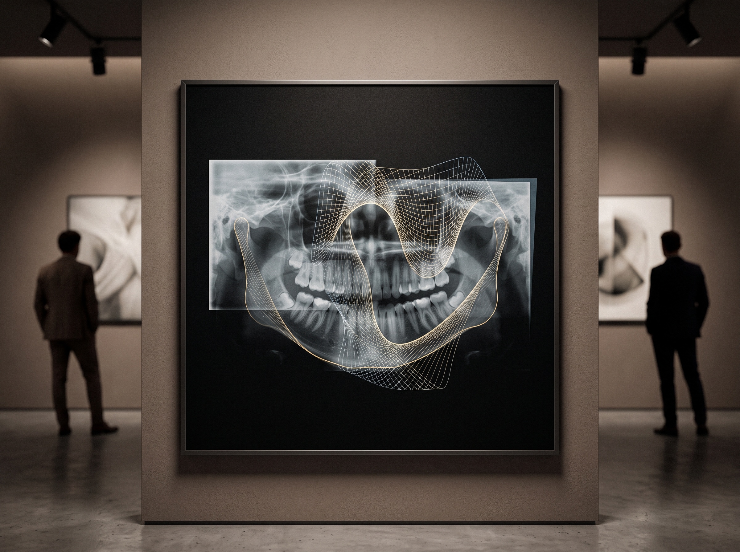
What has changed is that the alignment no longer has to happen at the moment of exposure. Fully digital sensors put both images into a form software can manipulate directly, and registration algorithms can now take a second image that was acquired at a slightly wrong angle and warp it – translating, rotating, and subtly stretching it – until its stable landmarks lie exactly over those of the first. In effect, the geometry is reproduced after the fact, in the computer, rather than demanded of the patient and the operator in the room. This retrospective registration is what lifts the heaviest part of the old burden. The reference wedge and the principle of density calibration remain valuable, but the punishing requirement to physically reproduce a projection to within a fraction of a degree is relaxed, because the software closes the remaining gap.
When the Alignment Becomes Automatic
The current frontier hands even that step to a machine. Recent work on artificial-intelligence techniques for automatically detecting peri-implant marginal bone remodelling in intraoral radiographs points toward a workflow in which registration, change detection, and quantification happen without a human manually matching landmarks at all. An algorithm identifies the stable structures, aligns the serial images, isolates the region of change around an implant neck or a periodontal defect, and reports the magnitude – the difficult, error-prone craft of alignment absorbed into software and offered back as a finished result. It is of a piece with the broader movement toward AI that now reads and reports radiographs: the value is not a prettier image but a reliable, repeatable measurement of something the eye cannot be trusted to judge. The old trade-off – exquisite sensitivity purchased with impractical discipline – is precisely the trade-off automation is built to dissolve.
Future Developments
If automatic registration holds up in ordinary clinical conditions, digital subtraction radiography may finally become what its sensitivity always deserved: not a specialist research instrument but a routine chairside habit. Imagine every recall radiograph quietly subtracted against the patient’s prior image of the same site, with the software surfacing only the regions that genuinely changed and a number attached to each – bone lost here, bone gained there, stability everywhere else. The recall would shift from a clinician straining to remember how a tooth looked last year to an objective, longitudinal record of what has actually moved. There is an aesthetic truth buried in the technique as well. The subtraction image is the rarest kind of picture in all of imaging: one composed almost entirely of nothing, a deliberate field of grey silence whose only content is difference. It is imaging stripped to its most honest purpose – not to show what is there, which any exposure can do, but to show what has changed, which is usually the only thing a clinician truly needs to know. Teaching a machine to make that image reliably, from images taken carelessly months apart, is how one of imaging’s most elegant ideas finally steps out of the laboratory and into the operatory.
Sources & further reading:
Sorry, the comment form is closed at this time.