


September 13, 2026 One Mouth, Two Truths: The Craft of Fusing an Intraoral Scan With a Radiograph
Point an intraoral scanner at a tooth and you receive something close to perfect: a dense, colour-accurate surface, every ridge and stain and marginal line recorded in three dimensions. And then it stops. The scan knows the crown exactly and the root not at all; it sees the world of light, and light does not pass through enamel. Take a radiograph or a cone-beam volume of the same tooth and you receive the opposite gift – the root, the surrounding bone, the dark whisper of a lesion no eye could find – rendered by radiation that passes straight through the surface and so barely records it. Each image is true. Each is blind to precisely what the other sees best. The interesting work of modern dental imaging is not choosing between them; it is bringing the two into a single frame so exactly that they cease to be two images and become one picture of one mouth. Medit’s recent launch of a unified 3D intraoral-scan and X-ray visualization is only the latest sign that this fusion is becoming ordinary – which makes it a good moment to look at the craft underneath it.
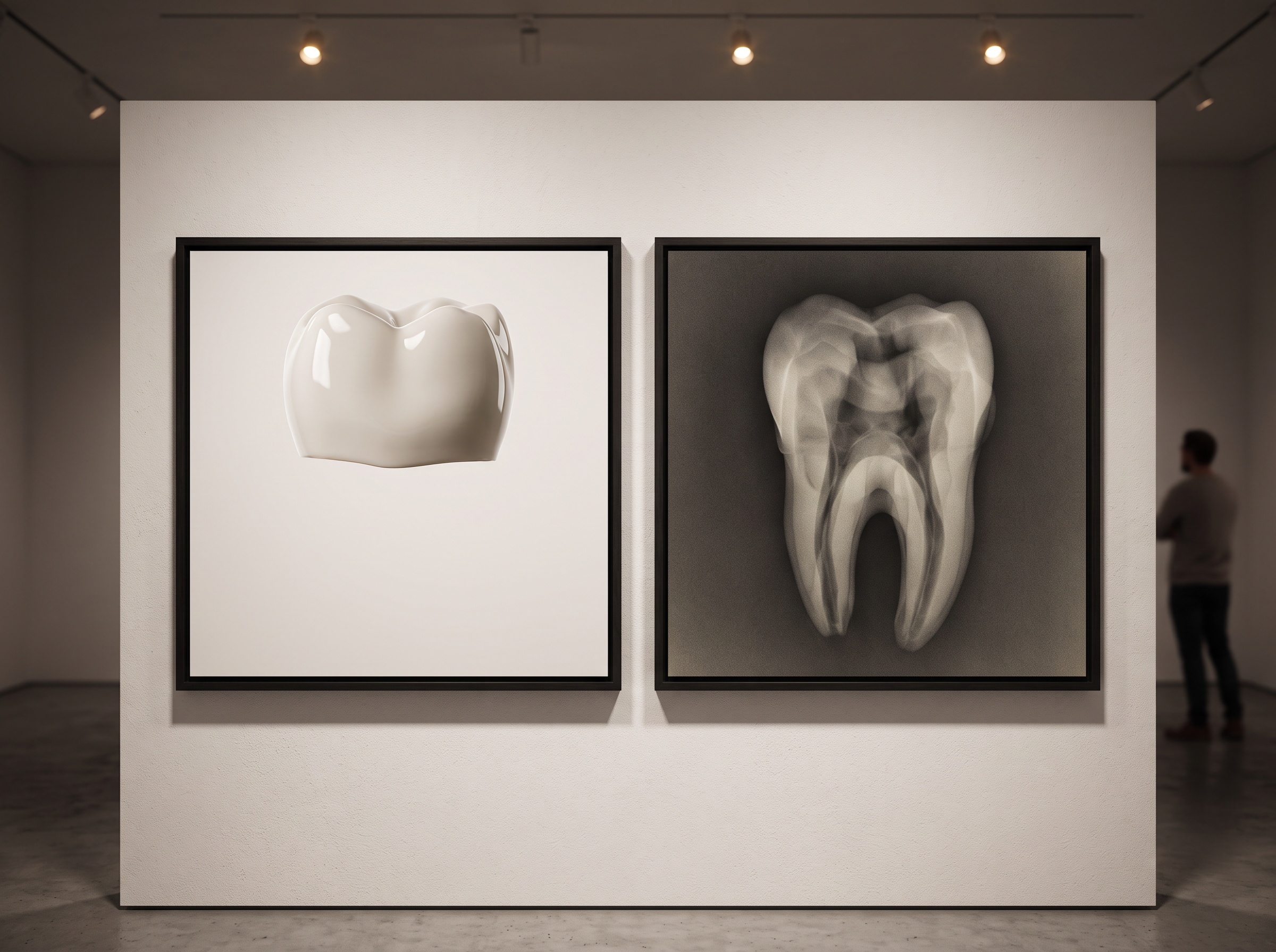
Start with the asymmetry, because everything follows from it. An optical scan is a surface mesh: millions of points describing where the outside of the tooth is, in colour, and nothing about what lies beneath. A radiographic dataset is a transmission image – or, in CBCT, a full volume of densities – that describes the interior beautifully and the true surface poorly or not at all. Neither is a complete portrait of the tooth. Fused correctly, they approximate one. The scanner supplies the skin; the volume supplies the skeleton; the fusion supplies the body.
Fusion Is Really a Problem of Registration
To lay one dataset over the other is not a matter of dragging two pictures together by eye. Each exists in its own coordinate space, captured by a different machine at a different moment, and the task is to find the single transformation – a rotation and a translation in three dimensions – that carries one perfectly onto the other. That is registration, and it is the whole game. Get the transformation right to a fraction of a millimetre and the surface mesh settles onto the volumetric data like a glove; get it wrong and you have two convincing images of a tooth that no longer agree about where that tooth is. This is the same fundamental discipline explored in digital subtraction radiography, where two images of the same site must be aligned before their difference means anything – fusion and subtraction are siblings, both built on the quiet precision of getting two pictures into the same frame.
Why Teeth Are Nearly Perfect Anchors
Registration succeeds or fails on the quality of its anchors – the features both datasets share and can be matched against. Here dentistry is quietly fortunate. Teeth are rigid: unlike soft tissue, a crown does not deform between the optical scan and the radiograph, so a single rigid-body transformation is enough to describe how the two align. Teeth are also uniquely shaped, no two arches quite alike, which gives an algorithm plenty of distinctive geometry to lock onto. And crucially, the crown appears in both worlds – as the enamel surface the scanner captured directly, and as the radiodense outline of that same enamel in the volume. The shared feature is real, not inferred. The mesh that the scanner builds from structured-light or confocal capture, stitched from thousands of frames, becomes the moving object; the crown surfaces extracted from the CBCT become the fixed target it is drawn toward.
The mechanics, stripped of jargon, are almost intuitive. Identify corresponding surfaces in both datasets. Let an algorithm of the iterative-closest-point family nudge one cloud of points against the other, at each step pairing every point with its nearest neighbour in the target and shifting to shrink the total distance between them, then repeating until the movement dwindles to nothing. When it converges, the surface mesh sits exactly atop the volumetric bone and root, and the two datasets finally speak in one coordinate language.
The Honest Difficulties
None of this is as clean in the mouth as it is on the page. The most common saboteur is the CBCT itself: metal restorations, posts, and implants throw scatter and streak artefact across the volume, and that glare falls precisely on the crown surfaces the registration wants to use as anchors. Corrupt the anchor and you corrupt the alignment. Patient motion is a second enemy – move between or during captures and the rigid-body assumption quietly breaks, because the tooth the scanner saw is no longer in the pose the volume recorded. And soft tissue must be treated with suspicion throughout: gingiva is not rigid, it is squeezed by the scanner tip and drifts between visits, so it can never serve as a trustworthy landmark. The discipline, as ever in this field, is knowing which parts of the image to believe. That judgement rests on the same foundation as everything else in imaging – if either source is degraded, the fusion inherits the flaw, a principle that runs through why image quality sets the ceiling on what any downstream process can achieve.
What the Fused Image Lets You See
When it works, the payoff is both clinical and, in PatientGallery’s sense of the word, aesthetic. An implant can be planned against the real bone volume while the exact soft-tissue emergence profile – captured optically, in colour – is visible in the same view, so the surgeon designs the restoration and the osteotomy against a single coherent object rather than juggling two screens. A restoration’s margin can be judged against the root and crestal bone beneath it. The whole tooth, surface and interior together, becomes legible at once – and the radiograph’s habit of revealing what lies beyond the teeth themselves is finally reunited with the surface reality the eye actually treats. For any of this to happen, of course, the two datasets have to reach the same software in compatible form, which is why the unglamorous plumbing of DICOM interoperability and open image exchange matters as much as the registration mathematics.
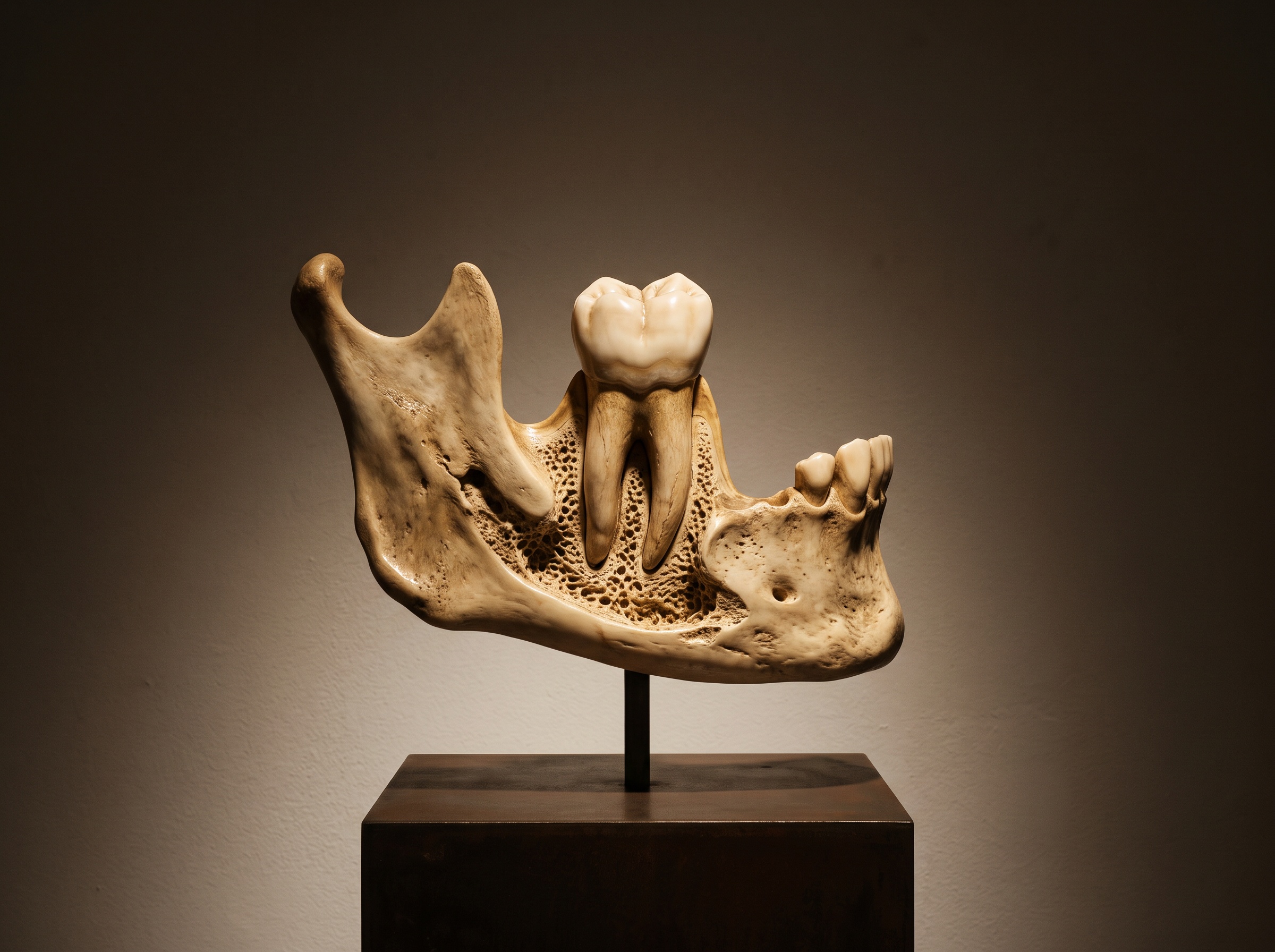
Future Developments
The direction of travel is toward fusion that is automatic, instant, and eventually radiation-free. Feature-based registration driven by machine learning is already replacing the manual selection of corresponding points, letting a network recognise the same anatomy in both datasets and propose an alignment in seconds rather than minutes – and doing it more robustly in the presence of artefact than a naive closest-point search can. Real-time fusion during the scan itself is the natural next step: the clinician watches the surface build up over a pre-loaded volume and sees the composite form as the wand moves. Deformable registration, which allows controlled, non-rigid warping, will begin to bring soft tissue honestly into the picture rather than excluding it. And further out sits the most elegant prospect of all – pairing the optical surface not with an X-ray but with a radiation-free volumetric source such as dental MRI, a composite portrait of the whole tooth built without a single photon of ionising radiation. Whatever the source, the essential act will remain what it is today: the reconciliation of two partial truths into one. The fused image is not a trick of the software. It is a new artefact of seeing – and, like the best of this craft, it is most convincing exactly when it stops looking like two things at all.
Sources & further reading:
Sorry, the comment form is closed at this time.