


August 5, 2026 What Your Dentist Actually Sees on Your X-Ray – and the 130 Years That Made It Possible
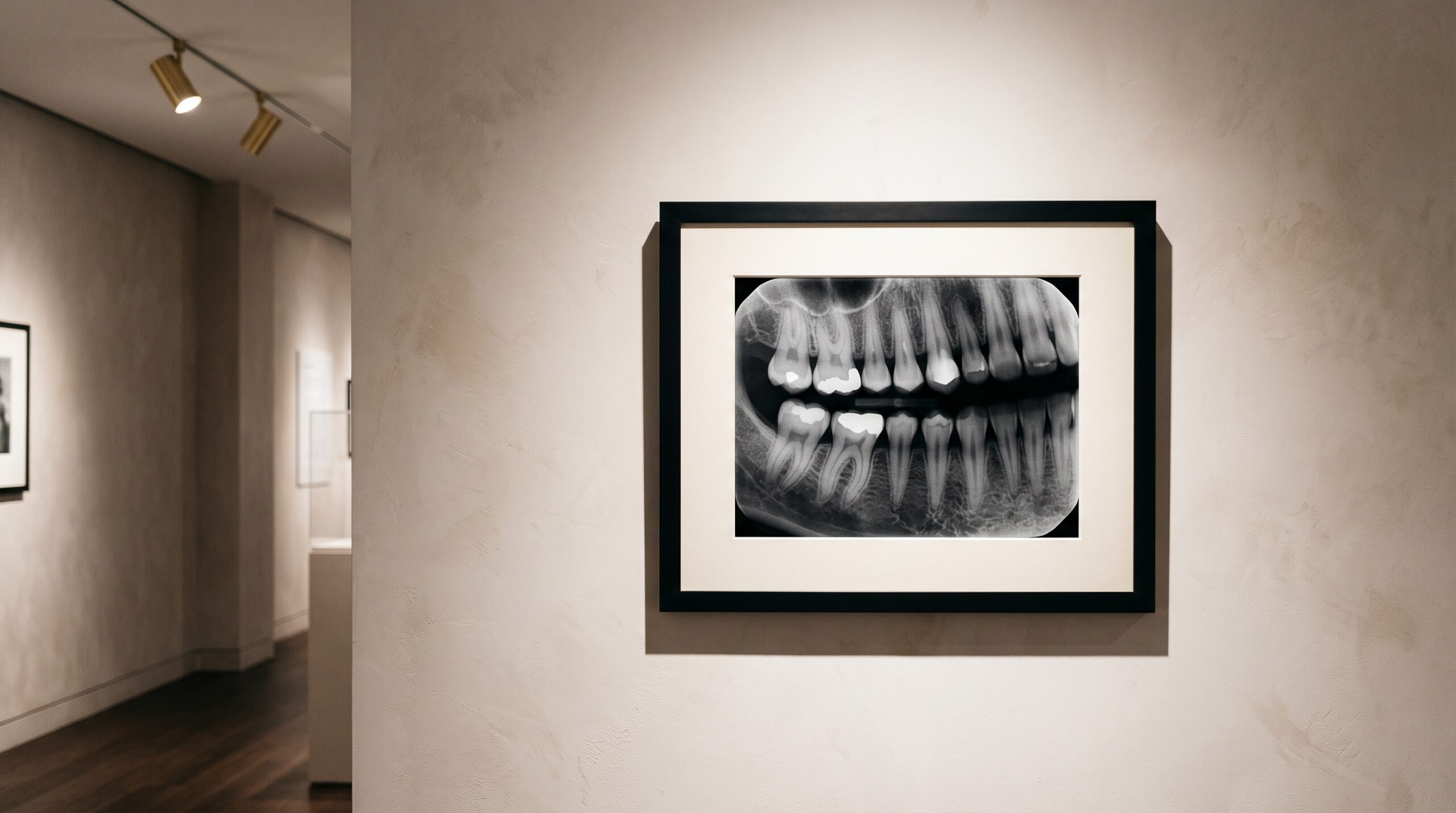
The dentist tilts the monitor, studies a small grey rectangle for a few seconds, and says something reassuring – or not. To you it looks like a smudged weather map. To a trained eye it is one of the most information-dense images in all of medicine, read in a visual language refined over a century and a third. This is a short guide to that language: what is actually on your dental X-ray, and how to look at it the way your dentist does.
Why it is grey, and what the greys mean
An X-ray image is a shadow picture. The machine sends a brief, invisible beam through your jaw to a sensor on the far side. Dense tissues – enamel, bone, metal fillings – absorb much of the beam, so little reaches the sensor there and those areas render pale, even bright white. We call them radiopaque. Softer or damaged tissue lets more of the beam pass, so more reaches the sensor and those areas render dark. We call them radiolucent.
That single rule is the whole grammar. Healthy enamel glows. A cavity, where the mineral has been eaten away, lets the beam through more easily and shows up as a darker shadow within the tooth. An infection at a root tip appears as a dark blot in the bright bone. Once you know that pale means dense and dark means loss, the smudged weather map starts to resolve into sentences.
The views, and the question each one answers
Your dentist does not order “an X-ray.” They choose a view to answer a particular question.
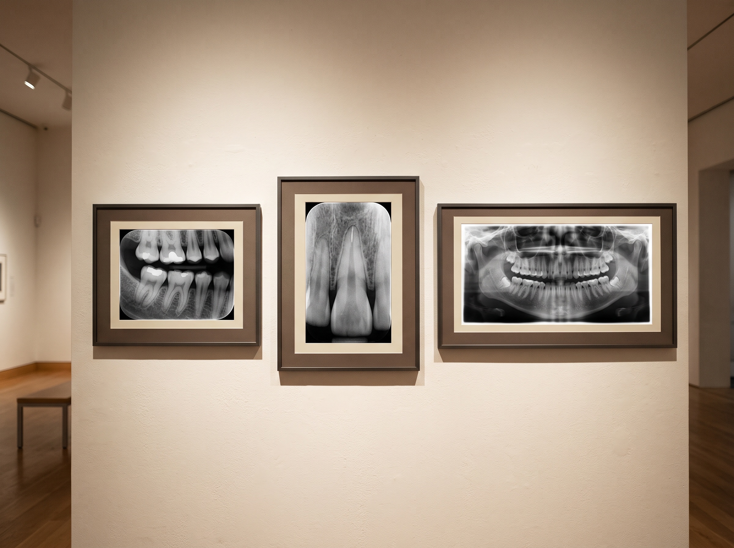
The bitewing – the one where you gently bite a small tab – frames the crowns of the upper and lower back teeth together. It is the specialist for two jobs: catching decay hidden between teeth, where no mirror or explorer can reach, and reading the level of the supporting bone. We devoted a whole piece to reading the bitewing, because dentistry’s most ordered image rewards a careful eye. The periapical view shows a whole tooth from crown to the very tip of its root and the bone around it – the image of choice when a specific tooth aches or an infection is suspected. The panoramic sweeps in one broad curve across the entire jaw, capturing every tooth, both joints, the sinuses and any teeth still buried in bone. It trades fine detail for the big picture: wisdom teeth, cysts, the overall architecture.
What the trained eye is hunting for
In those few quiet seconds, a dentist runs through a mental checklist far longer than most patients imagine.
First, decay between teeth – the interproximal cavities that are invisible to eye and mirror until they are large, but which announce themselves early as faint dark notches on a radiograph. Then bone levels: in health the bone rises to just below where the teeth emerge; when gum disease has eroded it, that line recedes, and the image measures the loss the eye cannot. They look for the dark halo of an abscess at a root tip, for impacted or extra teeth, for cysts and other shadows in the bone that should not be there. They check the fit of existing work – whether a filling or crown seats cleanly or hides a gap where new decay can start – and they note hardened deposits of calculus clinging beneath the gumline. One image, a dozen questions, answered almost at a glance.
One hundred and thirty years to make it look easy
That glance rests on an astonishing history. When Wilhelm Roentgen discovered the X-ray in 1895 – the accidental breakthrough we trace in The Invisible Light – dentists seized on it within weeks. The first dental radiographs were made on glass plates and, later, film, with exposure times measured in agonizing minutes and images developed by hand in a darkroom.
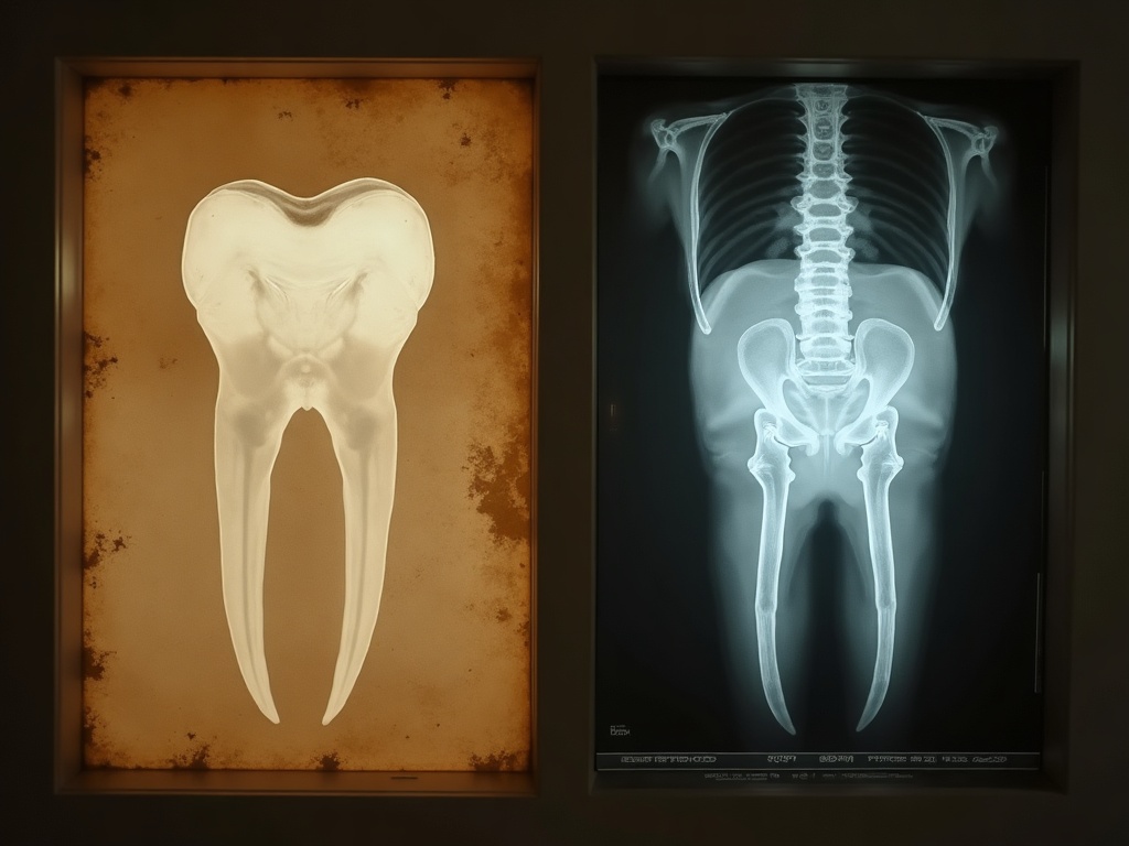
From there the craft advanced steadily: faster films that cut exposure and dose, standardized positioning, and finally the leap to digital sensors that turn the invisible beam into a picture on a screen in less than a second. The whole arc from that first glass plate to today’s instant image is a story we tell in full in our history of dental X-rays. What matters to you in the chair is the payoff: today’s dentist can zoom in, adjust the contrast, and measure a bone level to a fraction of a millimeter, all from an image made with a fraction of the radiation your grandparents received.
Where your image lives afterward
A digital radiograph is also a part of your medical record, and it is guarded like one. The moment it is captured it is filed to your chart in software built for privacy: access is limited to your care team, activity is logged, and the data is encrypted so it stays unreadable if a device is lost or a network is prowled. Nothing has to travel on a fragile glass plate or in a paper envelope; the image moves – to a specialist, or to your own patient portal – only when your care calls for it, over secure channels. The same century of progress that sharpened the picture also built the quiet infrastructure that keeps it yours.
Future developments
The craft is still moving. Artificial intelligence is beginning to sit alongside the dentist as a second reader, flagging faint early lesions and subtle bone changes for a human to confirm. Three-dimensional cone-beam scans are turning the flat shadow picture into a rotatable model of the jaw for complex cases. And each generation of sensor squeezes a clearer image from a smaller dose. The direction is clear: more information, more certainty, less radiation – the same invisible light Roentgen found by accident, read with ever greater artistry. The next time that grey rectangle appears on the monitor, you will know it for what it is: not a smudge, but a portrait, and one your dentist has spent years learning to read.
Sources & further reading:
The Story of the X-Ray, Across Our Network
The 130-year story behind your X-ray, told from other angles:
- How Marie Curie drove X-ray trucks into the First World War
- The equipment history from gas tubes to digital sensors
- From Roentgen’s lab to the practice server, as an IT discipline
Sorry, the comment form is closed at this time.