
14 Feb From Röntgen’s Discovery to Digital Imaging: The History of X-rays in Dentistry
When Wilhelm Conrad Röntgen accidentally discovered a mysterious new form of radiation in his Würzburg laboratory on November 8, 1895, he could never have imagined the profound impact his finding would have on dentistry. What began as a curious observation of fluorescent screens glowing in a darkened room would revolutionize how dental professionals diagnose and treat oral health conditions. Today, X-ray technology remains one of the most indispensable tools in every dental practice.
The Accidental Discovery That Changed Medicine
Röntgen was experimenting with cathode ray tubes when he noticed that a nearby fluorescent screen began to glow, even though it was shielded from the tube’s direct light. Intrigued, he spent weeks investigating this unknown radiation, which he dubbed “X-rays” — the “X” standing for the unknown. His first famous X-ray image, showing the bones of his wife Anna Bertha’s hand complete with her wedding ring, captivated the world when it was published in January 1896.
The medical community immediately recognized the diagnostic potential. Within weeks of Röntgen’s announcement, physicians and dentists around the world were experimenting with X-ray equipment. The speed at which this technology was adopted is remarkable — no regulatory hurdles, no clinical trials, just pure scientific enthusiasm driving immediate practical application.
Dentistry’s Early Adoption of X-rays
Dentistry was among the first medical specialties to embrace X-ray technology. In January 1896 — just weeks after Röntgen’s discovery was published — Dr. Otto Walkhoff of Braunschweig, Germany, made the first dental radiograph. He placed a glass photographic plate wrapped in rubber in his own mouth and exposed it to X-rays for an astonishing 25 minutes. The resulting image, though crude by modern standards, showed the outlines of his teeth and surrounding bone structure.
Shortly thereafter, Dr. C. Edmund Kells of New Orleans became one of the first American dentists to use X-rays clinically. Kells demonstrated dental radiography at a meeting of the Southern Dental Association in July 1896, stunning his colleagues with the ability to see inside a patient’s jaw without surgery. Tragically, Kells’ extensive unprotected exposure to radiation over the years led to progressive radiation injuries, and he eventually lost fingers and then his hand to radiation-induced cancer — a sobering reminder of the dangers that early pioneers faced.

The Wild West of Early Dental Radiography
The early decades of dental X-ray use were marked by both remarkable innovation and alarming recklessness. Exposure times in the early 1900s could range from several minutes to over twenty minutes for a single image. Patients and practitioners alike received radiation doses hundreds of times greater than what modern equipment delivers. There were no lead aprons, no thyroid collars, and no concept of “as low as reasonably achievable” (ALARA) radiation exposure.
Despite these dangers, the diagnostic benefits were undeniable. For the first time, dentists could detect cavities between teeth, identify infections at the root tips of teeth, evaluate bone loss from periodontal disease, and locate impacted wisdom teeth — all without exploratory surgery. X-rays transformed dentistry from a largely reactive profession into one capable of early detection and preventive planning.
By the 1910s and 1920s, dental X-ray equipment became more compact and purpose-built. The development of intraoral film packets — small, light-tight film holders that could be placed directly inside the mouth — made the process faster and more practical. Companies like Eastman Kodak began manufacturing standardized dental film, bringing consistency and reliability to dental radiography.
Mid-Century Advances: Panoramic Imaging and Safety Standards
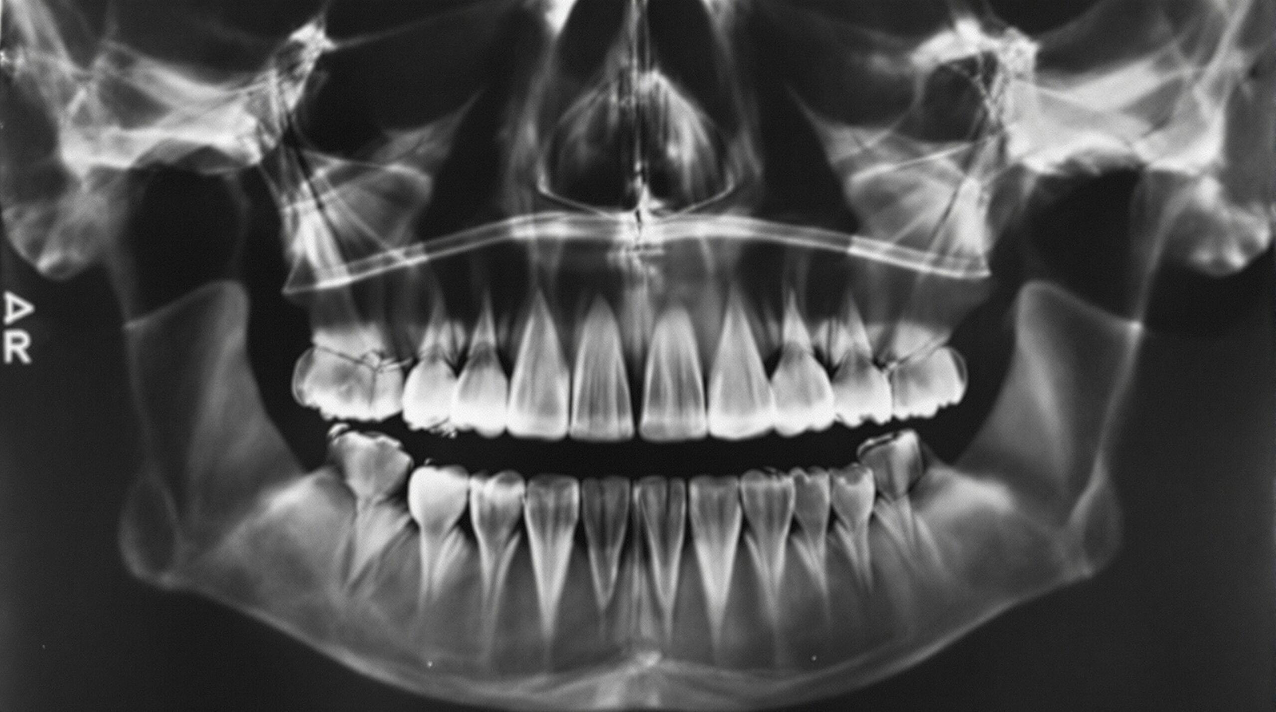
The mid-20th century brought two transformative developments to dental radiography: panoramic imaging and radiation safety standards. In 1949, Finnish researcher Yrjö Paatero developed the concept of panoramic radiography, which could capture the entire jaw in a single image. By the 1960s, commercial panoramic X-ray machines were becoming available in dental offices, offering a comprehensive view of all teeth, the jawbones, sinuses, and temporomandibular joints in one exposure.
Simultaneously, the growing understanding of radiation biology led to the establishment of safety protocols. The use of lead aprons became standard practice. Film speeds improved dramatically — what once required minutes of exposure could now be accomplished in fractions of a second. Collimation techniques narrowed the X-ray beam to expose only the area of diagnostic interest, and rectangular collimation further reduced patient dose.
The introduction of rare earth intensifying screens in the 1970s and faster film emulsions continued to drive radiation doses downward while improving image quality. Dental professionals also began adopting standardized techniques like the paralleling method, which produced more geometrically accurate images and reduced the need for retakes.
The Digital Revolution in Dental Imaging
The most dramatic transformation in dental radiography began in the late 1980s when Dr. Francis Mouyen developed the first direct digital dental X-ray sensor, marketed as the RadioVisioGraphy (RVG) system by the French company Trophy Radiologie in 1987. Instead of exposing photographic film, digital sensors converted X-rays directly into electronic signals that could be displayed on a computer monitor within seconds.
Digital radiography offered numerous advantages over traditional film. Radiation doses were reduced by up to 80 percent compared to D-speed film. Images appeared instantly — no more waiting for film processing in darkroom chemicals. Dentists could enhance, magnify, and adjust contrast on digital images to extract maximum diagnostic information. Images could be stored electronically, shared instantly with specialists or insurance companies, and never degraded over time.
Two main types of digital sensors emerged: charge-coupled device (CCD) sensors and photostimulable phosphor (PSP) plates. CCD sensors (and later CMOS sensors) provided instant images but required a cable connection to the computer. PSP plates offered a more film-like workflow — they could be placed in the mouth like traditional film and then scanned in a desktop reader — but required an additional processing step.
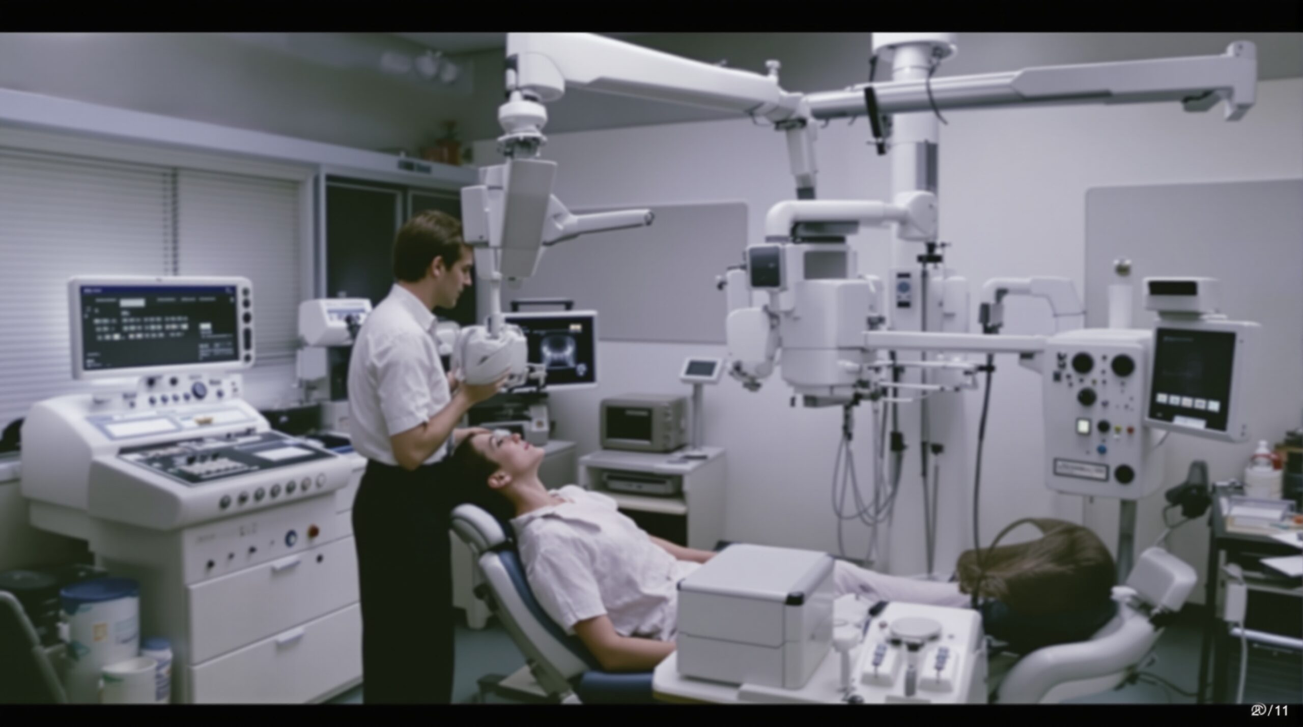
Cone Beam CT: Three-Dimensional Dental Imaging
Perhaps the most revolutionary advancement in dental imaging came with the introduction of cone beam computed tomography (CBCT) in the late 1990s and early 2000s. Unlike traditional medical CT scanners, which use a fan-shaped beam and produce high radiation doses, CBCT uses a cone-shaped X-ray beam that rotates around the patient’s head in a single pass, capturing a complete three-dimensional volume of data.
CBCT has transformed treatment planning for dental implants, allowing clinicians to precisely measure bone height, width, and density before surgery. It has revolutionized endodontics by revealing root canal anatomy in three dimensions. Orthodontists use CBCT to evaluate impacted teeth and airway dimensions. Oral surgeons rely on it for precise localization of pathology relative to vital structures like the inferior alveolar nerve.
Modern CBCT units designed for dental offices are compact, relatively affordable, and deliver radiation doses far lower than traditional medical CT scans — often comparable to a full-mouth series of conventional dental X-rays.
Where Dental Imaging Is Headed
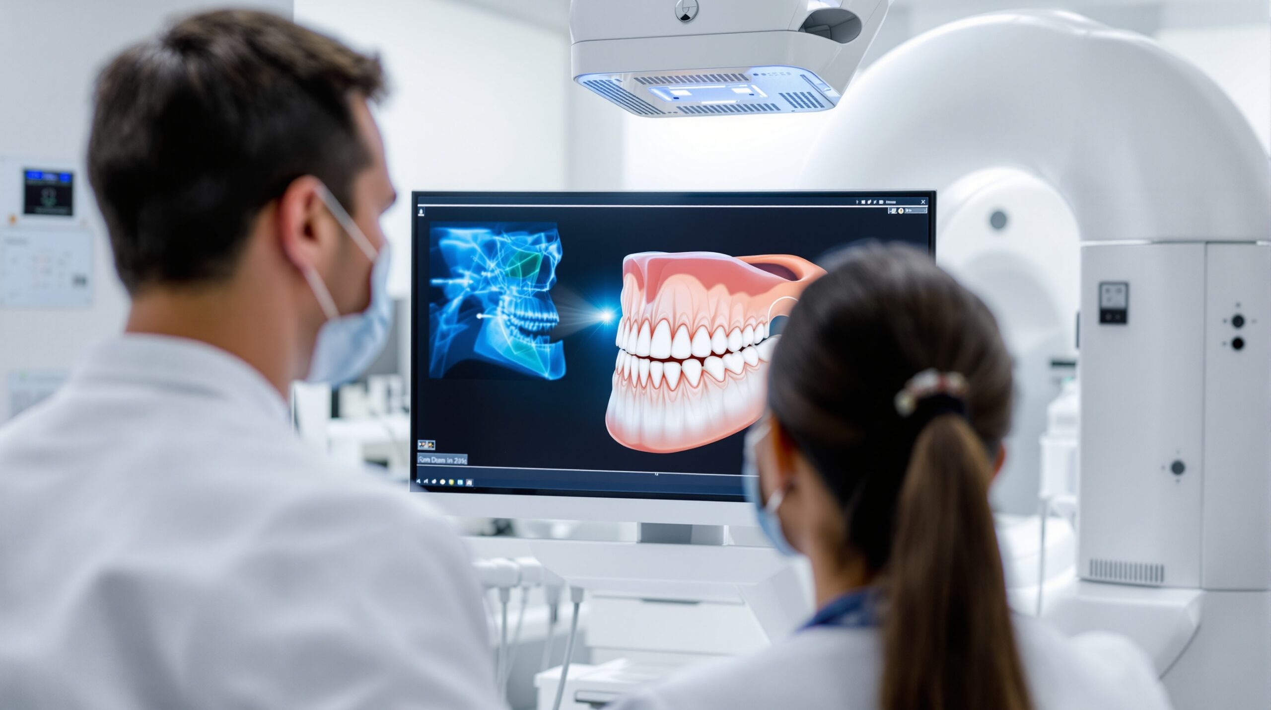
The future of dental X-ray technology is being shaped by artificial intelligence and continued hardware innovation. AI algorithms are now being trained to automatically detect cavities, periodontal bone loss, periapical pathology, and other conditions on dental radiographs with accuracy that rivals — and in some studies exceeds — that of experienced dentists.
Photon-counting detector technology promises even further radiation dose reductions while improving image quality. Integration of imaging data with digital impressions and CAD/CAM workflows is creating seamless digital ecosystems where diagnosis, treatment planning, and restoration fabrication all flow from the same dataset.
From Röntgen’s accidental discovery in a German physics laboratory to AI-powered diagnostic imaging in your neighborhood dental office, the history of X-rays in dentistry is a story of relentless innovation in service of patient care. Each generation of technology has made dental X-rays safer, faster, more informative, and more accessible — a trajectory that shows no signs of slowing down.
The next time your dental hygienist positions that small sensor in your mouth and asks you to bite down, take a moment to appreciate the 130-year journey that made that instant, low-dose, crystal-clear image possible. It all started with a curious physicist, a glowing screen, and an “unknown” ray.
Sorry, the comment form is closed at this time.