


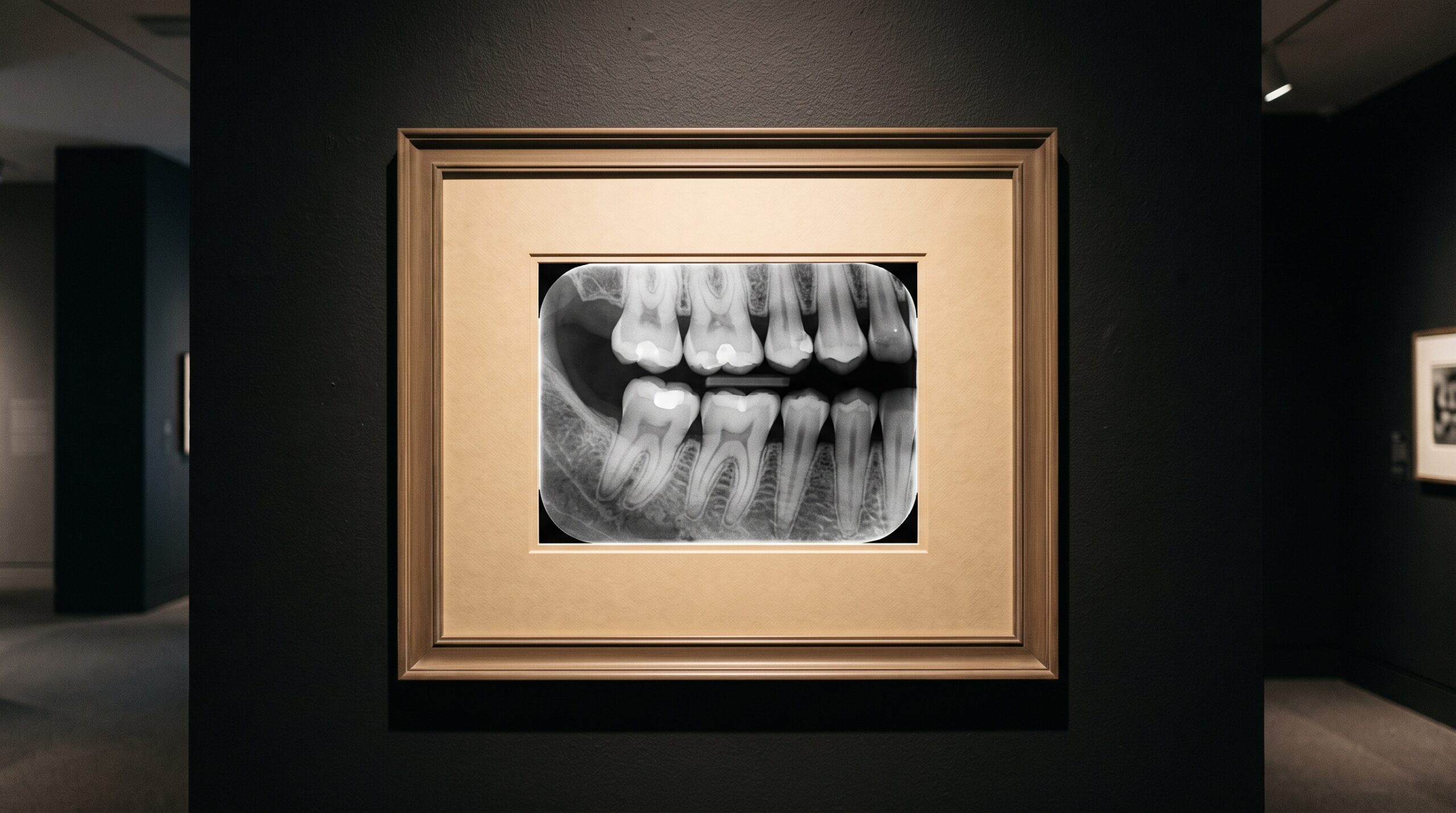
July 31, 2026 Reading the Bitewing: What Dentistry’s Most Ordered Image Reveals — and Quietly Hides
There is no image in dentistry more familiar than the bitewing. It is taken by the millions, clipped to the corner of a screen, glanced at, and moved past. Familiarity is its gift — every clinician knows how to read one — and also its trap. When you have seen ten thousand bitewings, it becomes easy to stop looking at them and start merely recognizing them. Yet a bitewing is not a window onto the teeth. It is a made object: a two-dimensional projection, sculpted by beam angle and geometry, that offers a curated version of the truth. Learning what it hides is how you learn, again, to read it.
Why the bitewing endures
The bitewing survives every advance in imaging because it does a few things better than anything else, cheaply and in seconds. It opens the interproximal contacts — the spaces between adjacent teeth where the eye and the explorer cannot reach — and lays them flat for inspection. It captures the crestal bone at exactly the level where early periodontal change first shows. It shows the crowns of upper and lower teeth in a single frame, ideal for reviewing existing restorations and the decay that likes to hide beneath and beside them. For caught-early interproximal caries, it remains the single most productive image a practice takes.
The image is a projection, not a photograph
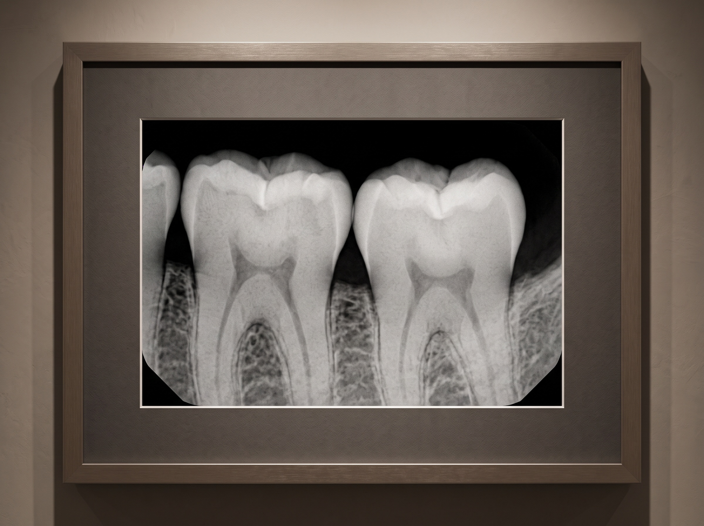
Here is the idea most worth holding onto: a radiograph is a shadow. X-rays pass through a three-dimensional structure and cast a flattened silhouette onto a sensor. Everything the tube passes through — enamel, dentin, bone, a metal margin — stacks into a single plane of greys. That flattening is why a bitewing can be simultaneously precise and deceptive. A lesion is only visible when enough mineral has been lost to change how much radiation reaches the sensor, and when the geometry lets that change fall in a place you can see it.
Which means the honest bitewing and the misleading one can be the same tooth, photographed a few degrees apart.
What angle gives, angle can take away
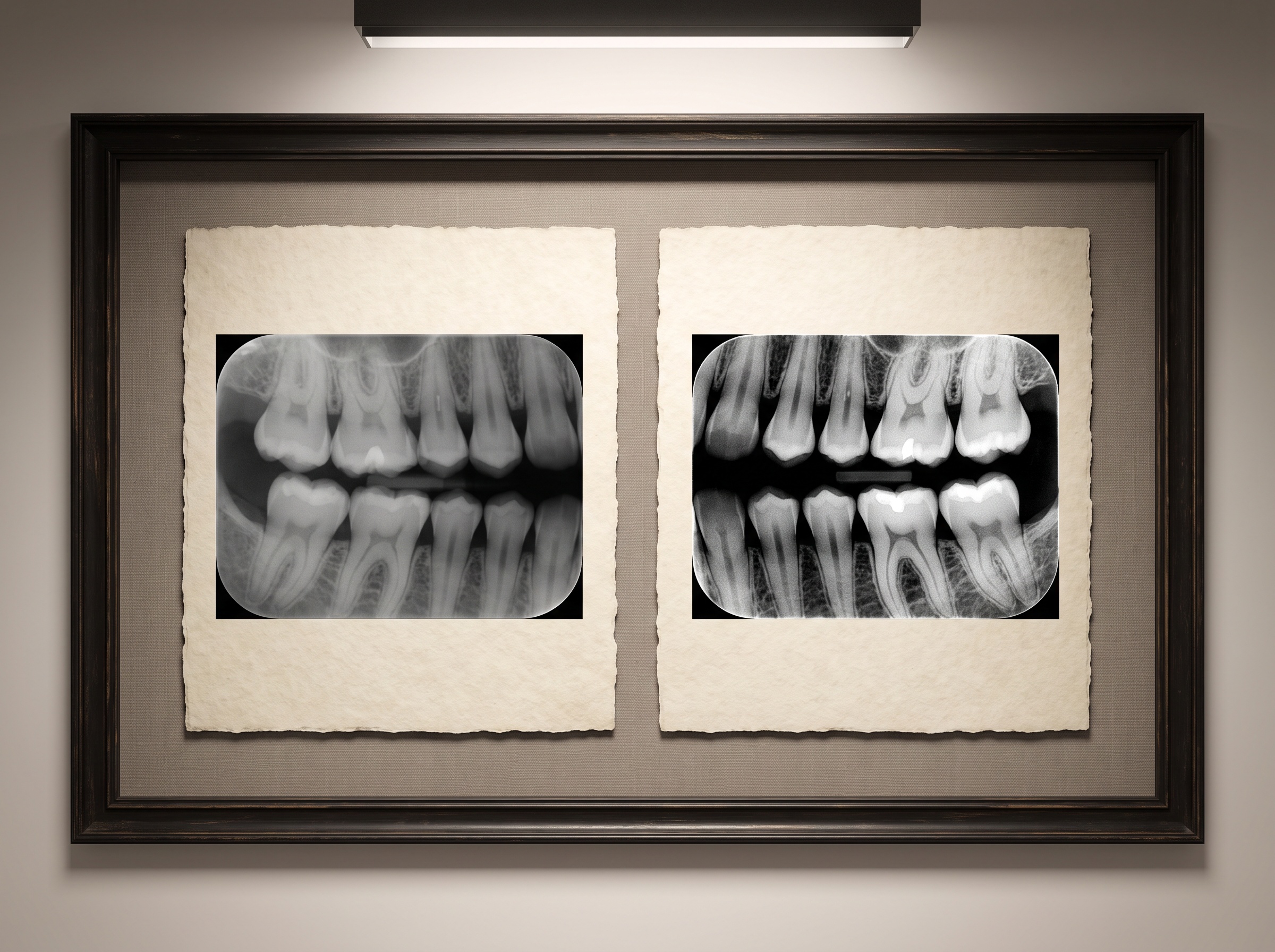
Horizontal angulation is the quiet author of the interproximal story. Aim the beam squarely through the contacts and they open cleanly; the enamel of one tooth ends and the next begins with a visible gap between them, and any demineralization in that zone stands out as a notch or a grey wedge. Aim a few degrees off, and the contacts overlap — the teeth superimpose, and a beginning lesion vanishes into the merged shadow. It is still there in the mouth. It is simply no longer in the image. Overlap is not a cosmetic flaw; it is a diagnostic one, and it is the most common reason a bitewing lies by omission.
Vertical angulation and cone position write their own footnotes — foreshortening, elongation, the pale scythe of a cone-cut where the beam missed the sensor entirely. Each is a reminder that the clinician’s hand, before it ever reaches for a diagnosis, has already shaped what there is to diagnose.
Early decay speaks in whispers
The dramatic cavity — the black cavern breaking into dentin — reads itself. The lesions that matter clinically are the ones caught before that, and they do not announce themselves. Early interproximal caries appears as the faintest loss of density: a slight greying at the outer enamel just below the contact, a subtle change in an otherwise uniform white. This is where interpretation becomes genuinely a craft of seeing. It rewards a calibrated monitor, controlled ambient light, and the discipline to zoom and compare rather than glance. The information is present in the pixels; whether it reaches the clinician depends on how the image is displayed and how carefully it is read.
The story beneath the teeth
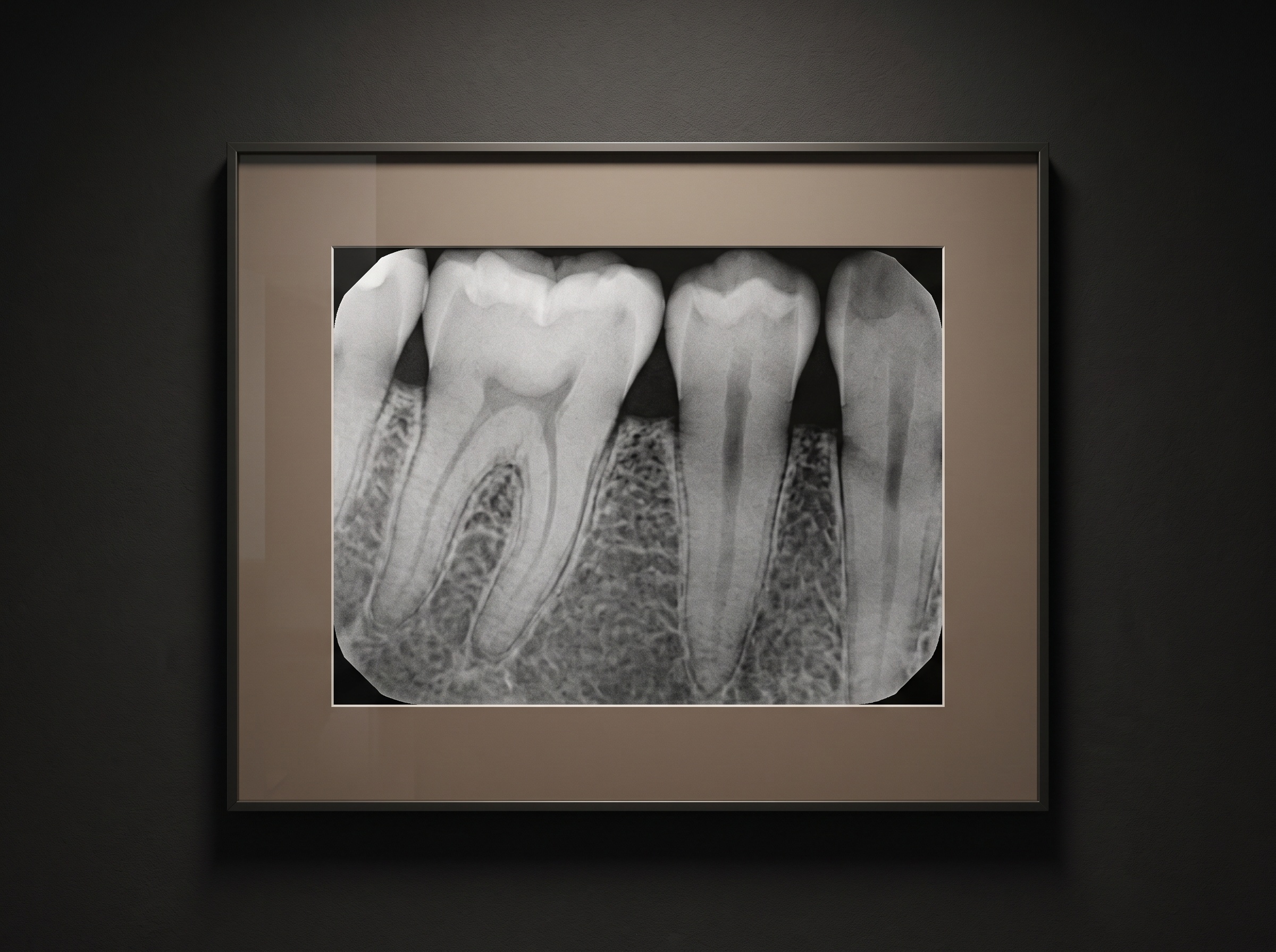
A bitewing read only for cavities is a bitewing half-read. The same frame records the height and contour of the alveolar crest, the first quiet evidence of bone loss long before mobility or pocketing make it obvious. It shows calculus as small radiopaque spurs clinging to root surfaces. It reveals the fit of old restorations — open margins, overhangs, recurrent decay tucked beneath a crown. The most valuable clinicians treat every bitewing as a short paragraph about the whole patient, not a single sentence about a single tooth.
Reading as an act, not a reflex
To read a bitewing well is to hold two ideas at once: trust the image enough to act on it, and distrust it enough to remember what it cannot show. It is flat where the mouth is round. It is silent about the buccal and lingual surfaces the beam passed straight through. It is only as honest as the angle that made it. The skilled reader carries those limits as context, the way a curator knows the lighting under which a painting was meant to be seen.
Future developments
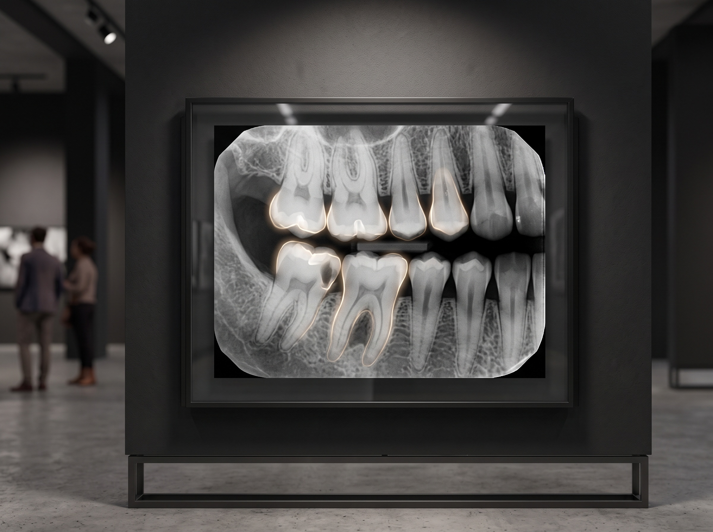
The bitewing is changing in two directions at once. Higher-fidelity sensors and better displays are widening the greyscale the image can hold, so that the faint whisper of an early lesion survives all the way from capture to the clinician’s eye. And AI-assisted overlays are beginning to read alongside us — flagging the subtle density changes and marginal shadows that fatigue and familiarity conspire to skip. Neither replaces interpretation; both raise its ceiling. The most ordered image in dentistry is quietly becoming the most intelligently read, and the clinician who understands what the bitewing has always concealed will be the one who gets the most from what it is about to reveal.
Sorry, the comment form is closed at this time.