


September 1, 2026 How AI Reads a Bitewing: Caries and Bone-Loss Detection, Explained
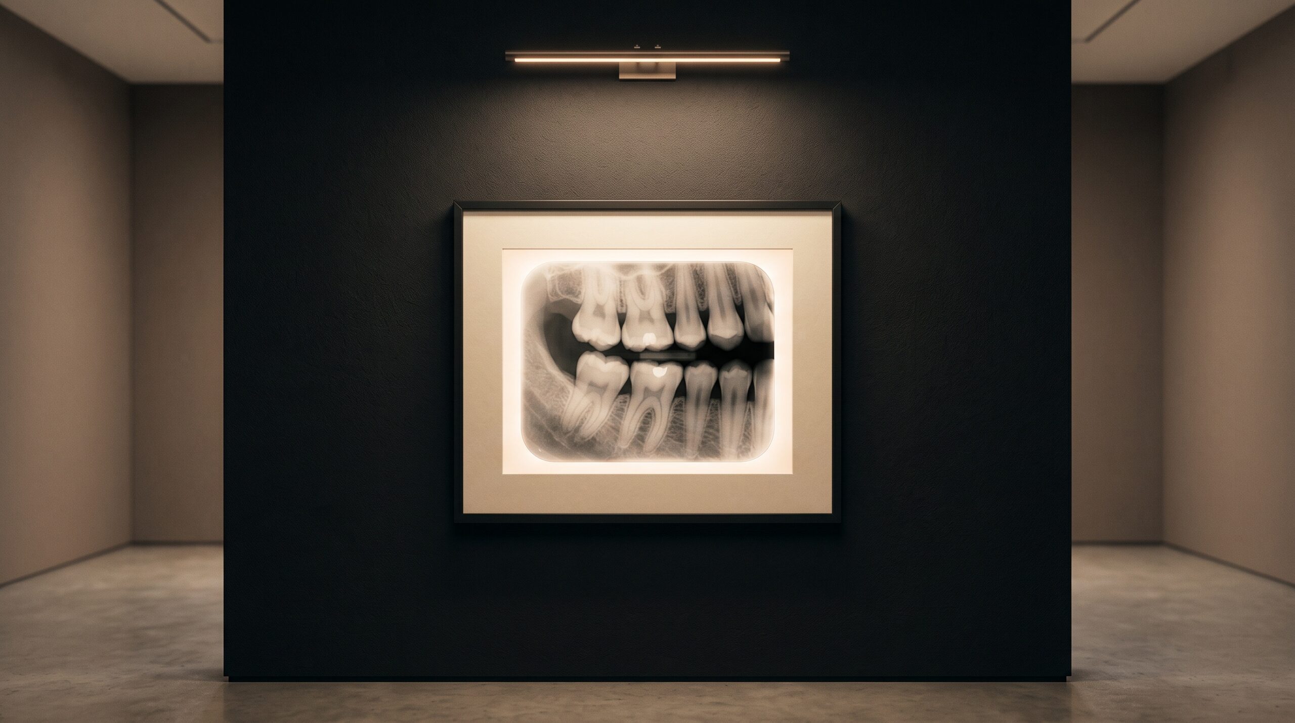
Hang a bitewing on the wall and it looks almost austere – a narrow rectangle of greys, a few crowns meeting in the middle, roots fading into the parchment darkness at the edges. But every diagnosis it contains is written in shadow, and shadow is precisely what the human eye reads least reliably. A lesion in enamel is not a line or a mark; it is a faint loss of density, a place where a few more photons passed through than should have. Two experienced clinicians can look at the same interproximal space and disagree. This is the quiet problem that dental AI was built to address – and to understand what it does, you have to stop thinking about teeth and start thinking about pixels.
An AI system does not know what a molar is in any sense you would recognize. It knows that this radiograph is a grid of intensity values, and that certain arrangements of those values – learned from tens of thousands of annotated images – correspond to conditions a dentist would name. The reading it produces is a translation, from the language of density gradients back into the language of caries and crestal bone. What follows is how that translation actually happens.
The Bitewing as a Field of Intensities
Strip away the clinical meaning and a bitewing is a two-dimensional map of X-ray attenuation. Enamel is dense and stops photons, so it renders bright. Dentin is softer and renders a shade darker. A carious lesion is demineralized tissue – it has lost the mineral that made it opaque, so it lets more radiation through and appears as a subtle darkening within an otherwise bright structure. The entire diagnostic task, for machine and clinician alike, is discriminating meaningful darkening from the ordinary noise of anatomy, overlap, and exposure.
A convolutional neural network approaches this the way it approaches any image. Its earliest layers detect nothing more sophisticated than edges and gradients – the boundary where enamel meets the interproximal space, the direction in which brightness falls away. Deeper layers assemble those primitives into shapes: the triangular radiolucency of a proximal lesion, the flat horizontal line of a healthy bone crest, the fuzzy margin of a recurrent lesion beneath a restoration. No single neuron holds the concept of “caries.” The concept is distributed across the pattern of activations the image produces.
Reading Caries: The Interproximal Shadow
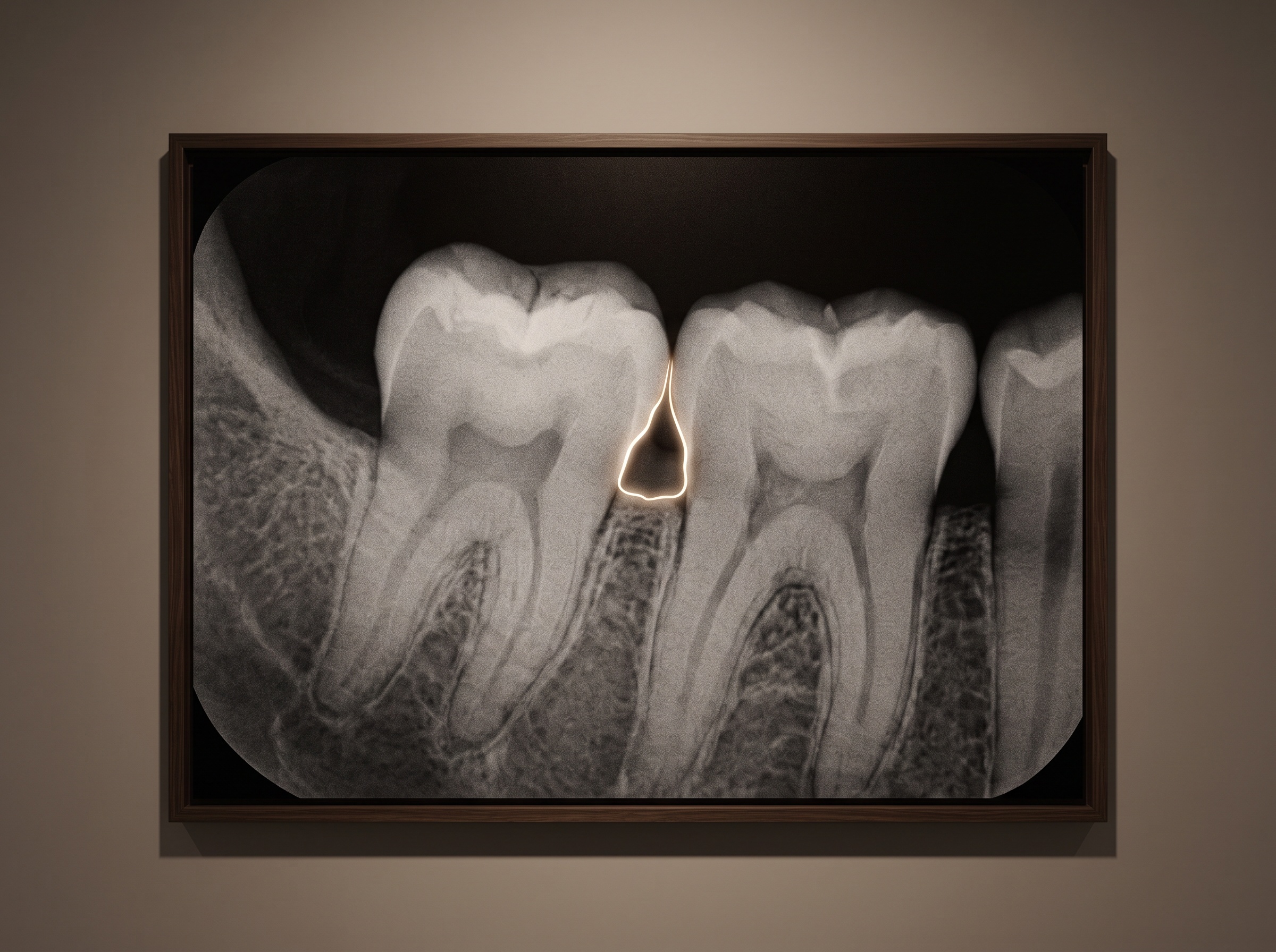
Interproximal caries is the classic case, and the reason bitewings exist. The contact point between two teeth is invisible to direct inspection and to the explorer; the radiograph is the only honest witness. A lesion begins as a faint notch in the enamel just below the contact, a shallow radiolucent triangle whose apex points toward the pulp. Early, it is a whisper – a handful of grey levels of difference across a few dozen pixels.
The network has seen this whisper repeatedly during training, always paired with a human annotation marking the lesion’s outline. So it learns the signature: not the absolute darkness, which varies with every exposure, but the local contrast – the way the density dips relative to the sound enamel bracketing it. When it flags a lesion, it is not classifying the tooth; it is outlining a region and assigning it a confidence score. That distinction matters. A good system tells you where it is suspicious and how much, and leaves the diagnosis to you. This is the same detection-and-outline behavior we described in Pearl on the Radiograph, and it is why the overlay, not a verdict, is the honest form of the output.
How well does it work? In validation studies of FDA-cleared caries-detection systems, reported sensitivities have landed roughly in the low-to-high seventies percent with specificities above 98% – meaning the machine misses some real lesions but very rarely invents one. Independent research has gone further: a 2024 study cited in the recent literature found that convolutional networks outperformed dental students at spotting non-cavitated proximal lesions, the very cases the human eye finds hardest. The AI is not infallible. It is consistent, and consistency at the threshold of visibility is worth a great deal.
Reading Bone Loss: Measuring the Crest
Periodontal assessment is a different kind of task – less detection, more measurement. Here the network is not hunting for a shadow but locating landmarks: the cemento-enamel junction (CEJ), the crest of the alveolar bone, and the root apex. From those three points it computes a proportion – how far the bone has receded from the CEJ toward the apex, expressed as a percentage of root length or as a linear distance in millimetres.
This turns a subjective impression – “the bone looks a little low here” – into a repeatable number. Clinically meaningful bone loss is often defined at thresholds such as 33% of root length or roughly 5 mm of attachment loss, and a system that measures the same way on every image, every visit, gives you something powerful: a stable baseline against which future radiographs can be compared. The measurement is only as good as the network’s ability to place those landmarks precisely, which is why crestal blur and overlap are the enemies of accurate bone reading.

The Overlay Is the Interface
What the clinician sees is not the network’s raw output but a rendered layer over the radiograph – a colored outline around a lesion, a line marking the bone level, a percentage or a confidence figure. This overlay is where the craft of imaging meets the mathematics. A well-designed one respects the image: it is translucent, it can be toggled off, and it never obscures the very anatomy you need to judge its claim. A confidence score is not a diagnosis; it is an invitation to look harder at a specific place. The best systems are calibrated so that a stated confidence roughly matches real-world hit rate – a claim worth verifying rather than trusting, because a miscalibrated overlay that always says “90%” is worse than none at all.
Where It Fails: The Image Beneath
Every claim above collapses if the radiograph is poor. A CNN cannot recover information the exposure never captured. Overlapping contacts hide the interproximal surfaces where lesions live; a cone-cut erases a region entirely; over-exposure burns out early demineralization into apparent health; motion or a bent sensor smears the crestal line the bone algorithm depends on. The network will still return an answer – that is the danger – but it is answering a corrupted question. Garbage in, confident garbage out. This is why AI has not made technique obsolete; it has raised the value of a clean, well-angulated, properly exposed bitewing, because the machine, like the eye, can only read what the image actually holds. The same principle governs the acquisition path we traced in VideaHealth and TWAIN: the AI is downstream of your imaging discipline, never a substitute for it.
Future Developments
The bitewing has been read the same way for a century – a clinician, a lightbox, and an act of interpretation performed alone and unrecorded. What is changing is not the image but the second reader standing permanently beside it, one that never tires, never rushes the last patient of the day, and measures the bone the same way every single time. The near future points toward longitudinal reading – AI comparing today’s crest to last year’s automatically, surfacing the slow lesion that grows a fraction of a millimetre between recalls, the kind of change no memory can hold. Further out, the same networks now confined to 2D film are learning the volumetric reasoning of CBCT, a lineage we followed in Diagnocat on CBCT Volumes. But the humblest truth endures: the algorithm is a reader, not a radiographer. It elevates a good image and is defeated by a poor one. The craft of making the picture – the angulation, the exposure, the honest geometry of the shot – remains, as it always has, the foundation on which every reading, human or machine, is built.
Sources & further reading:
Sorry, the comment form is closed at this time.