


August 29, 2026 Diagnocat on CBCT Volumes: 60+ 3D Findings and the API Path
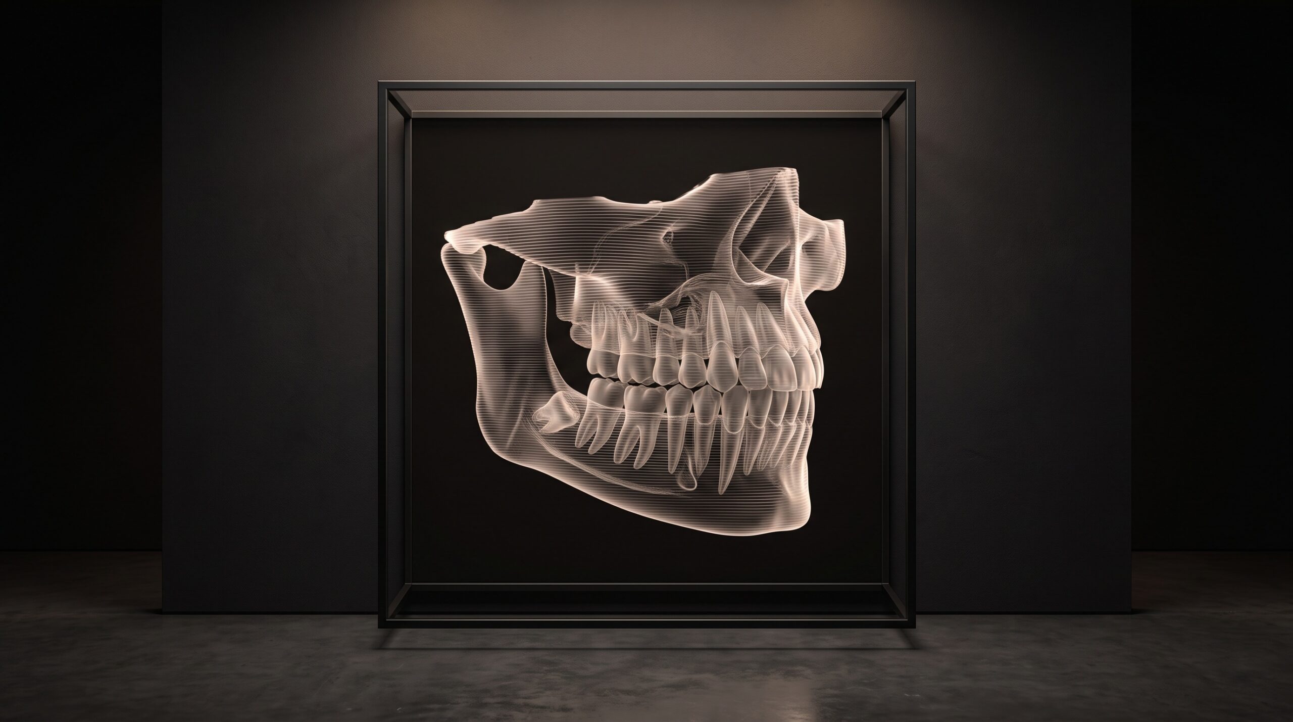
A cone-beam volume is the most generous image in dentistry and the least forgiving. Generous, because a single CBCT acquisition holds hundreds of thin slices – a quarter of a million voxels of jaw, root, canal, and sinus rendered in true three dimensions. Unforgiving, because almost all of that information stays latent: the eye can only travel one plane at a time, and the findings that matter most often hide in the slices no one scrolls to. Diagnocat’s proposition is to read the whole volume at once, turn it into a navigable 3D object, and surface what is there rather than what happened to be on screen. It is a beautiful idea. It is also, as ever, downstream of a far more literal question – how does the volume get to the AI in the first place?
What Diagnocat Is Cleared to Do in 3D
Precision matters here, because CBCT is squarely in medical-device territory and hand-waving does real harm. In September 2025, the FDA granted 510(k) clearance to Diagnocat’s Segmentron Viewer (submission K251072, by DGNCT LLC), classified under CFR 892.2050 as a Class II medical image management and processing system, product code QIH. The cleared indication is specific and modest in its language: the device performs automated 3D segmentation of a CBCT scan and generates a segmentation report for a medical professional – a dentist or radiologist – to further evaluate a patient’s teeth and anatomy, in patients fourteen years and older with permanent teeth.
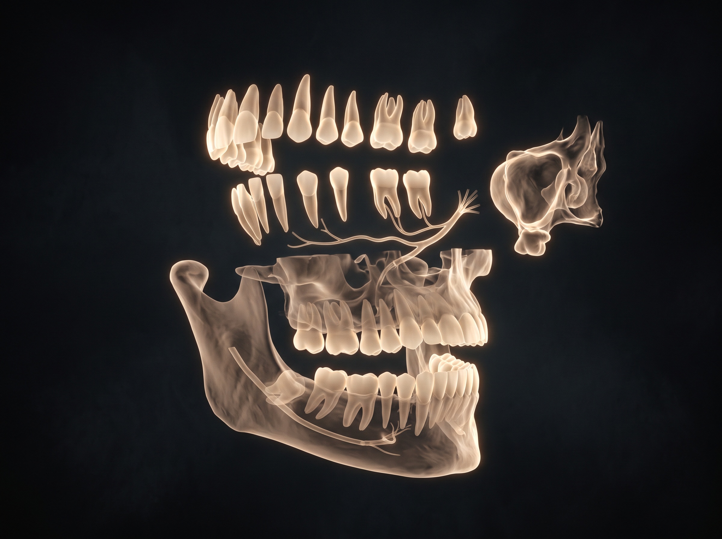
That clearance is quietly significant for imaging. Diagnocat is, at the time of writing, the only tool with US regulatory clearance for automated CBCT segmentation – the step that separates a raw volume into discrete, labelled anatomical objects. The practical payoff is that segmented structures can be exported as STL models for implant planning, guided surgery, and endodontic assessment, and that a clinician gets a navigable 3D reading instead of a stack of grey slices. The honest framing, and the one the clearance itself insists on, is assist: it reports and visualises for a professional to evaluate. It does not diagnose alone.
Sixty-Five Findings in a Single Volume
Beyond segmentation, Diagnocat’s broader platform is built to read across the volume for a wide roster of conditions – the company describes coverage of more than sixty-five dental and maxillofacial findings on CBCT. That range is what distinguishes a volumetric reader from a flat-film caries detector. On a bitewing, the question is essentially two-dimensional. On CBCT, the same engine is asked to weigh periapical lesions, root fractures, canal anatomy, bone levels, impactions, and – notably – findings that are not strictly dental at all: airway and sinus observations, and structural anomalies that live at the edges of the scanned field.
This is where volumetric AI earns its keep. Incidental findings are the classic failure mode of CBCT reading, precisely because the general dentist’s attention is trained on teeth while the scan happily captures the maxillary sinus and a good deal of the skull base. A model that reviews every slice with the same patience does not get bored at the sinus floor. For an imaging craft that has always prized the discipline of reading the whole film, an engine that reads the whole volume is less a novelty than a natural continuation.
The Round Trip You Do Not See
Here is the detail imaging people should sit with, because the marketing tends to go quiet at exactly this point: Diagnocat is cloud-based. The analysis does not happen on the operatory workstation. The CBCT volume – a substantial DICOM dataset, often hundreds of megabytes – is transmitted to Diagnocat’s infrastructure, segmented and analysed by the models there, and the structured reading is returned to a web browser for the clinician to navigate. Nothing about the intelligence occurs until the scan has successfully left the building and come back.
That round trip promotes the transport layer from footnote to precondition, and for a CBCT volume the stakes are heavier than for a single radiograph. The dataset is large, so upload time and connection quality are real clinical friction. The data is unambiguously PHI in transit, so the security of that channel is not optional. And the returned reading has to reunite with the exact study it came from. It is the same infrastructural truth we traced through a flat-film vendor in VideaHealth and TWAIN and through a detection engine in Pearl on the Radiograph: the image is only as useful as its ability to travel.
DICOM In, Reading Out: The Real Integration Story
Diagnocat’s saving grace on the send-out question is that it is deliberately hardware-agnostic. It does not care which CBCT unit produced the scan; it consumes standard DICOM, the universal currency of volumetric imaging. Any scanner or imaging platform that can export a clean DICOM volume already speaks the language Diagnocat requires. In practical terms, the dominant integration path is a DICOM upload to the cloud – whether initiated from a web portal, a desktop uploader, or the vendor’s practice-management overlay – with results returned in the browser.
That overlay is the second, deeper channel. Diagnocat offers a Panel that sits over supported practice-management systems, letting newly acquired images be saved directly into Diagnocat and read within the existing workflow. This is the smoother, more native experience – but it is a partner-gated integration, available where Diagnocat and a PMS have done the work, not a universal door. It is worth being candid about what does not appear to be prominently published: a broad, open, self-serve developer API for arbitrary third-party software to pipe volumes in and pull structured findings out programmatically. The reachable, documented surface today is DICOM-based cloud upload plus specific PMS panel integrations. For anyone evaluating who they can actually wire into, that distinction – open standard versus partner-gated – is the whole ballgame.

Why the Pipe Decides Who Gets the AI
For those of us thinking about imaging software as a platform rather than a single product, Diagnocat is an instructive case. Its clearance and its finding-count are genuinely leading; its reachability profile is specific and worth cataloguing precisely because it is not interchangeable with a TWAIN-fed flat-film reader. A CBCT engine that consumes standard DICOM is, in principle, reachable by any imaging system disciplined enough to export clean, complete volumes. A system that hoards its scans in a proprietary container, or that cannot cleanly hand off a DICOM study, faces a bespoke bridge for every AI it wants to touch.
This reframes the “adding AI” conversation entirely. As cleared reading edges toward commodity, the differentiator quietly becomes the send-out: can a platform get its acquired volume to a chosen AI at all, over a secure channel, and bring a structured reading home to the right study without dragging the clinician out of their workflow? DICOM-fluency is most of that answer for a tool like Diagnocat. Partner-gated panels are the smoother-but-narrower remainder. For the plain-language, consumer-facing view of what this same tool does, our companion piece Diagnocat: Cloud AI for 2D X-rays and 3D CBCT covers the experience; this article is about the conduit beneath it.
Future Developments
The trajectory of volumetric reading is easy to sketch: more conditions, finer segmentation, and steady movement from a report a clinician opens toward findings that live inside the operatory’s own viewer. The more interesting frontier, as always, is access. The first regulatory milestone for automated CBCT segmentation has been passed; the next contest is over how effortlessly a volume can reach a reader and return as something the clinician can turn in their hands. The likely winners are the imaging platforms fluent in the standard that already moves volumes – clean, complete DICOM – and honest enough to treat security of the round trip as a feature rather than fine print. A CBCT scan has always been a made thing, an act of imaging craft performed in three dimensions. What the AI era adds is a second craft laid over the first: the quiet discipline of the handoff, the pipe that carries the volume out and brings the reading back to the exact slice where it belongs.
Sources & further reading:
- Segmentron Viewer 510(k) Summary K251072 – U.S. FDA (accessdata.fda.gov)
- It’s official! Diagnocat is now FDA-cleared and available in the USA – Diagnocat
- AI Diagnostic Dental Solutions – Diagnocat (US)
Sorry, the comment form is closed at this time.