Logo Logo Logo Logo Logo
  • Home
  • Download
  • About
  • Blog
  • Home
  • Download
  • About
  • Blog
A translucent molar seen in three dimensions with its branching internal canal system glowing faintly inside, a delicate extra canal becoming visible behind a larger one

14 Sep The Hidden Architecture of a Tooth: What Cone-Beam CT Reveals That a Flat Radiograph Cannot

Posted at 10:37h in Dental Imaging by Raymond 0 Comments
0 Likes

For a century the radiograph told us almost everything about a tooth - and quietly kept one secret. Pressing a three-dimensional root system flat onto a single plane, it overlapped canals, hid the ones tucked directly behind others, and turned a fused, ribbon-like root into...

Read More
A single translucent tooth shown as a colour surface shell perfectly overlaid on a faint glowing interior of root and structure, aligned as one object

13 Sep One Mouth, Two Truths: The Craft of Fusing an Intraoral Scan With a Radiograph

Posted at 10:37h in Dental Imaging by Raymond 0 Comments
0 Likes

The intraoral scanner records the mouth as it looks - a flawless colour surface, and nothing beneath it. The radiograph records the mouth as light passes through it - the roots, the bone, the hidden lesion - and almost nothing of the surface. Each is...

Read More
A single empty ornate frame on a gallery wall, evoking the radiograph deliberately not taken

09 Sep The Discipline of Restraint: The New Imaging Guidance and the Art of the Radiograph Not Taken

Posted at 10:39h in Dental Imaging by Raymond 0 Comments
0 Likes

The most revealing decision a clinician makes about a radiograph is often whether to take it at all. The first ADA and AAOMR patient-selection recommendations in more than a decade - and the first to address cone-beam CT - reframe imaging not as a schedule...

Read More
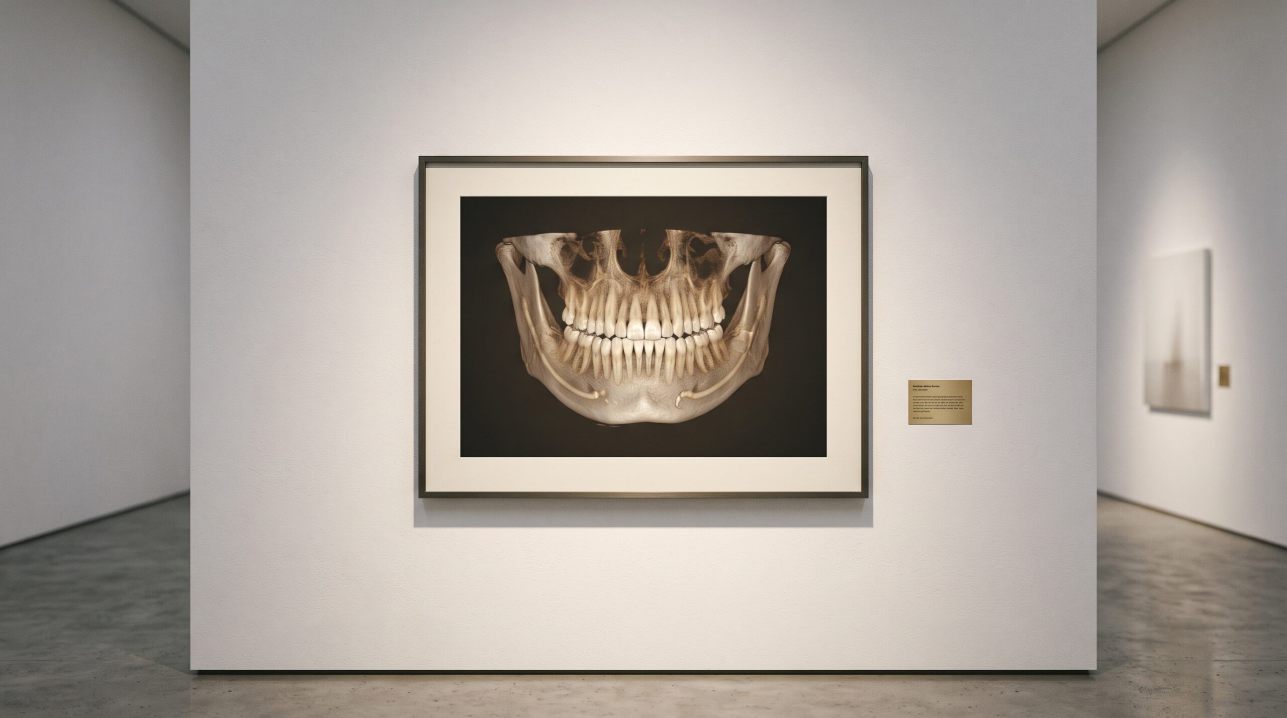
A panoramic dental radiograph framed as fine art, the frame deliberately wider than the teeth to include the surrounding anatomy

05 Sep The Whole Picture: What a Dental X-Ray Reveals Beyond the Teeth

Posted at 10:38h in Dental Imaging by Raymond 0 Comments
0 Likes

A dental radiograph is framed for your teeth, but the exposure does not stop at the enamel. In the same grey field sit the sinuses, the jaw, the airway, the soft tissues of the neck - and now and then, something no one was looking...

Read More
A single pale cube on a brass museum plinth engraved with a number that appears to shimmer and double, presented as an artwork whose value cannot be trusted.

03 Sep The Number That Lies: Why a CBCT’s Gray Values Are Not True Hounsfield Units

Posted at 10:37h in Dental Imaging by Raymond 0 Comments
0 Likes

On a medical CT, the number in a voxel is a promise: water reads zero, cortical bone reads a thousand, and the scale means the same thing tomorrow as today. On a cone-beam scan, the same-looking number keeps no such promise - and mistaking one...

Read More
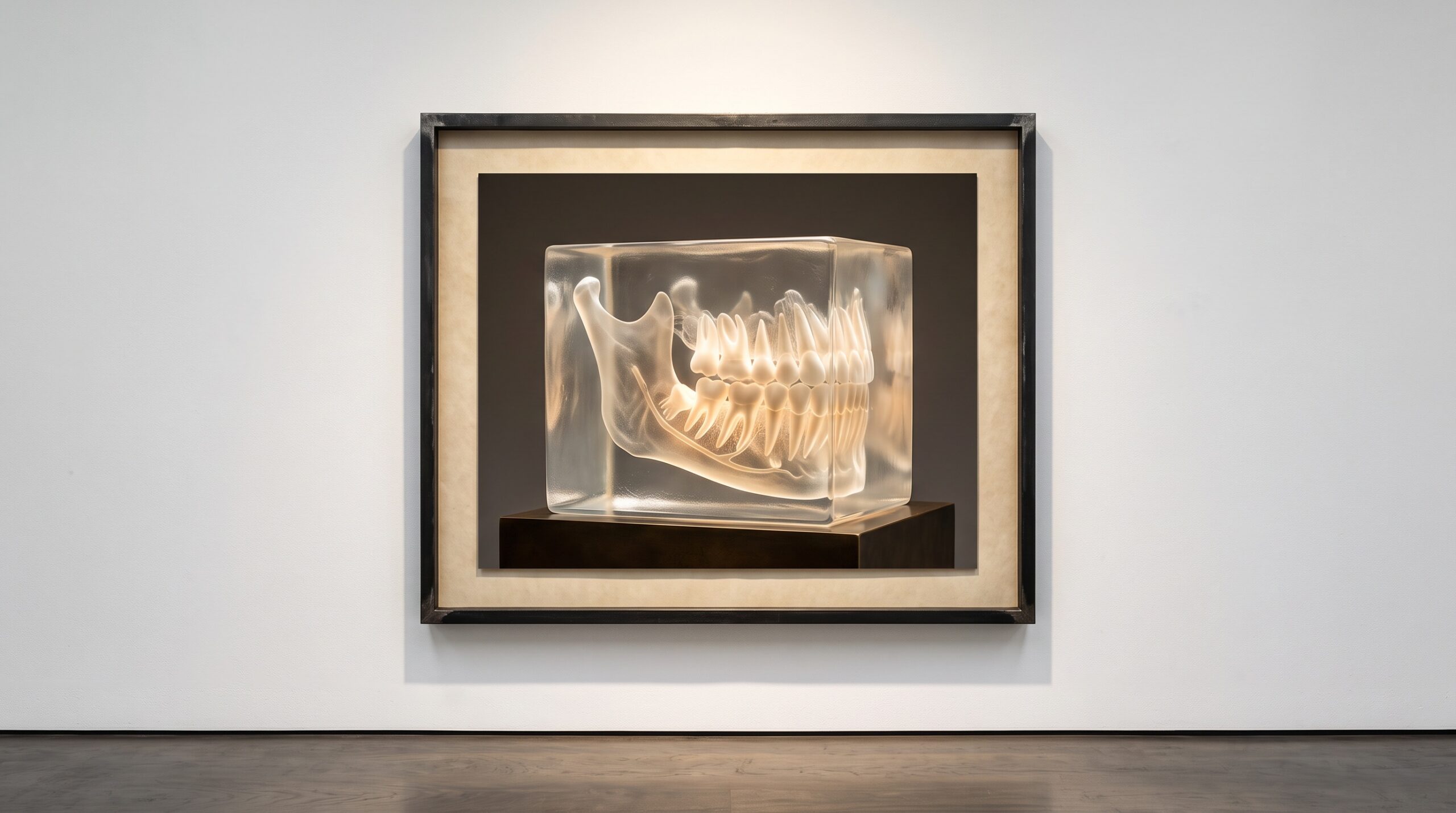
A translucent 3D CBCT volume of a jaw presented as a sculptural exhibit on a gallery plinth

02 Sep AI on CBCT: What 3D Segmentation Actually Finds

Posted at 09:44h in Dental Imaging by Raymond 0 Comments
0 Likes

A CBCT scan arrives as a fog of grey voxels. Segmentation is the act of naming them - drawing the boundary between a tooth and the bone that holds it, between the nerve canal and the space around it. Here is what the machine actually...

Read More
A gallery wall of small framed dental images arranged in a grid, progressing from faint and grainy to crisp and luminous

30 Aug Two Hundred Patents Deep: What a Two-Decade Portfolio Reveals About How the Dental Image Learned to See

Posted at 10:37h in Dental Imaging by Raymond 0 Comments
0 Likes

A single vendor recently tallied two decades of dental-imaging patents - hundreds of filings, a couple hundred granted. Read as more than a press release, it is a map of how the dental image was engineered, invention by patient invention....

Read More
A volumetric CBCT render of a jaw and teeth presented as suspended sculpture, its internal cross-section planes faintly visible

29 Aug Diagnocat on CBCT Volumes: 60+ 3D Findings and the API Path

Posted at 09:44h in Dental Imaging by Raymond 0 Comments
0 Likes

A CBCT scan is a quarter-million slices of latent information. Diagnocat turns that volume into a navigable 3D reading of 65+ conditions - but only after the DICOM leaves the operatory for the cloud. The findings are the art; the path is the plumbing....

Read More
A framed panoramic dental radiograph on a gallery wall beside a softly glowing museum placard, with faint threads of light connecting picture to words

26 Aug The Radiograph That Talks Back: When Vision-Language AI Learns to Describe a Dental Scan

Posted at 11:39h in Dental Imaging by Raymond 0 Comments
0 Likes

For years, dental AI pointed: a ring here, a shaded region there. A new generation of vision-language models does something stranger and more human - it describes the radiograph in sentences. The portrait of the mouth is learning to narrate itself....

Read More
A framed dental bitewing radiograph on a gallery wall with faint glowing rings marking two suspected findings between the teeth

25 Aug Pearl on the Radiograph: FDA-Cleared Detection and How the Image Gets There

Posted at 09:44h in Dental Imaging by Raymond 0 Comments
0 Likes

Pearl's Second Opinion is the most-cleared name in dental AI - intraoral, panoramic, and now CBCT. The detections are the spectacle; the plumbing that carries the image out of the operatory and back is the part that actually decides who can use it....

Read More
  • 1
  • 2
  • 3
© Copyright 2020, Raster Builders Inc. All Rights Reserved.