


September 26, 2026 The Image Leaves the Screen: Augmented Reality and the Craft of Seeing the Scan Inside the Patient
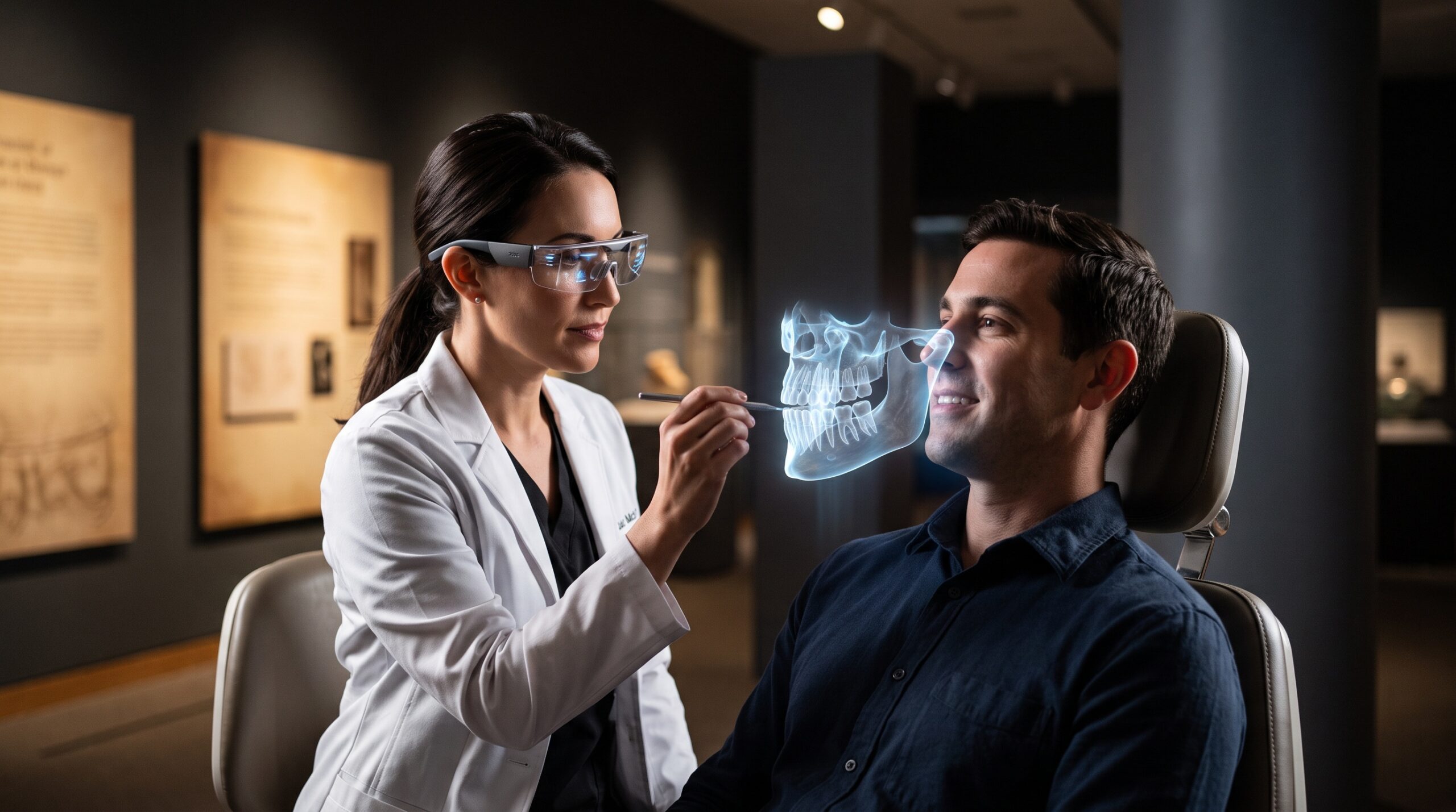
For as long as there has been dental imaging, the image has lived behind glass. Röntgen’s first shadows were fixed on photographic plates; film hung on a lightbox; the digital sensor delivered its picture to a monitor bolted to the operatory wall. In every case the clinician performs the same small, unremarked choreography—eyes down to the patient, eyes up to the screen, and back again—translating in the mind’s eye a flat picture on a wall into the three-dimensional anatomy under their hands. Augmented reality proposes to end that translation by doing something that would have seemed like sorcery a generation ago: lifting the scan off the screen and hanging it, weightless and precisely aligned, inside the patient’s own head.
It is the most literal thing imaging has ever tried to do. Not to render the anatomy more beautifully, not to detect what the eye would miss, but to put the picture back where the anatomy actually is.
The problem AR is trying to solve
Consider what a surgeon placing an implant is really asked to do. The plan lives in a rendered cone-beam volume on a screen—a beautifully reconstructed picture of bone, nerve, and sinus. The patient lies a meter away. Between the two sits the surgeon’s imagination, holding the geometry of the scan in memory while working blind inside living tissue. Static surgical guides help, but they occlude the site, cannot be adjusted mid-procedure, and still force the eye away to a monitor for confirmation.
The gap is not one of image quality; it is one of location. The best radiograph in the world is in the wrong place—on the wall, not in the wound. Augmented reality closes that distance by projecting the imaging data into the clinician’s line of sight through a transparent head-mounted display or, more simply, through the screen of a camera-equipped tablet, so the plan appears to float exactly over the surgical field.
What it means to register the invisible
The entire discipline of AR overlay rests on a single, unforgiving word: registration. To make a virtual implant appear to sit inside real bone, the software must know, continuously and to within a fraction of a millimeter, exactly where the patient is in space and exactly how the clinician is looking at them. Get it right and the hologram becomes a window into the body. Get it wrong by a degree or a millimeter and it becomes a confident, beautiful lie hovering slightly off the mark.
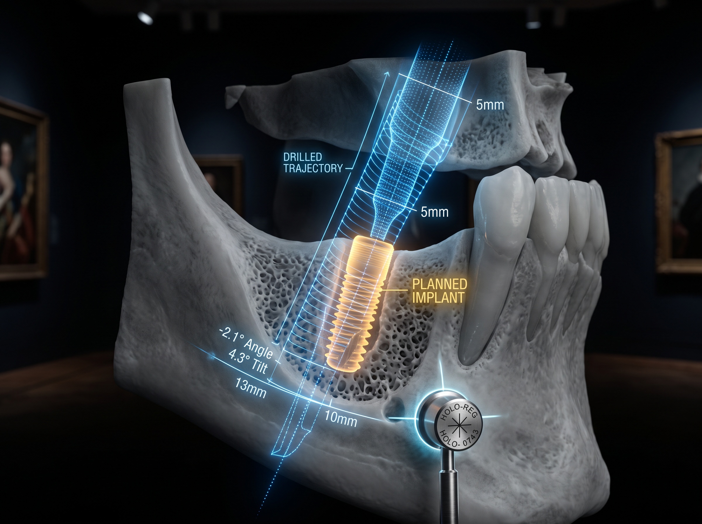
Most systems solve this the way film once solved exposure—with a physical reference. A small fiducial marker frame is clipped to the teeth or fixed to the arch, its shape known precisely to the software; cameras track it many times a second and lock the virtual scan to it. Marker-free approaches, which recognize the patient’s own dentition or facial contours directly, are the harder and more coveted goal, because every added appliance is one more thing between clinician and patient. The published accuracy studies—a growing body of systematic reviews and clinical reports—are cautiously encouraging: wearable AR navigation for implant placement is beginning to approach the deviations achieved by static guides, though the technique is still maturing from experimental models toward routine clinical use.
The image as something you stand inside
There is a quieter shift underneath the surgical use case, and it is the one that matters most to how we see. A radiograph on a monitor is a flat object we look at. A registered AR hologram is a spatial object we look into—we can lean, tilt our head, and walk around it, and the anatomy behaves as anatomy should, revealing its depth through the same parallax and motion cues the brain uses for every real object. The lingual concavity that is a subtle grey gradient on a screen becomes, in space, a hollow you can plainly see behind the ridge.
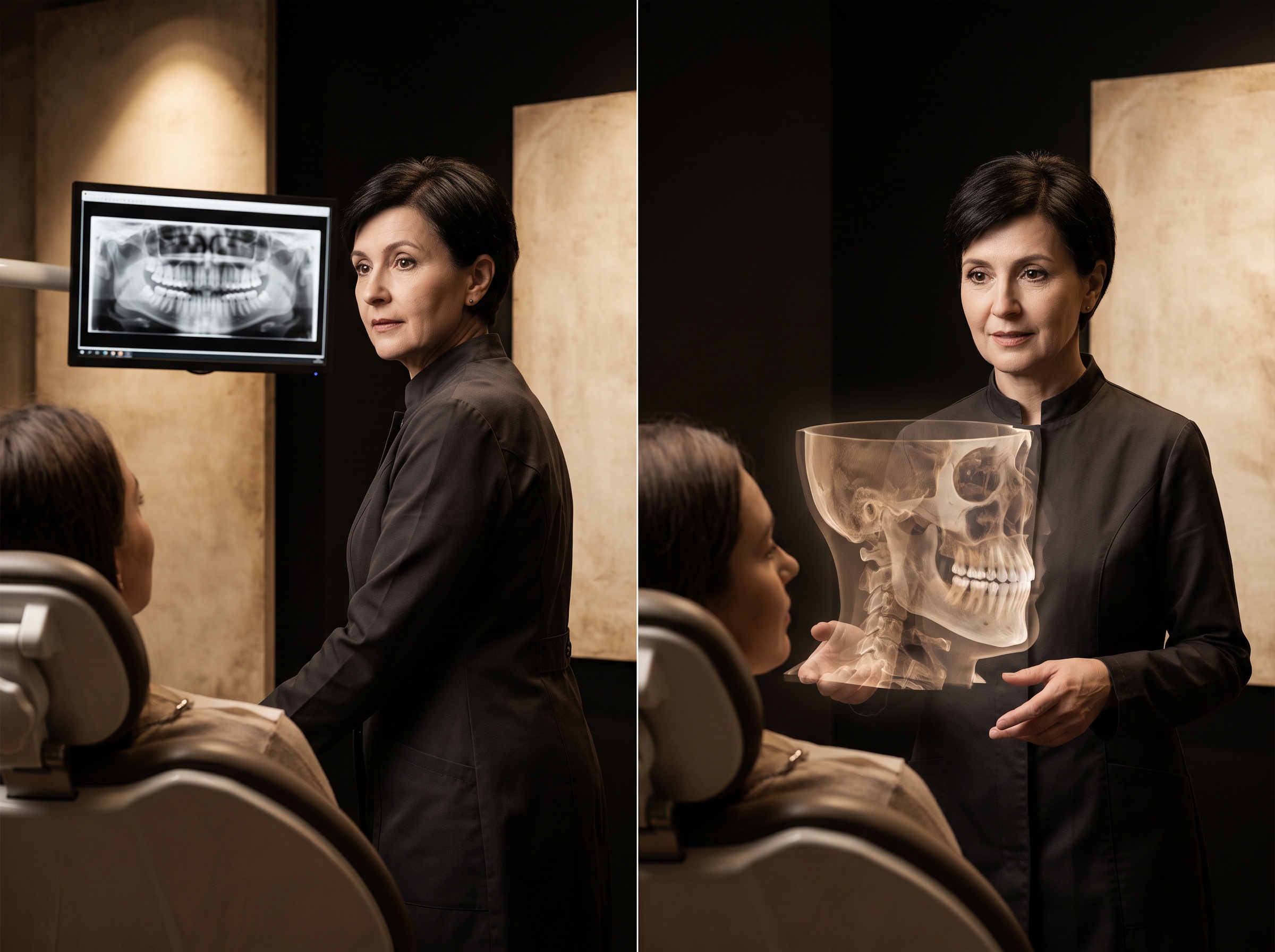
This is the same craving that drove the field from flat film toward true three-dimensional imaging of structures like the temporomandibular joint, and toward the volume-rendering techniques that turn a stack of slices into a sculpture. AR is the logical end of that trajectory: not a better picture of three-dimensional anatomy, but the anatomy presented as three-dimensional, at true scale, in the very place the hands will work. The scan stops being a representation and starts behaving like a presence.
Where the craft still lives
None of this removes the older disciplines; it depends on them utterly. An AR overlay is only ever as honest as the data feeding it. If the underlying cone-beam volume carries metal artifact, if its gray values are misread, or if the segmentation that separates nerve from bone is wrong, the hologram will render those errors in flawless, persuasive three dimensions—floating them directly into the surgeon’s confidence. The techniques that let us tell bone from metal in the scan itself matter more, not less, once that scan is projected into a living head, because the display no longer offers the reassuring frame of a monitor that says this is only a picture.
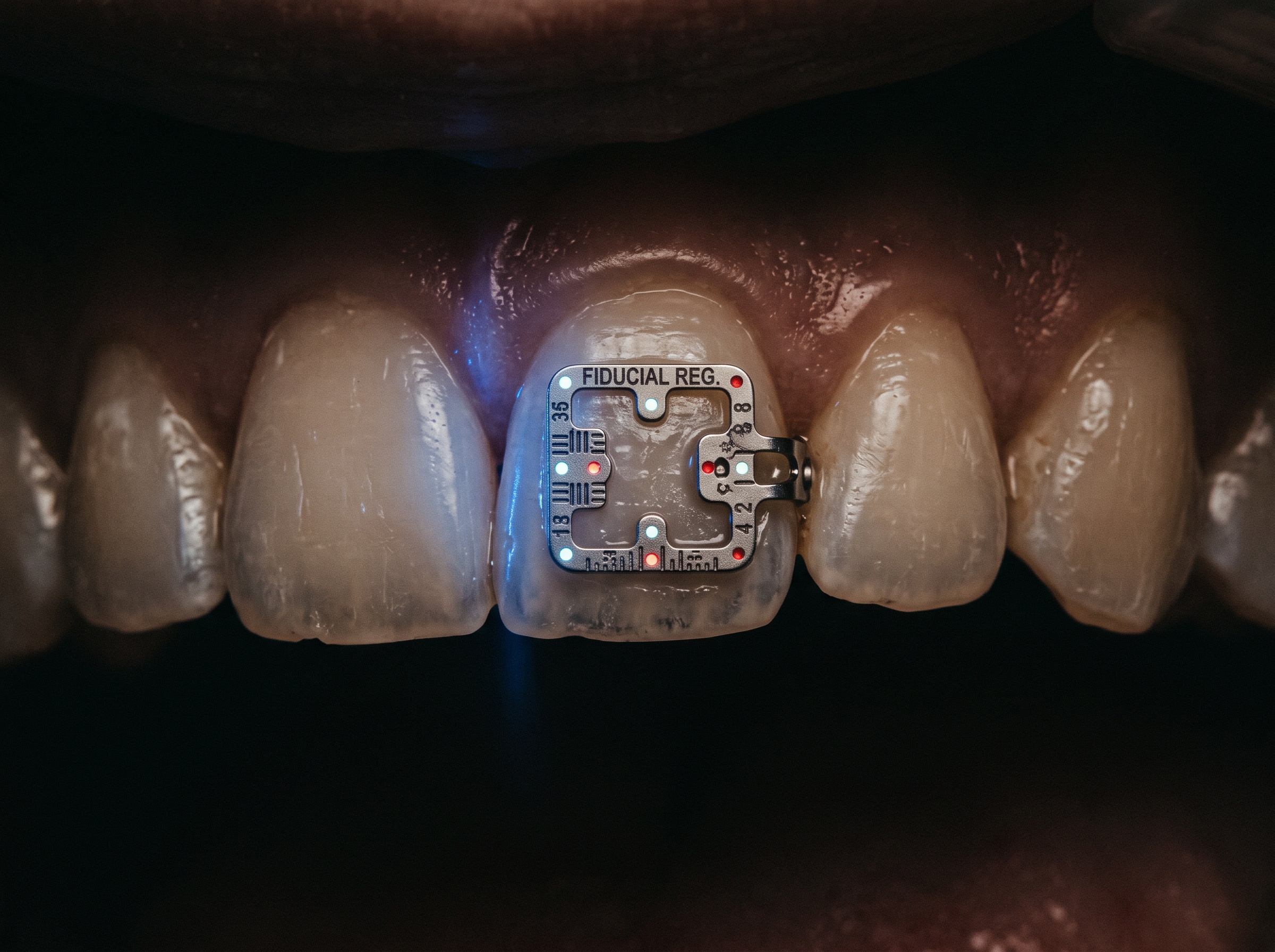
The new craft, then, is a craft of trust and calibration. It is knowing how to verify registration before the first incision, how to recognize the subtle drift of an overlay that has come loose from its anchor, and—hardest of all—how to keep looking at the patient rather than the compelling ghost laid over them. Immersive systems introduce their own failure modes: attention captured by the hologram, depth misjudged when virtual and real light do not agree, fatigue from a headset worn through a long procedure. A clinician who cannot mistrust a beautiful image has no business projecting one into a wound.
Future Developments
The direction of travel is toward disappearance. The marker frames clipped to the teeth will give way to marker-free registration that reads the patient’s own anatomy; the tethered, weighty headsets will slim toward ordinary-looking glasses; and the same artificial intelligence now learning to segment and interpret scans will increasingly drive the overlay itself, flagging the inferior alveolar canal in glowing relief and warning when an instrument strays toward it. Early mixed-reality systems for patient-specific implants and orthognathic surgery, reported through 2026, are the first crude drafts of a future in which the boundary between the image and the body simply dissolves.
It is worth pausing on how strange, and how fitting, that ending is. Dental imaging spent its first century moving the picture ever closer to the eye—off the plate, off the lightbox, onto a screen an arm’s length away. Augmented reality reverses the last step and moves the image back onto the patient, where the anatomy was all along. The gallery, in the end, was never really the wall. It was the person on the chair; we were only ever learning better ways to look.
Sources & further reading:
- Implementation of Augmented, Mixed and Virtual Reality in Implant Dentistry (ITI Forum Implantologicum)
- Wearable Augmented Reality for Dental Implant Navigation: From Experimental Models to Clinical Application—A Scoping Review (PMC)
- Mixed Reality-Guided System for Patient-Specific Implants (J Oral Maxillofac Surg, 2026)
Sorry, the comment form is closed at this time.