


September 21, 2026 Sculpting the Volume: Maximum-Intensity Projection, Volume Rendering, and the Craft of Turning a CBCT Into a Picture
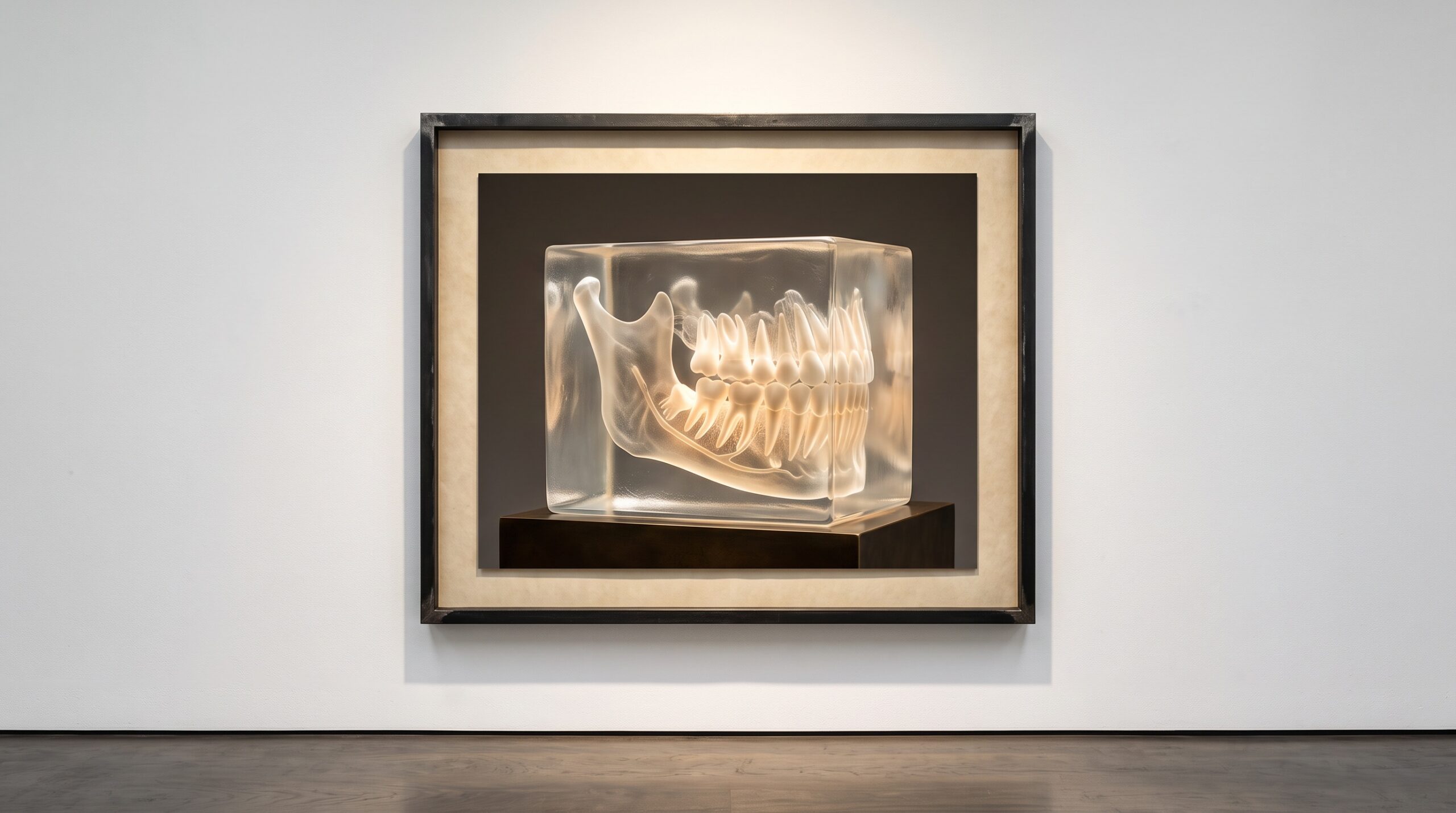
A cone-beam scanner does not, strictly speaking, take a picture. It fires a diverging cone of X-rays through the jaw from hundreds of angles, and from those projections it reconstructs something stranger and more useful than a photograph: a dense three-dimensional grid of voxels, each holding a single number that records how strongly the tissue at that point absorbed the beam. That grid – the volume – is the real product of the scan. Every flat image a clinician actually looks at is a decision made afterward about how to collapse that block of numbers onto a screen. Between the raw volume and the picture on the monitor sits a set of choices as consequential, and as quietly aesthetic, as anything in photography. To read a CBCT well is to understand that you are not looking at what the scanner saw. You are looking at what someone chose to show you.
A Volume Is Not a Picture
The difficulty, and the opportunity, is that a screen is flat and a volume is not. There is no single correct way to render a three-dimensional block of attenuation values into two dimensions, only a family of methods that each keep some information and throw the rest away. A conventional radiograph made this choice for you the instant the beam hit the sensor: everything along each ray was summed into one shadow, depth flattened irretrievably. A volume keeps the depth. It lets you defer the flattening, and then make it deliberately, differently, and as many times as the question requires. That deferral is the whole point of volumetric imaging, and the reason the same dataset can answer questions a flat film never could – the kind of hidden three-dimensional anatomy explored in what cone-beam CT reveals about fused roots and extra canals.
Slicing: The Simplest Translation
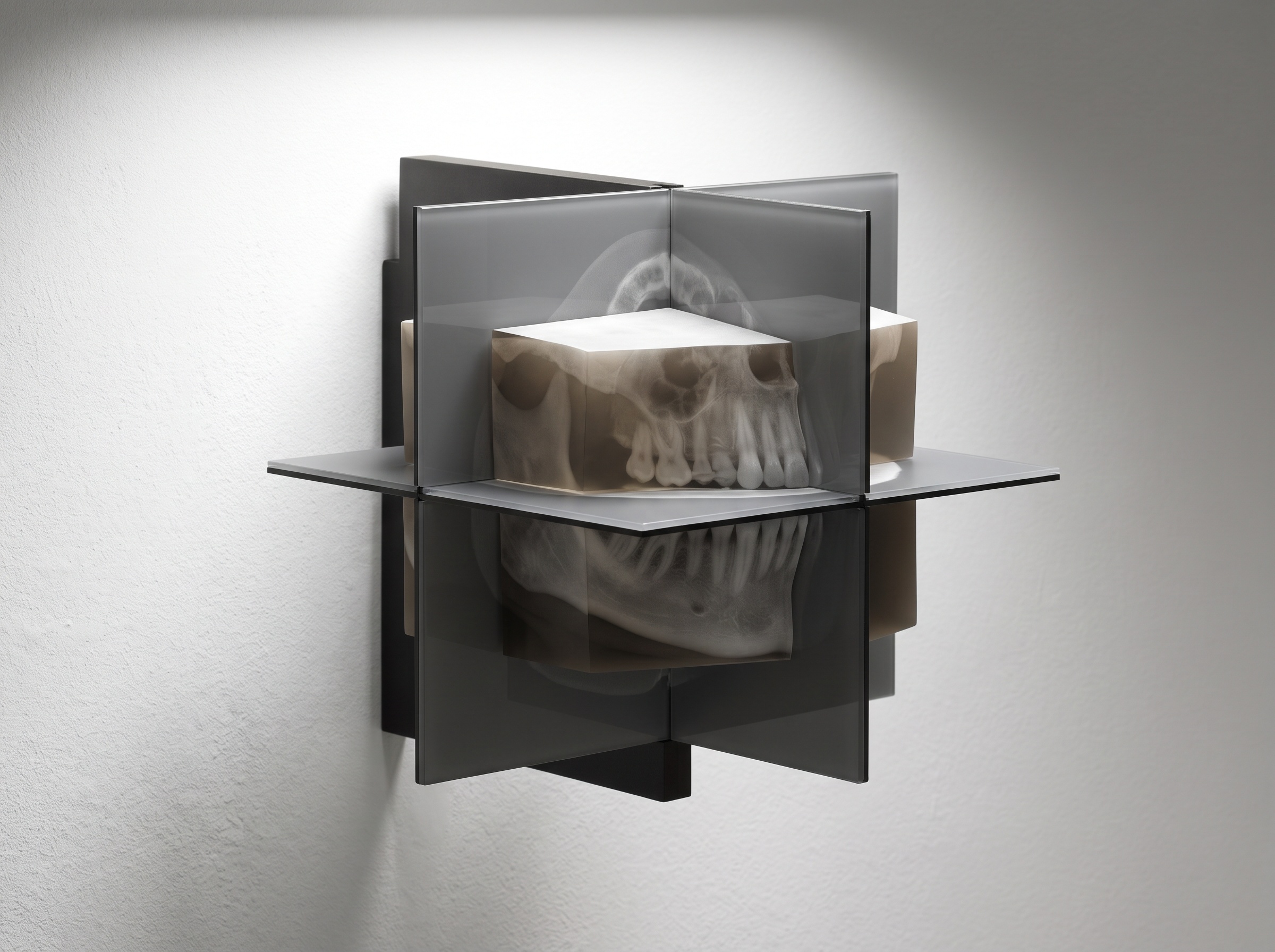
The most restrained way to look into a volume is simply to cut it. Multiplanar reconstruction resamples the grid along any plane the clinician cares to define – the familiar axial, sagittal, and coronal views, but also oblique planes angled to follow a canal or a nerve. Because the volume is isotropic, a slice through it is as sharp in one direction as another, and the plane can be swept through the anatomy interactively, a moving window rather than a fixed frame. A slab view thickens that plane, averaging or maximizing across a few millimeters of depth to gather a structure that wanders in and out of a single thin slice. Slicing discards nothing permanently; it only chooses where to stand. It is the quiet, honest baseline against which the more dramatic renderings should be judged.
Maximum-Intensity Projection
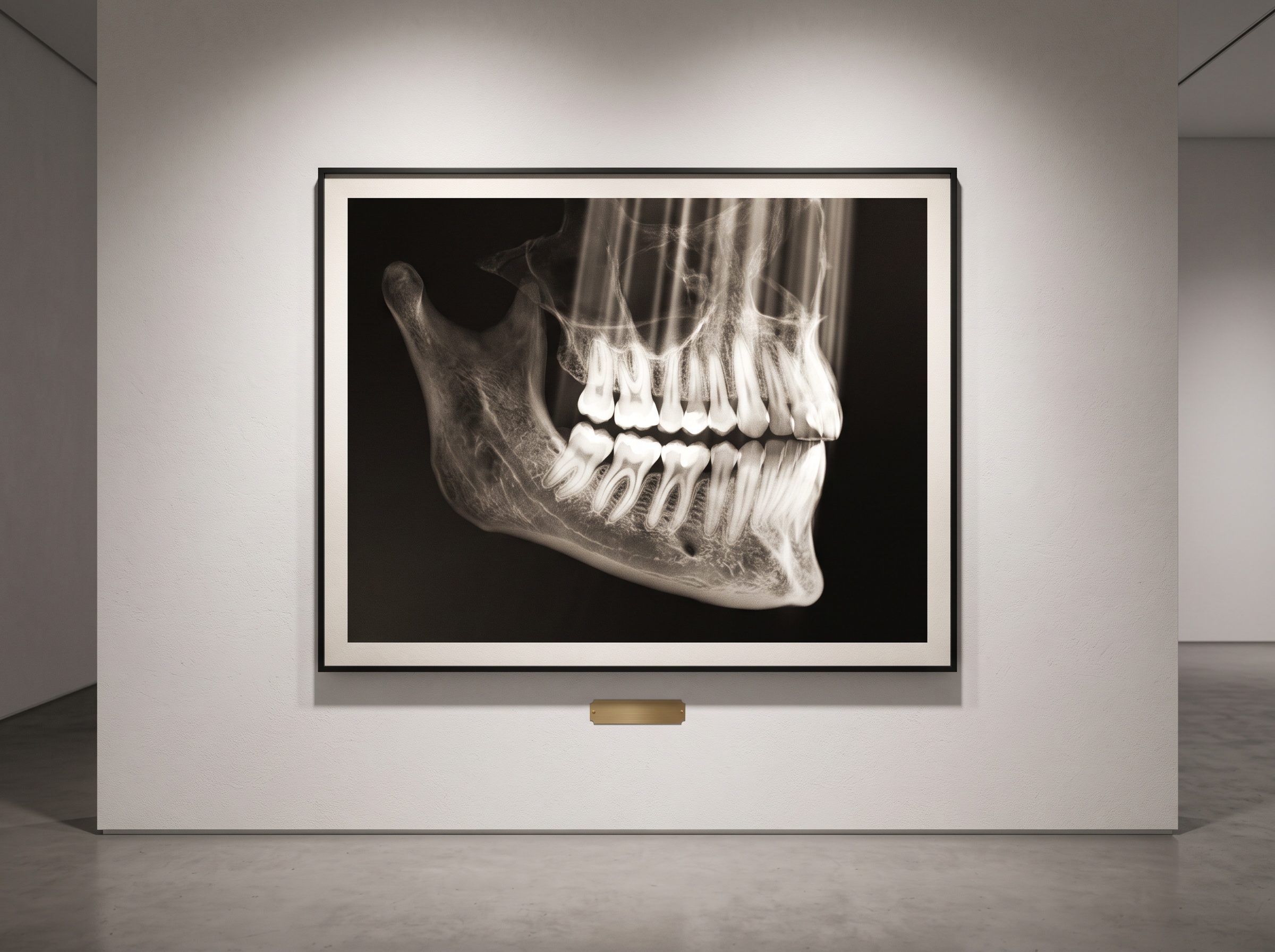
To make the whole depth of the volume speak at once, the simplest projection casts a ray through the block for every pixel on the screen and keeps only the single brightest voxel that ray encounters. This is maximum-intensity projection, and its effect is striking: dense, high-attenuation structures – cortical bone, root canals filled with gutta-percha, metal restorations – leap forward as if lit from within, while soft, low-attenuation tissue simply falls away and vanishes. For tracing a bright, well-separated structure through three dimensions, nothing is faster or cleaner. But the trade is severe and must be understood. By keeping only the peak along each ray, the projection discards all sense of what lies in front of or behind that peak; two structures at very different depths collapse onto the same spot with no cue to tell them apart. A maximum-intensity projection is superb at showing you that something dense is there and hopeless at telling you exactly where. It is a map of intensity, not of space.
Volume Rendering and the Transfer Function
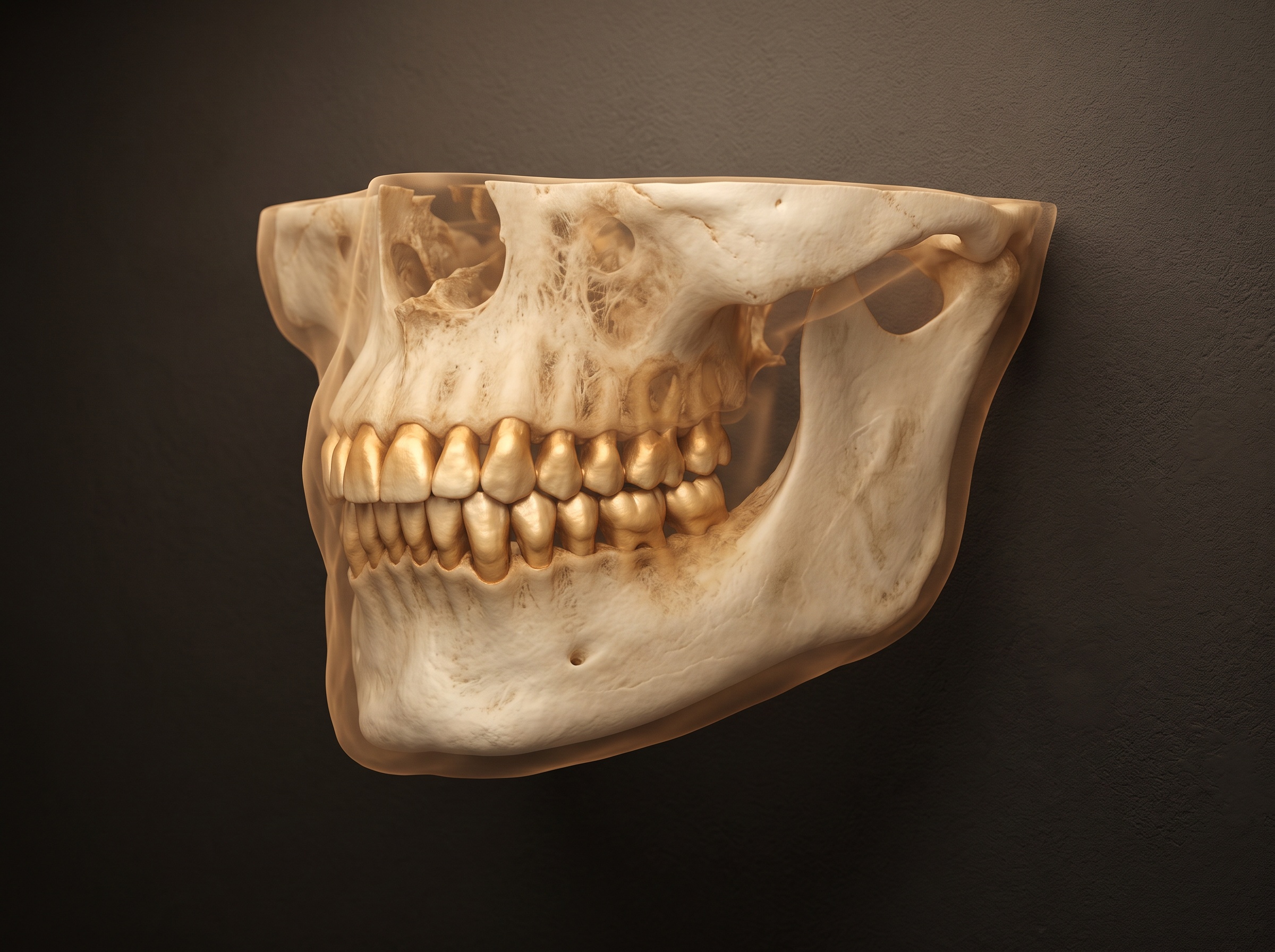
Full volume rendering is more ambitious and more expressive. Instead of keeping one voxel per ray, it accumulates along the entire ray, letting each voxel contribute a little color and a little opacity to the final pixel, as though light were passing through a block of tinted, semi-transparent glass. The instrument that governs all of this is the transfer function: a lookup that maps every possible attenuation value to a color and, crucially, to an opacity. Set enamel’s values to opaque and warm, and the crowns render as solid ivory. Set soft tissue near-transparent, and the lips become a faint veil you can see the teeth through. Set air to zero opacity, and it disappears entirely. The transfer function is where the volume becomes a picture with intent – the operator is, quite literally, deciding what the object is made of and what light is allowed to pass through it. Distinguishing one material from another is exactly the problem that dedicated hardware attacks in spectral and dual-energy CBCT, where two energies tell bone from metal; the transfer function is the software counterpart, sculpting the same distinction out of a single grey volume.
The Hard Shell and the Translucent Body
There is a blunter cousin to volume rendering that trades subtlety for solidity. Surface rendering picks a single attenuation threshold, declares everything above it “object” and everything below it “empty,” and drapes a hard polygonal shell over the boundary. The result is a crisp, sculptural model – excellent for surgical guides, printed replicas, and any workflow that needs a watertight mesh to hand to another system. But a threshold is a violent simplification: a thin cortical plate hovering just below the cutoff can disappear from the model entirely, and a hair fracture whose density sits between object and void may never form a surface at all. Translucent volume rendering keeps that ambiguity visible, showing the faint and the uncertain as faint and uncertain rather than deciding, silently, that they do not exist. Each approach buys clarity by discarding information; the craft is in knowing which information the clinical question can afford to lose. That same tension – what to keep, what to fuse, what to set aside – runs through any attempt to combine datasets, as in registering an intraoral scan onto a radiograph to hold two truths in one frame.
The Ethics of the Chosen View
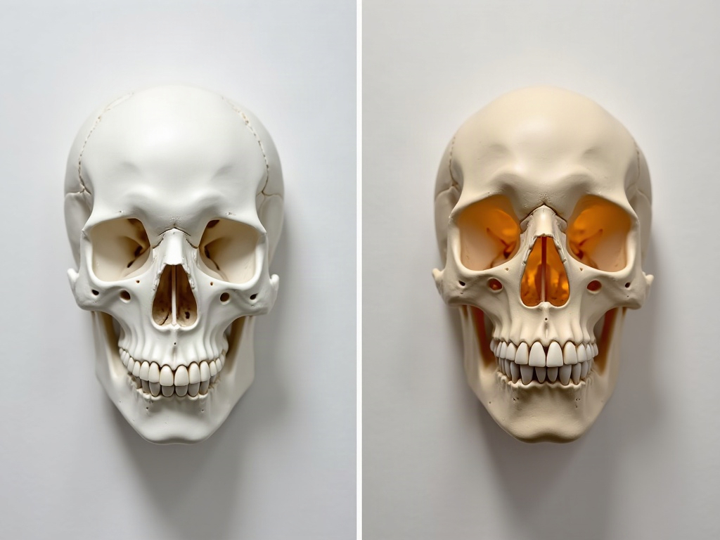
All of this leads to an uncomfortable and important truth: because the same volume can be made to show or hide a thin plate of bone, a periapical lucency, or a fracture line purely by nudging a transfer function or moving a threshold, the rendered image is not a neutral record but an argument. A window setting can flatter a diagnosis into existence or quietly erase a finding that a plainer slice would have shown. The discipline, then, is to treat the render as a claim that must be justified: to choose settings appropriate to the question being asked rather than to the answer one hopes to find, and to return to the honest, unglamorous multiplanar slices whenever a striking three-dimensional view starts to feel too persuasive. A beautiful volume rendering is a beginning, not a verdict.
Future Developments
The next chapter is already rendering. Cinematic, physically based volume rendering – the same light-transport mathematics that produces film-quality computer imagery – is arriving in medical viewers, casting soft shadows and realistic scattered light through the volume so that a jaw looks less like a diagram and more like an object you could pick up. AI-learned transfer functions promise to pre-select an appropriate render for a stated clinical question, sparing the clinician the slow manual tuning that good volume rendering still demands, while real-time interactive volumes let the whole block be turned, peeled, and re-lit at the speed of thought. The trajectory is consistent with everything imaging has always done here: it moves the picture closer to the object and puts more of the composition in the clinician’s hands. At PatientGallery we have always held that an image of the body is a made thing, lit and framed with intent. The CBCT simply hands us the raw block of stone. What we carve from it, and how honestly we light it, remains the work.
No Comments