Logo Logo Logo Logo Logo
  • Home
  • Download
  • About
  • Blog
  • Home
  • Download
  • About
  • Blog
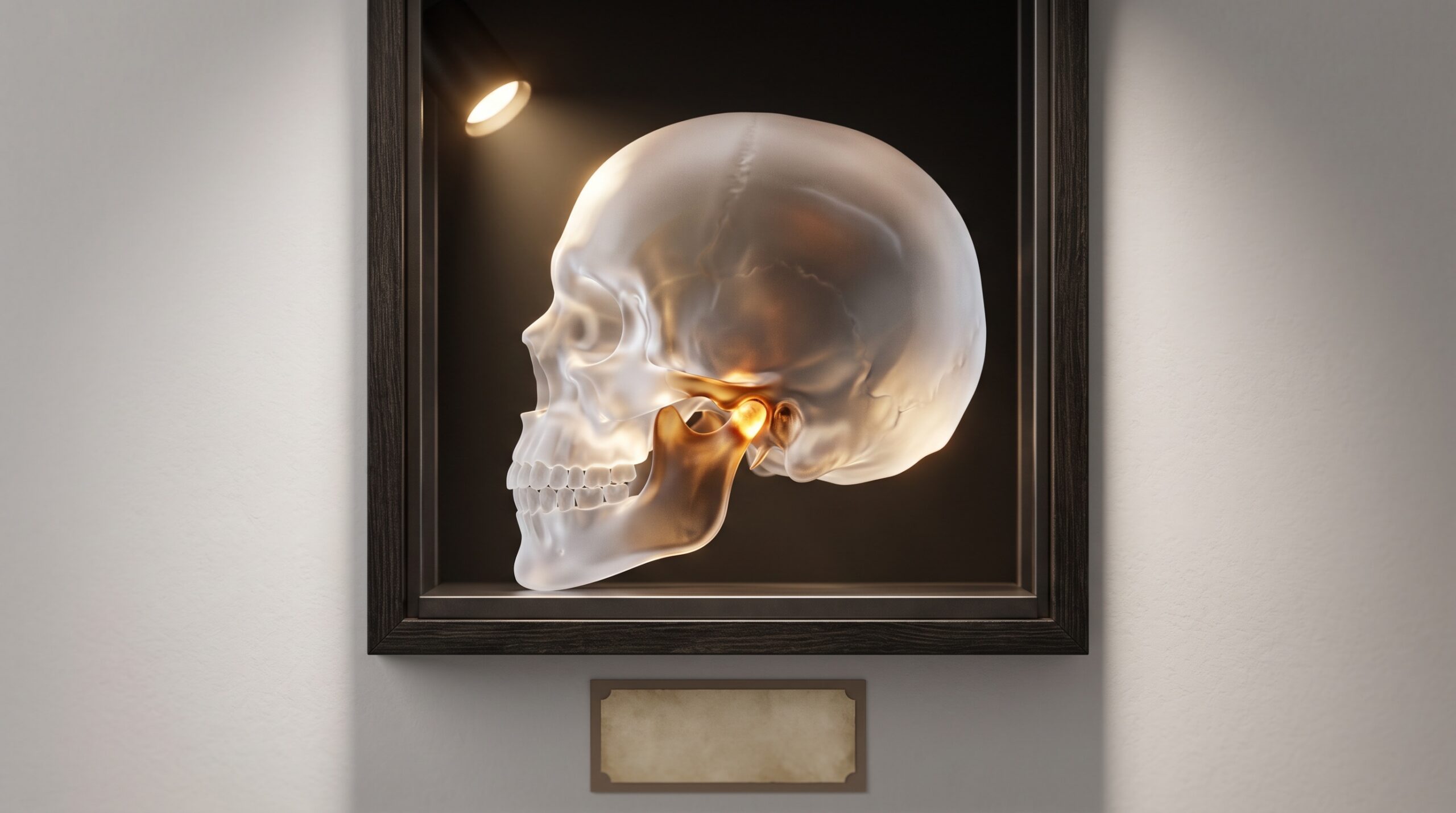
A translucent glass skull in profile with the jaw joint glowing warm and solid where the lower jaw meets the skull, presented as framed gallery art

22 Sep Imaging the Joint That Moves: MRI, CBCT, and the Craft of Picturing the Temporomandibular Joint

Posted at 11:30h in Dental Imaging by Raymond 0 Comments
0 Likes

The temporomandibular joint is the one structure in the mouth that refuses to sit still for its portrait. It is part bone and part soft cartilage, it works as a matched pair, and it is defined by movement - three properties that no single imaging...
Read More
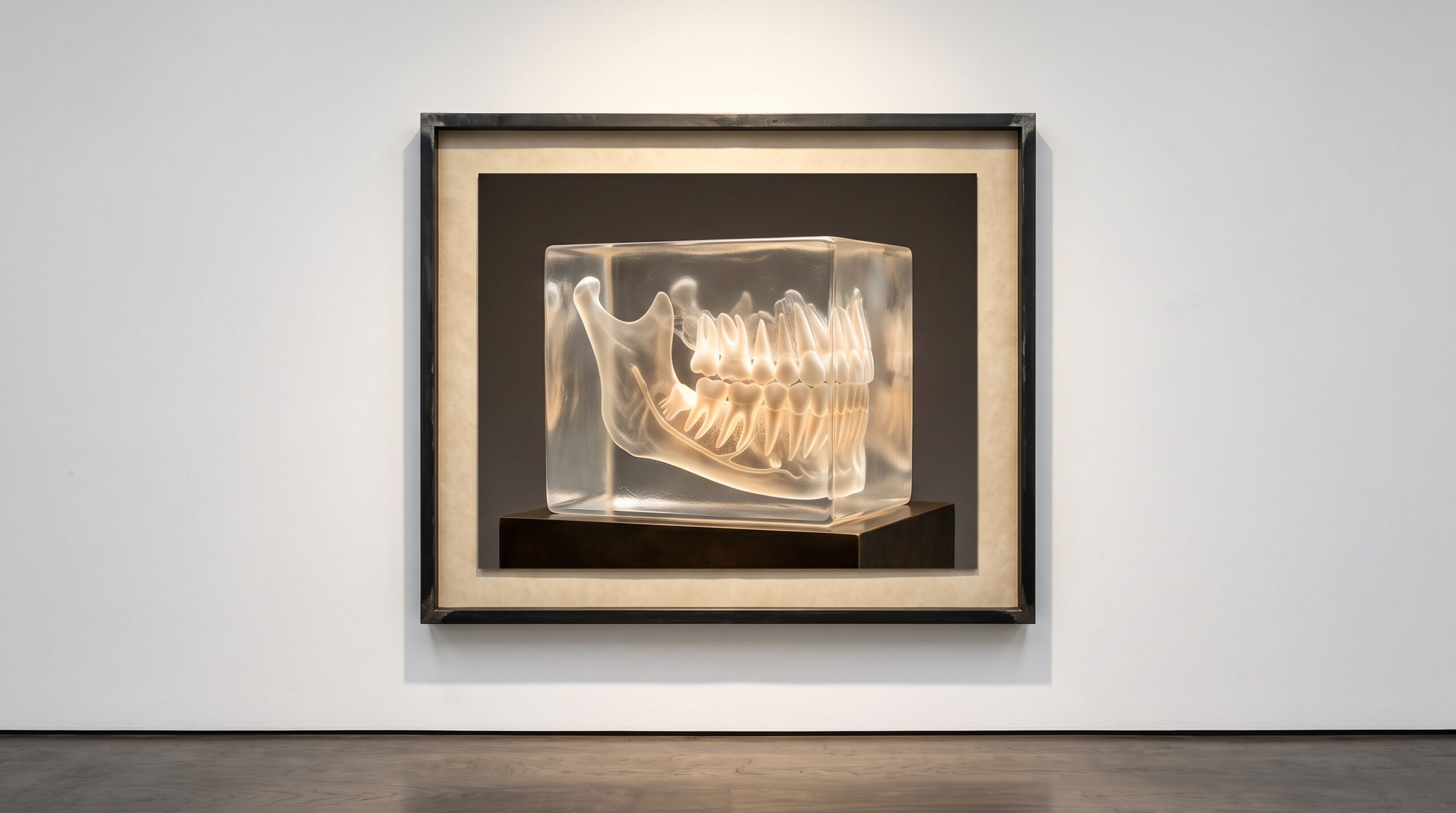
A jaw and teeth rendered as a translucent luminous volume with dense roots and bone glowing solid inside, presented as framed gallery art

21 Sep Sculpting the Volume: Maximum-Intensity Projection, Volume Rendering, and the Craft of Turning a CBCT Into a Picture

Posted at 10:37h in Dental Imaging by Raymond 0 Comments
0 Likes

A cone-beam scanner does not, strictly speaking, take a picture. It measures a dense three-dimensional block of numbers - a volume - and every flat image a clinician actually looks at is a decision made afterward about how to collapse that block onto a screen....

Read More
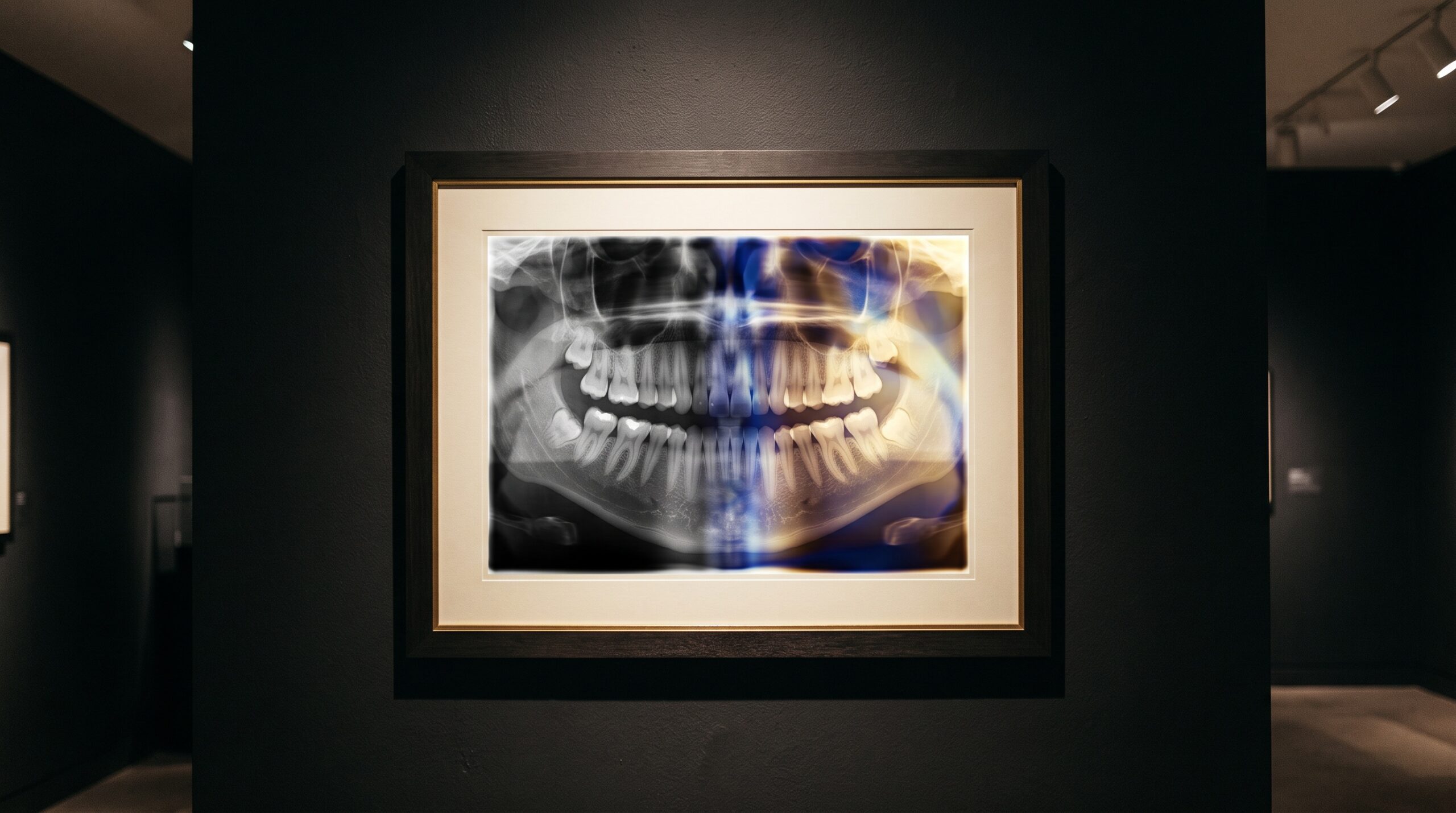
A framed dental radiograph split down the middle, neutral grayscale on the left dissolving into a smooth luminous color gradient on the right

28 Aug The Color of Grayscale: Pseudocolor, Colormaps, and the Quiet Editorial Hidden in a False-Colored Image

Posted at 11:38h in Dental Imaging by Raymond 0 Comments
0 Likes

A radiograph is data wearing gray. Map that gray to color and hidden structure leaps out - but the wrong colormap can paint boundaries that were never there. Pseudocolor is where imaging becomes both revelation and editorial....

Read More
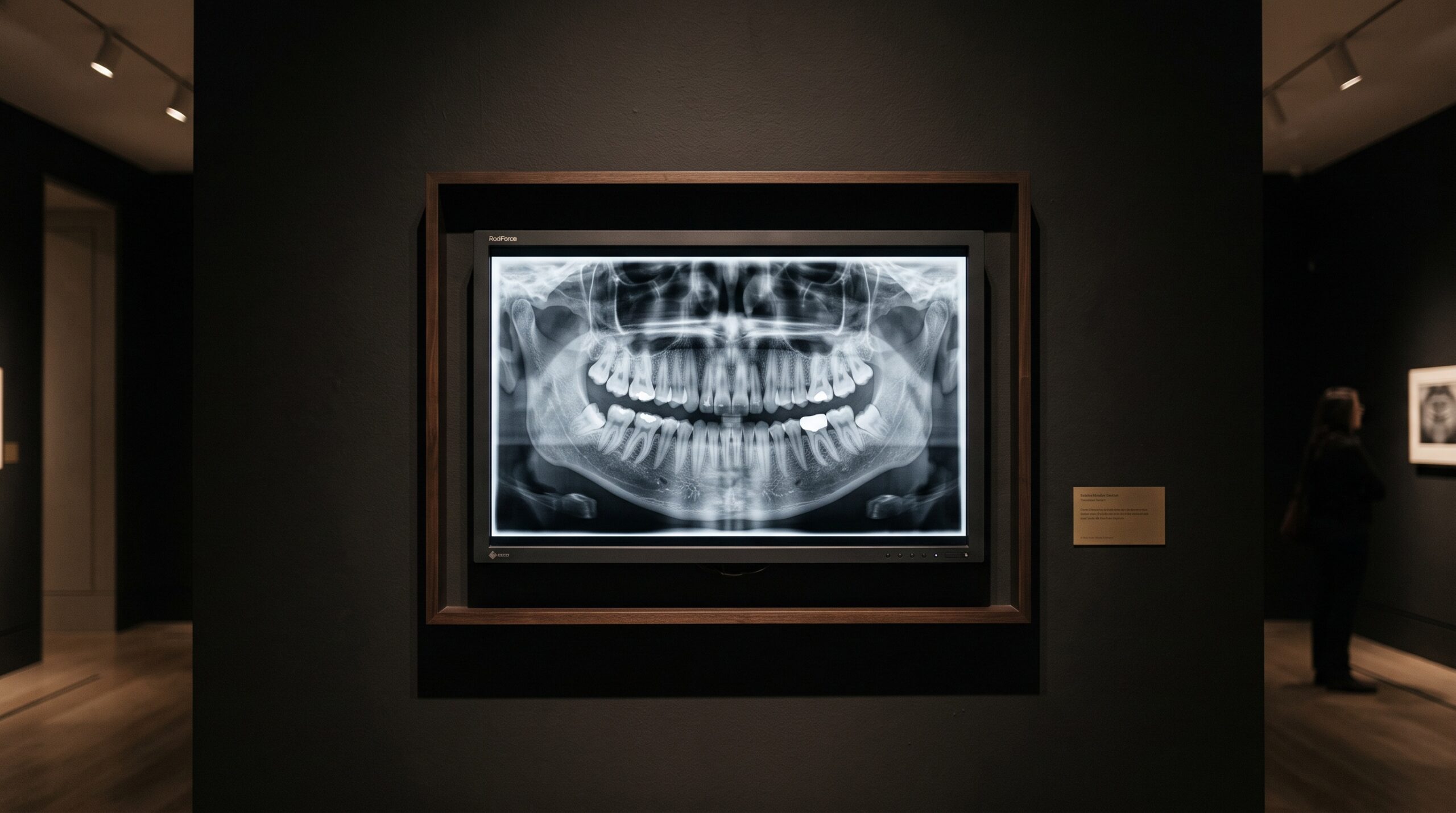
A diagnostic display monitor shown as framed gallery art, its screen holding a dental radiograph in a smooth ramp of grays

17 Aug The Last Instrument: Diagnostic Display Calibration and the DICOM Grayscale Standard for Reading a Radiograph

Posted at 11:38h in Dental Imaging by Raymond 0 Comments
0 Likes

Every radiograph ends its journey on a screen, and the screen is the one instrument in the imaging chain that most practices never calibrate. The DICOM grayscale standard exists because the human eye reads brightness unevenly — and because a monitor that does not account...

Read More
© Copyright 2020, Raster Builders Inc. All Rights Reserved.