


August 17, 2026 The Last Instrument: Diagnostic Display Calibration and the DICOM Grayscale Standard for Reading a Radiograph
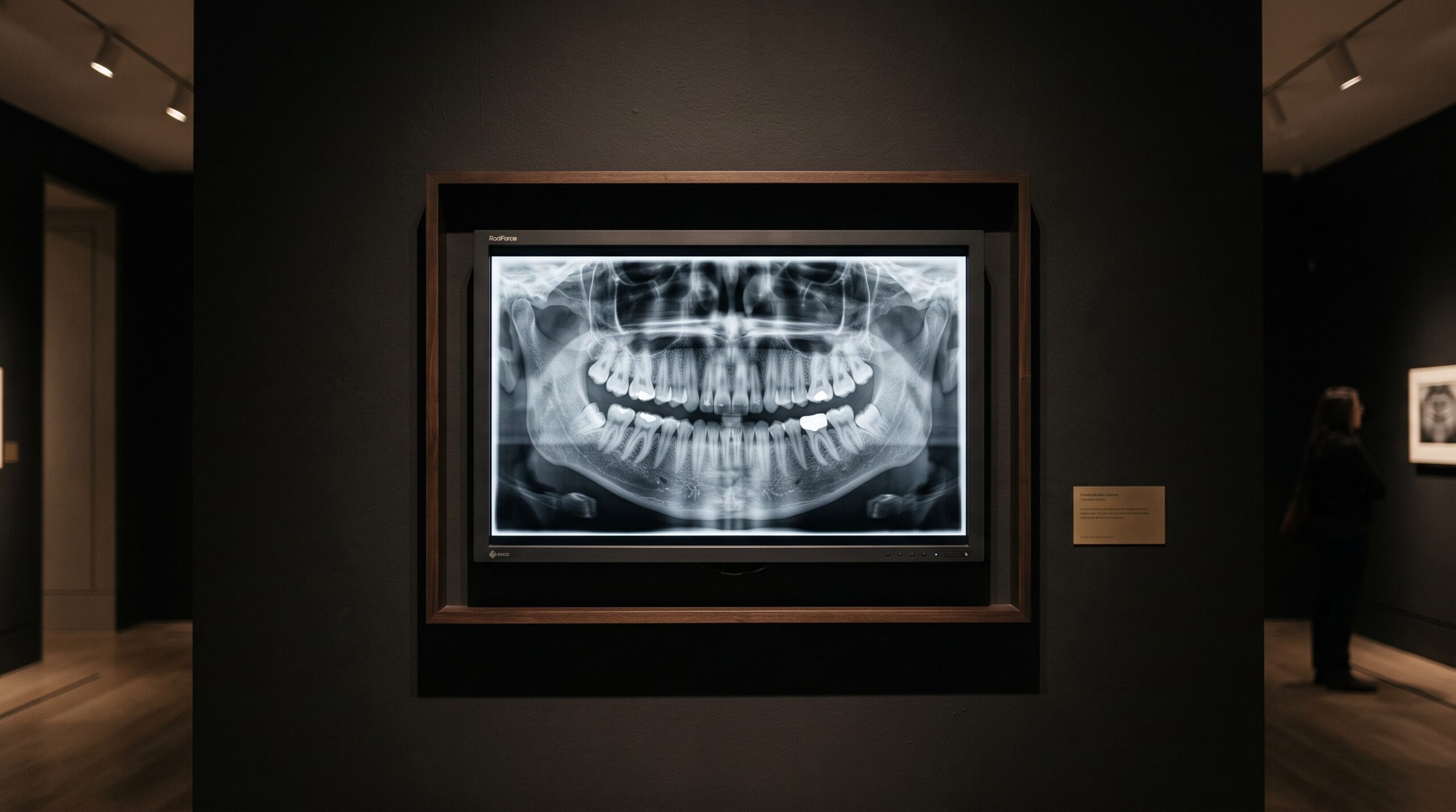
Consider the long journey of a single radiograph. A current is driven through a tube; a cascade of X-ray photons is shaped, filtered, and sent through the tissue; a sensor catches the survivors and renders their pattern into numbers; algorithms sharpen and scale those numbers into an image. And then, at the very end of this exquisite chain, the picture is thrown onto whatever monitor happens to sit on the desk — often the same commodity screen used for email and appointment books — and there, in the last handspan of its journey, much of the diagnostic subtlety so carefully gathered can quietly vanish. The display is the final instrument in imaging, and it is very nearly the only one that most practices never calibrate.
The Last Instrument in the Chain
We are accustomed to thinking of image quality as something the tube and the sensor decide. And they do decide a great deal of it — the true measure of what a detector can resolve is a genuinely physical quantity, as explored in the story of detective quantum efficiency. But a faithful signal is only as good as its final rendering. A radiograph that carries a real, faint radiolucency in its darker tones can arrive at a screen that cannot separate those tones from one another, and the finding is lost — not because the beam failed, not because the sensor failed, but because the last instrument in the chain was never tuned. The monitor is where physics finally becomes perception, and perception has rules of its own.
The Eye Is Not a Photometer
The whole problem begins with a quirk of human vision: we do not perceive brightness in equal, linear steps. Present the eye with a smooth ramp of luminance from black to white and it will discriminate the middle tones easily while struggling to tell one deep shadow from the next, or one near-white from its neighbour. A fixed increase in luminance is vividly obvious in the mid-grays and nearly invisible at the extremes. The smallest brightness difference a viewer can just detect — the just-noticeable-difference — is not a constant; it grows and shrinks across the tonal range. This is why a naive display, one that spreads its grays out in even physical steps, spends far too much of its scale where the eye cannot use it and far too little where the eye is keen. The diagnostic tones a radiograph lives in are precisely the subtle ones, and a screen indifferent to human contrast sensitivity buries them.
The Grayscale Standard That Bends the Curve
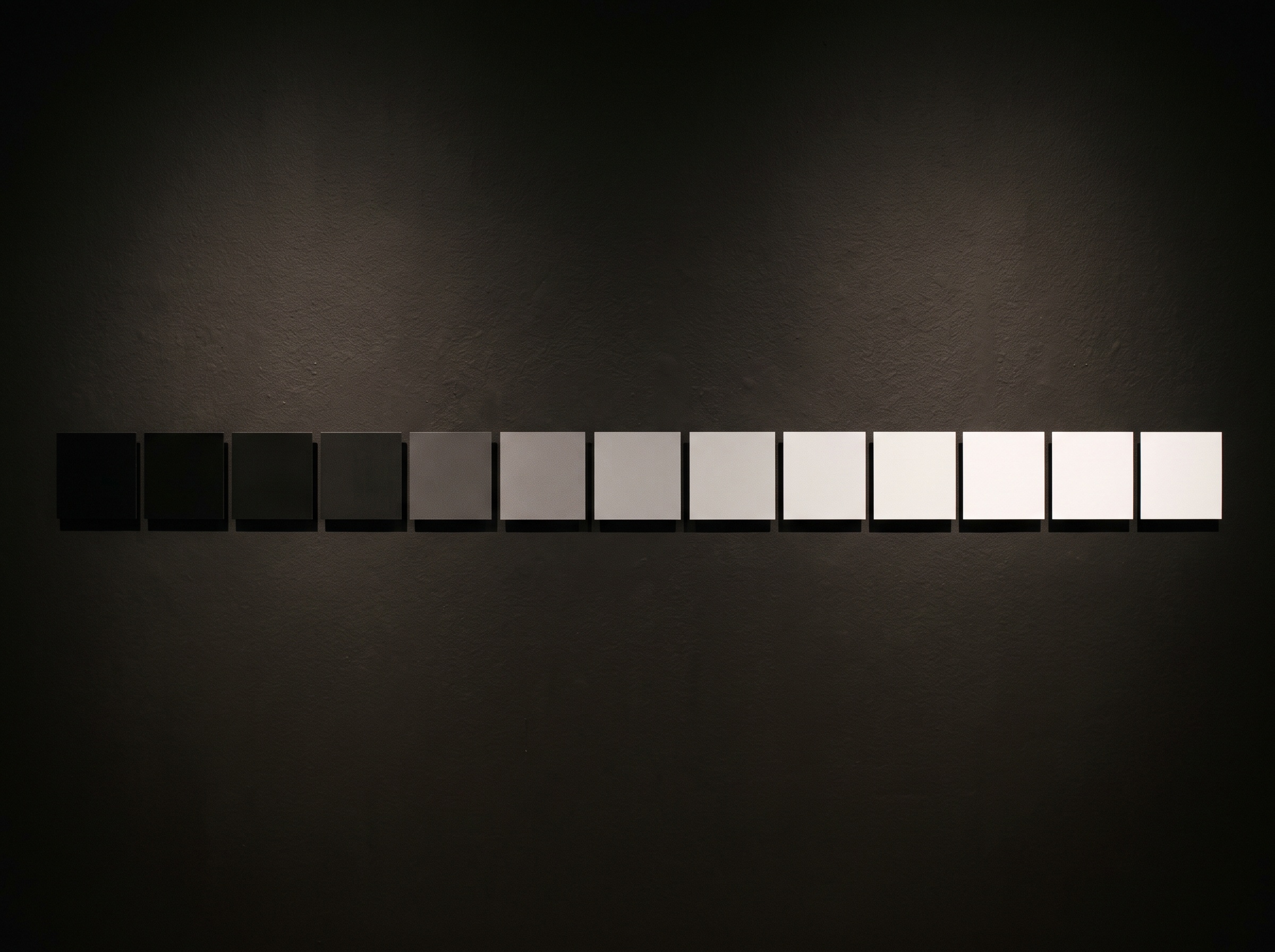
The answer, codified in the DICOM standard’s Grayscale Standard Display Function, is to stop treating the display as a physical device and start treating it as a perceptual one. Rather than assigning its brightness levels in equal luminance steps, a conformant display arranges them so that each successive step is a roughly equal perceptual step — one just-noticeable-difference apart, all the way from its darkest black to its brightest white. The characteristic curve of the monitor is deliberately bent into an S, lavishing extra separation on the shadow and highlight tones the eye finds hard and easing off in the middle where the eye is already sharp. The effect is that a given change in the underlying image value produces a similarly visible change wherever it falls on the scale. Two monitors calibrated to this standard, though built quite differently, will show the same radiograph with the same distribution of visible contrast — a consistency that matters enormously when one clinician’s finding must be trusted by another’s screen.

Luminance, Contrast, and the Room
A perceptual curve is only half the craft; the endpoints matter as much as the shape between them. A display’s maximum luminance sets how bright its brightest white can be, and the ratio between that white and its darkest black defines the tonal range available for the whole grayscale to inhabit. A dim consumer panel with a shallow luminance ratio simply has less room in which to lay down distinguishable grays, so its shadows crowd together no matter how the curve is shaped. Diagnostic displays are specified to reach and hold far higher luminance, and to sustain it as the backlight ages, because a display that dims over its life slowly erases the faint end of the scale. And none of it survives a badly lit room: ambient light falling on the screen adds a veiling glow that lifts the blacks, collapses contrast, and drowns exactly the low tones the calibration worked to reveal. The reading environment — subdued lighting, no reflections across the glass — is not a nicety but part of the instrument itself. Brightness written into the image can still be read away by the room, in a manner not unlike how post-processing can deceive on a digital radiograph.
Why the Office Monitor Quietly Fails
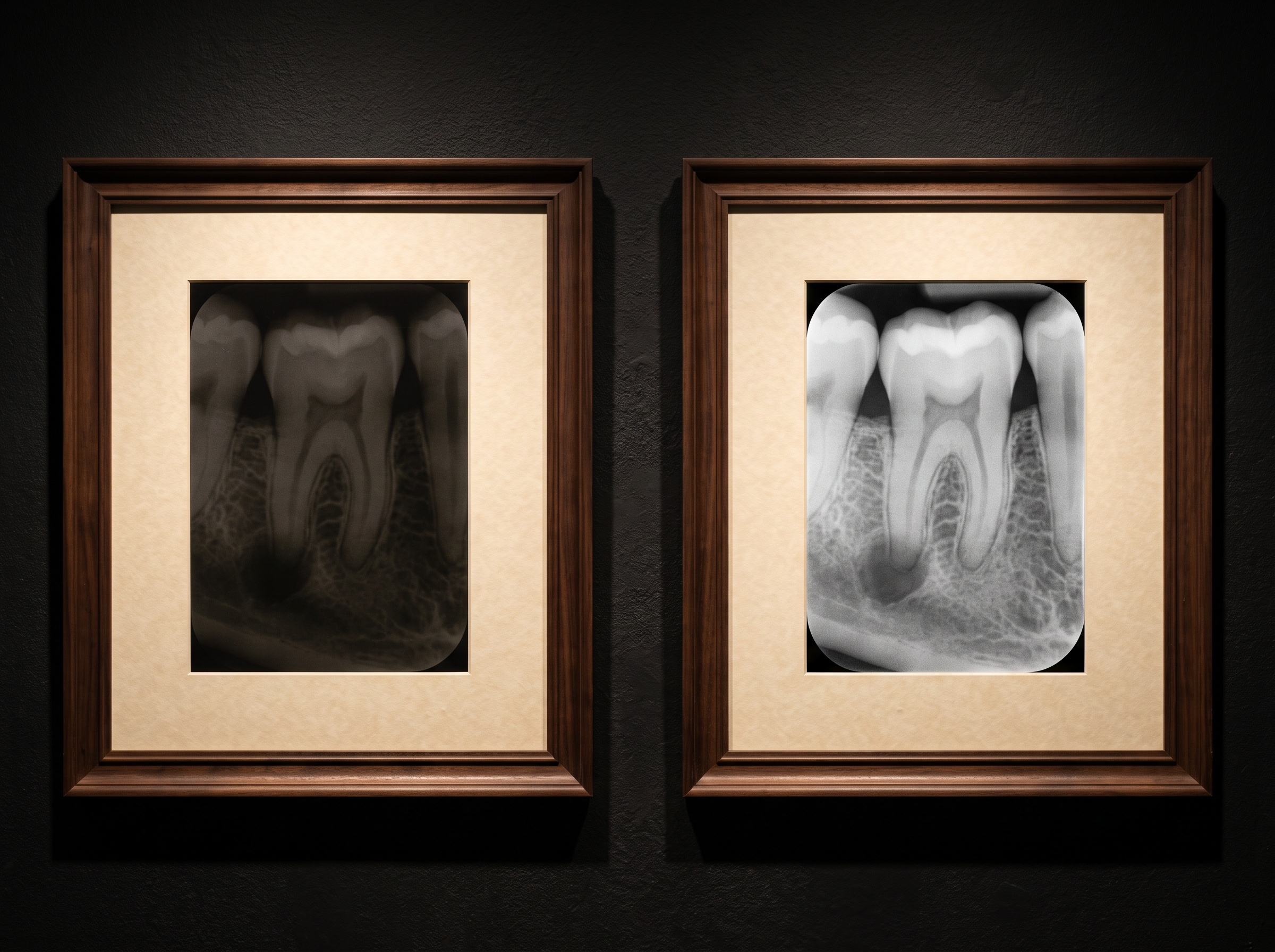
The everyday screen fails not dramatically but silently, and that is what makes it dangerous. It renders a radiograph that looks perfectly acceptable — clean, contrasty, even pleasing — while having compressed the darkest and lightest tones into indistinguishable blocks. The pathology that hides in a subtle shift of gray, an early interproximal shadow or a faint change at a root, is not flagged as missing; it is simply not there to be seen, indistinguishable from the tone beside it. This is a distinct concern from color fidelity — the difficulty of reproducing a tooth’s true hue on any screen is its own separate problem of gamut and metamerism — because the radiograph asks nothing of color and everything of grayscale separation. Consumer displays are engineered for vivid photographs and text legibility, tuned to look attractive rather than to be honest, and their generous factory contrast is often the very thing crushing the diagnostic shadows. A screen that flatters the image is not the same as a screen that tells the truth about it, and the grammar of that truth is the disciplined handling of grays described in the craft of contrast and density.
Keeping the Display Honest
Because a display drifts as it ages, conformance is not a one-time purchase but an ongoing discipline. The standardized approach is to verify the monitor against known test patterns — the AAPM’s TG18 patterns being the enduring reference — which make grayscale conformance visible to the eye and measurable by a photometer. In a well-kept pattern, a low-contrast target sitting in the deepest shadows should be as perceptible as one sitting in the brightest highlights; if the dark target has vanished, the curve has drifted and the display is lying by omission. A periodic check, a luminance measurement, an honest look at whether the darkest steps are still separable — these are the quiet quality-assurance habits that keep the final instrument trustworthy. It is unglamorous work, but it is of a piece with everything a gallery does: the frame, the glass, and the light are never allowed to distort the work they present.
Future Developments
The display is beginning to grow as sophisticated as the sensor that feeds it. Self-calibrating diagnostic monitors now carry built-in photometers that measure their own output and re-tune the grayscale curve automatically, silently correcting the drift of an aging backlight without a technician’s intervention. Ambient-light sensors are learning to compensate the black level for the room’s changing brightness, narrowing the gap between the ideal reading environment and the real one. And as machine-learning tools take up residence in the interpretation of radiographs, a subtle new alignment problem appears: the algorithm reads the raw image values directly, unbothered by any monitor, while the clinician reads a rendered picture whose faint tones may or may not be visible on the screen in front of them — a discrepancy that display calibration quietly closes by ensuring the human sees what the data actually holds. The horizon points toward displays that guarantee, moment to moment, that no diagnostic shade is lost between the sensor and the eye. For now, the lesson is older and simpler. An image is not finished when it is captured; it is finished when it is seen. The monitor is where seeing happens, and a picture is only ever as honest as the last surface it is shown upon.
Sources & further reading:
Sorry, the comment form is closed at this time.