Logo Logo Logo Logo Logo
  • Home
  • Download
  • About
  • Blog
  • Home
  • Download
  • About
  • Blog
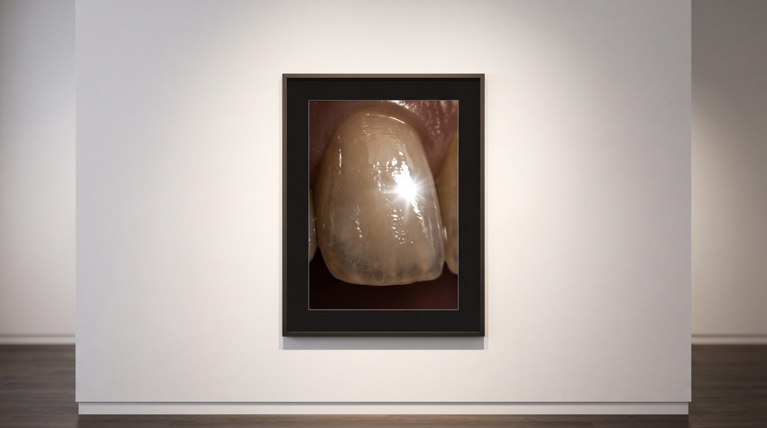
A luminous tooth casting a soft graduated shadow with a fading penumbra edge, lit by a small glowing rectangular source above

12 Sep The Blur Built Into Every X-Ray: Focal Spot Size, Penumbra, and the Geometry of a Sharp Radiograph

Posted at 10:38h in Dental Imaging by Raymond 0 Comments
0 Likes

Every dental radiograph carries a blur that no detector, no software, and no amount of retaking can fully remove - because the X-ray source is not a point of light but a small glowing area on a metal target, and every point on that area...

Read More
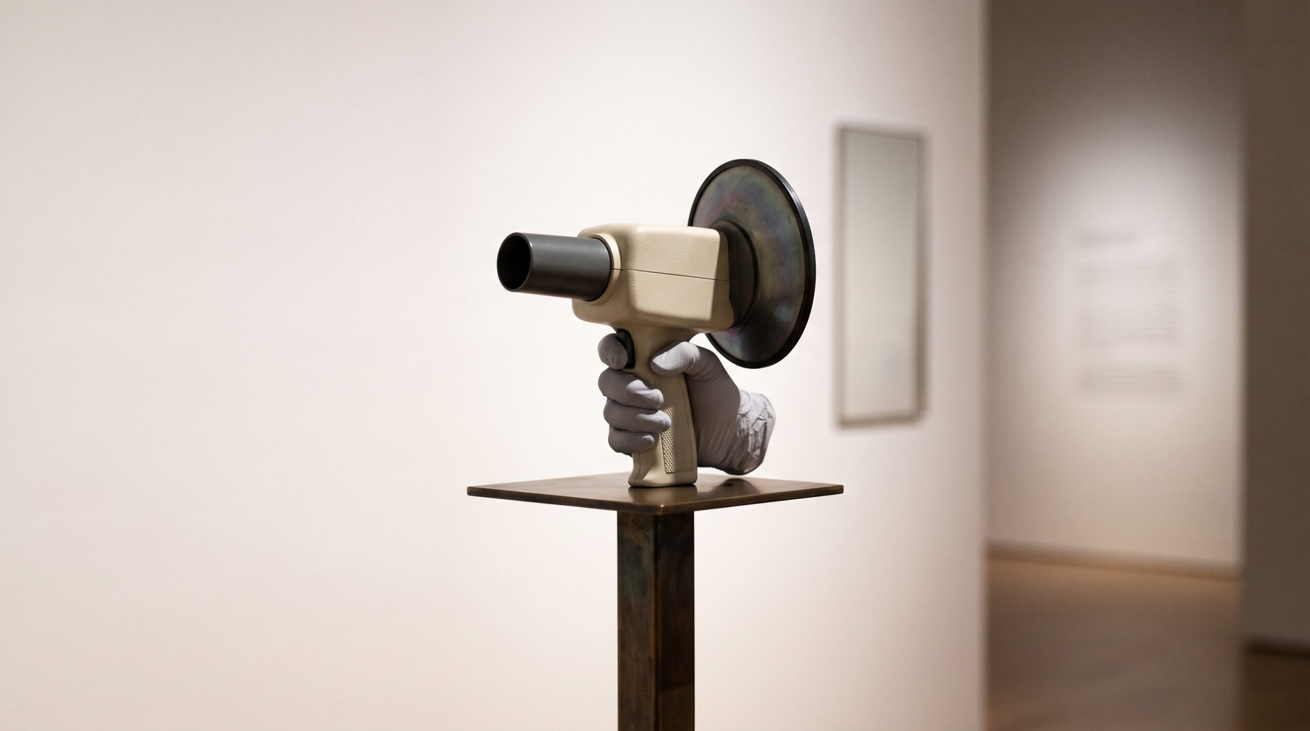
A handheld dental X-ray unit with a round backscatter shield at the cone, held as if it were a piece of gallery sculpture

07 Sep Can a Handheld Radiograph Match the Wall-Mount? The Physics of Image Quality in Portable Dental X-Ray

Posted at 10:38h in Dental Imaging by Raymond 0 Comments
0 Likes

A wall-mounted X-ray head is a fixed instrument, aligned by a stable arm and switched from behind a wall. A handheld unit puts the same tube in one gloved hand and asks the clinician to be both aimer and tripod. The device has become common...

Read More
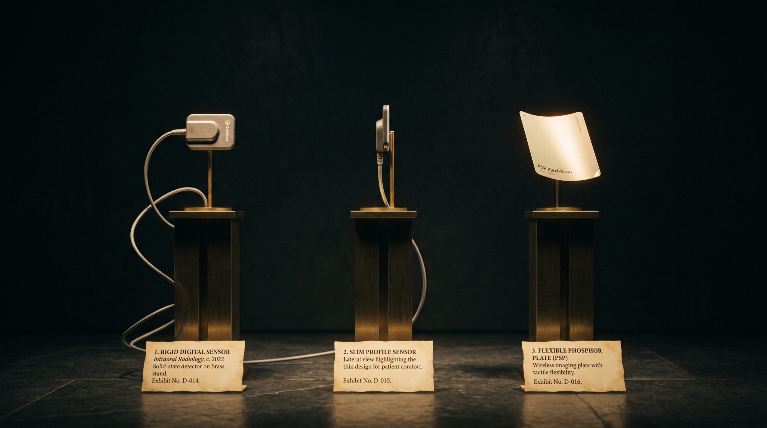
Three dental x-ray detectors displayed as artworks on brass museum plinths: two rigid corded solid-state sensors and one thin flexible phosphor plate.

04 Sep Three Ways to Catch a Photon: CCD, CMOS, and Phosphor Plates Compared

Posted at 10:39h in Dental Imaging by Raymond 0 Comments
0 Likes

Every digital intraoral image begins with a decision made in silicon or phosphor: how to turn an x-ray photon into a number. There are three answers in common use, and each one quietly shapes what the radiograph can and cannot show....

Read More
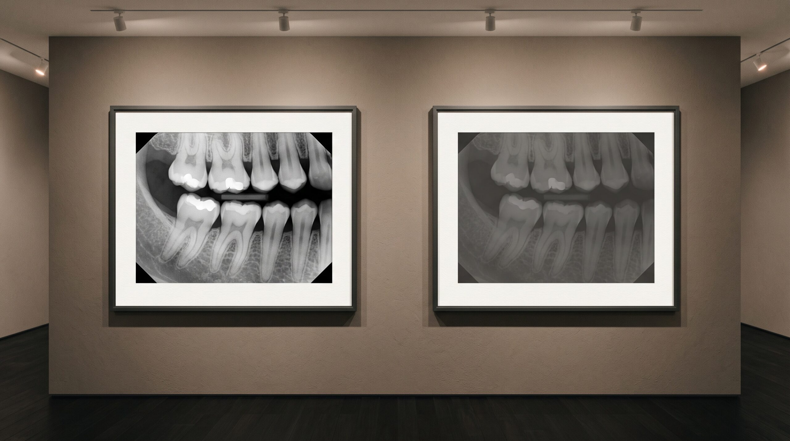
Two framed versions of the same dental radiograph on a gallery wall, one crisp and one muddy and underexposed

04 Sep Garbage In, Garbage Out: Image Quality and Why AI Still Needs a Good Radiograph

Posted at 09:44h in Dental Imaging by Raymond 0 Comments
0 Likes

A detection model will read an underexposed, cone-cut, motion-blurred bitewing without hesitation and hand you a confident, tidy result. That composure is the danger. Everything AI concludes about a radiograph is downstream of how well the image was made - and the machine cannot see...

Read More
A blank digital x-ray sensor panel presented like a primed empty canvas on a brass easel under museum lighting, its surface a faint grain of countless cells.

01 Sep The Sensor’s Fingerprint: Dark Current, Fixed-Pattern Noise, and the Flat-Field Correction That Makes a Detector Honest

Posted at 10:38h in Dental Imaging by Raymond 0 Comments
0 Likes

No two pixels on a digital sensor are born equal. Each carries its own faint bias, its own gain, its own quiet defect - and unless the detector first learns and subtracts that fingerprint, the pattern it prints onto every image is its own, not...

Read More
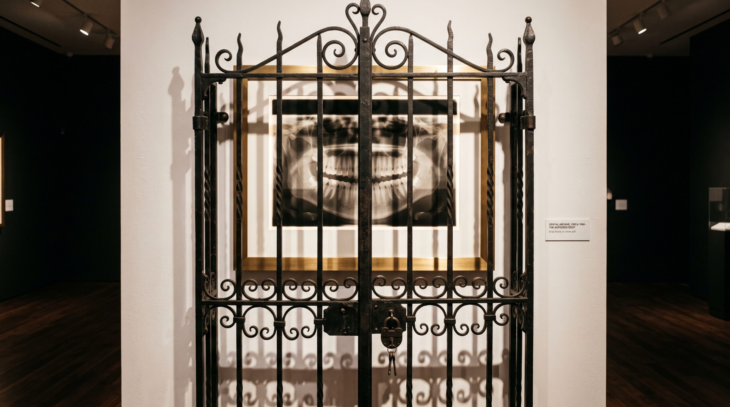
A framed dental radiograph glowing behind a closed, locked iron gate on a gallery wall - the image visible but unreachable.

31 Aug The Image That Cannot Travel: DICOM, Interoperability, and the Quiet Cost of a Radiograph That Will Not Move

Posted at 10:38h in Dental Imaging by Raymond 0 Comments
0 Likes

The finest radiograph in the world is worthless to the specialist who cannot open it cleanly. Between one practice and the next stands the quiet, unglamorous problem of interoperability - and the images that arrive flattened, stripped, or not at all....

Read More
A framed dental radiograph split down the middle, neutral grayscale on the left dissolving into a smooth luminous color gradient on the right

28 Aug The Color of Grayscale: Pseudocolor, Colormaps, and the Quiet Editorial Hidden in a False-Colored Image

Posted at 11:38h in Dental Imaging by Raymond 0 Comments
0 Likes

A radiograph is data wearing gray. Map that gray to color and hidden structure leaps out - but the wrong colormap can paint boundaries that were never there. Pseudocolor is where imaging becomes both revelation and editorial....

Read More
A framed extreme close-up of pink gingival tissue around a single restored tooth, lit like a museum artwork with a thread of light tracing its contour

27 Aug Scoring the Soft Frame: When a Neural Network Learns to Read the Pink Esthetic

Posted at 10:38h in Clinical Photography by Raymond 0 Comments
0 Likes

The Pink Esthetic Score turns the gingival frame into a number. Now a neural network is learning to read that number straight from the photograph - and to preview it before the implant is ever placed....

Read More
Two framed images of the same molar side by side - a color surface scan and a grey radiograph - with light suggesting they are merging

21 Aug Two Ways of Seeing, One Frame: Fusing the Intraoral Scan and the Radiograph Into a Single Image

Posted at 10:38h in Dental Imaging by Raymond 0 Comments
0 Likes

One instrument captures the surface and refuses to look beneath it. The other looks straight through and forgets the surface exists. For decades the clinician fused them in the mind. Now the software does it - and the craft is changing....

Read More
A framed dental radiograph on a gallery wall with a small brass placard beneath it bearing a single glowing number

20 Aug The Dose You Cannot See: Exposure Index, Dose Creep, and the Number a Forgiving Sensor Hides

Posted at 11:39h in Dental Imaging by Raymond 0 Comments
0 Likes

Film punished overexposure by going dark. Digital rewards it with a cleaner picture - which is exactly how dose creeps upward, invisibly. Here is the number that fixes it....

Read More
  • 1
  • 2
  • 3
© Copyright 2020, Raster Builders Inc. All Rights Reserved.