


September 18, 2026 Composing the Clinical Portrait: Framing, the Midline, and the Craft of the Retracted View
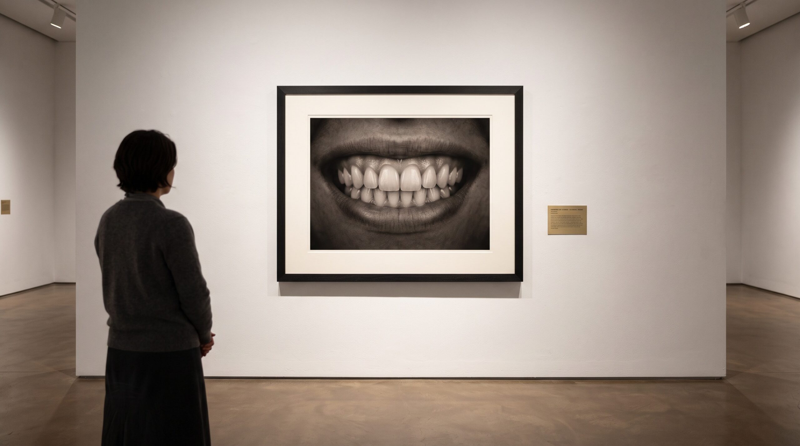
We tend to praise a clinical photograph for the things that are easy to measure. It is sharp, or it is not; the exposure is clean; the color is true. Yet all of those virtues are downstream of a decision made before the shutter ever moves – the decision of where to put the frame. Composition is the part of dental photography least often taught and most quietly consequential, because it is not something the camera does. It is something the operator chooses. What the picture includes, what it leaves out, how it is centered and leveled and how much room the subject is given to breathe – these choices are made by a person, and they are the difference between a diagnostic record and a snapshot. It is also the part of the craft that most resembles the discipline of the gallery, where a curator’s first act is not to light the work but to decide where its edges fall.
The Frame Is a Decision
A frame is a boundary, and every boundary is an argument about what matters. Point a camera at a retracted smile and you must immediately settle a dozen questions: how much of the lip and soft tissue to keep, where the crop lands relative to the incisal edges, how tightly to fill the frame with the subject versus how much context to preserve. Fill it too loosely and the diagnostic detail shrinks into a corner; crop too aggressively and you lose the landmarks that let anyone orient the image later. Magnification and working distance are compositional tools as much as optical ones, because they decide the scale at which the subject is rendered. And the retracted intraoral view brings its own adversary – specular reflection from wet enamel and metal that can wash out the very structure you are trying to record. Managing that glare is a compositional concern as much as a lighting one, and it is the reason the craft of cross-polarization in clinical dental photography earns its place in the standardized workflow: it lets the composed frame show true surface texture instead of a scatter of highlights.
The Midline as Plumb Line
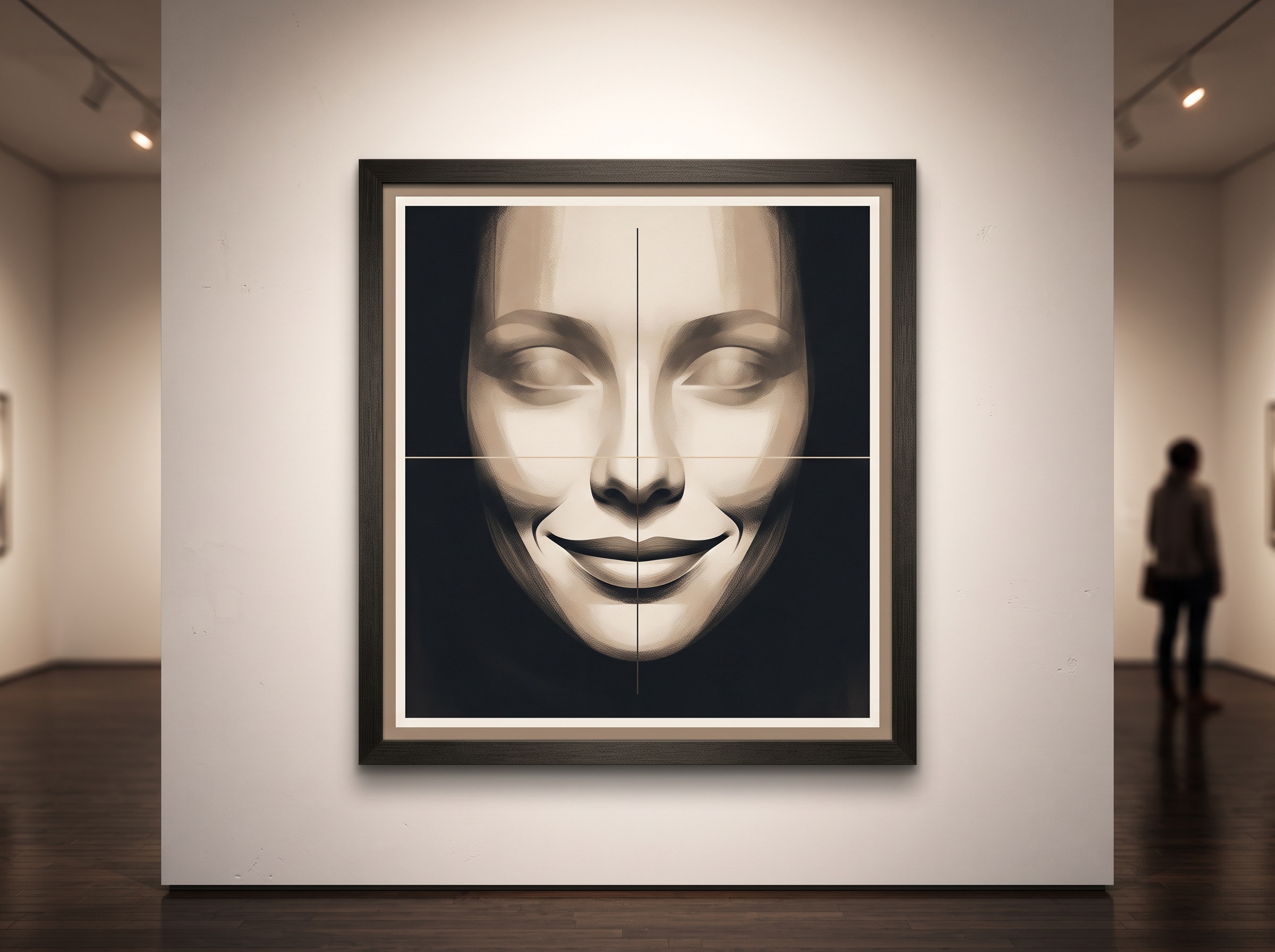
A painter hangs a canvas level; a photographer of the mouth has two references that serve the same purpose. The facial midline is the frame’s plumb line, and the occlusal plane is its horizon. Get either one wrong and the image lies in a specific, insidious way. Rotate the sensor a few degrees off the patient’s true axis and the picture manufactures asymmetries – a cant, an uneven gingival display, a midline that appears to deviate – none of which belong to the patient. They are artifacts of the frame. This is why squaring the camera to the facial midline and leveling the occlusal plane is not a nicety but the compositional act that keeps a clinical portrait honest. When a real asymmetry does appear in a well-composed image, you can trust it, because you have removed the camera as a suspect. The same discipline governs whether the picture preserves true proportion at all, which is exactly the dimensional-fidelity question weighed in our comparison of whether a smartphone can frame the esthetic zone as faithfully as a DSLR – geometry and framing decide whether a millimeter on the sensor is still a millimeter in the mouth.
Retraction, Negative Space, and the Gallery Wall
A museum does not crowd its walls. It gives a single canvas a broad field of quiet around it so the eye has somewhere to rest and nothing to compete. The clinical frame asks for the same restraint by different means. Full retraction pulls the lips and cheeks clear of the field; a dry, isolated tooth surface removes the distraction of saliva and reflection; a clean contrastor or a neutral, uncluttered background behind an anterior shot suppresses the busy shadows of the posterior mouth. And negative space – the deliberate emptiness around the subject – does real work rather than wasting the frame. It isolates the structure, sets a consistent scale, and leaves margin for the crop to be adjusted later without amputating a landmark. A composition that respects negative space reads instantly; one that jams the subject edge-to-edge forces the viewer to hunt. The gallery wall and the retracted, dried, well-backed clinical field are solving the same problem: how to let the subject be seen without argument.

The Standardized Series as a Body of Work
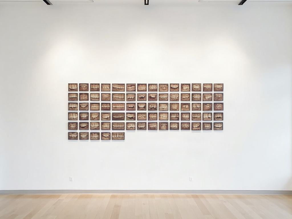
No single clinical photograph carries the full weight of the record. The value lives in the series – the coordinated set of extraoral portrait, intraoral frontal, right and left laterals, upper and lower occlusals, and the anterior close-ups – taken together as one coherent body of work. And what makes a series worth anything is consistency so strict it approaches ritual: the same framing, the same magnification, the same orientation, the same retraction, visit after visit. The point of that rigor is comparability. Only when the frame is held constant can two images taken months apart be laid side by side and read as a change in the patient rather than a change in the photographer. The moment framing drifts between sessions, the comparison is contaminated – you can no longer tell recession from a tighter crop, or wear from a closer lens. A standardized series is a promise that the frame will not become a variable, and it is the compositional habit that turns a folder of pictures into a longitudinal document.
What the Composition Protects
Discipline in framing is not aesthetics for its own sake; it protects several things at once. It protects reproducibility, so before-and-after comparisons are honest. It protects the medico-legal record, because a consistently composed, well-oriented image is a defensible one – it shows what it claims to show, from a known vantage, without the ambiguity that a careless crop invites. It protects communication, letting a laboratory technician or a specialist read the frame the same way the clinician did. And it protects the aesthetic reading of the case – the balance of the soft-tissue frame around the teeth that determines whether a restoration will look alive, a reading that machine vision now formalizes, as in the work on a neural network learning to score the pink esthetic from a clinical photograph. None of that scoring means anything on a poorly composed frame. Faithful color is the companion virtue that composition sets the stage for, since a beautifully framed image still fails the shade discussion if the hue is wrong – the reason we keep returning to the limits of gamut, metamerism, and why a tooth refuses to be perfectly reproduced. Compose well and you give every downstream judgment – clinical, legal, aesthetic – a frame it can actually stand on.
Future Developments
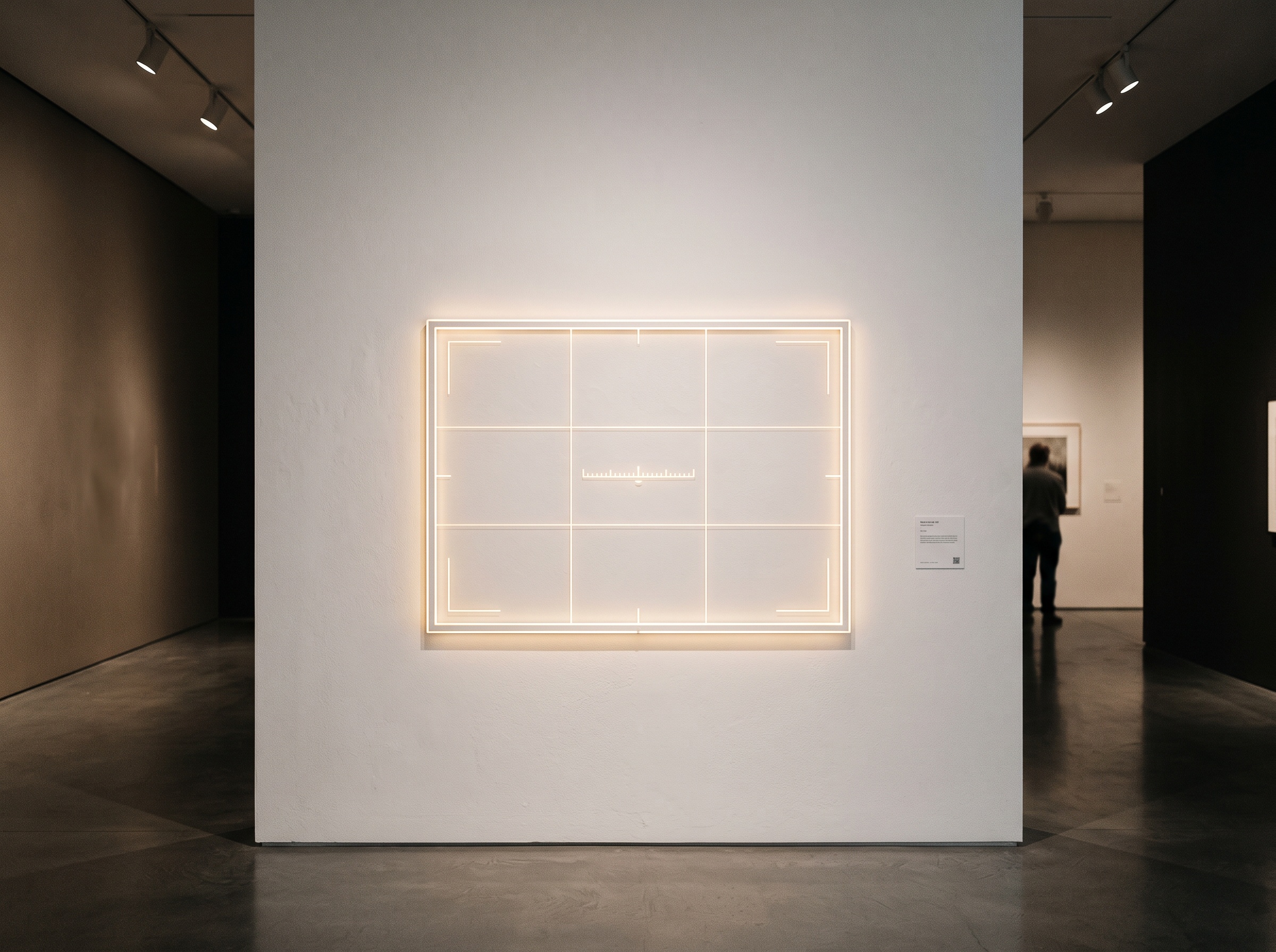
The compositional eye has always been a trained, human thing, and it will remain so. But the camera is beginning to help keep the standard the eye already knows. Real-time framing guides, midline and occlusal-plane overlays projected into the viewfinder, and level indicators that warn of an off-axis capture are moving from consumer photography into clinical systems, and behind them sits the more interesting prospect: software that recognizes a poorly cropped or rotated frame at the instant of capture and asks for the shot again before the patient has left the chair. This is not automation replacing the composer. It is automation enforcing a discipline that has always been easy to know and hard to keep, especially in a busy operatory where the temptation is to accept the image that is merely good enough. As those guides mature, the gallery’s oldest lesson becomes something the instrument itself defends – that a picture worth trusting begins not with light or color but with the quiet, deliberate placing of the frame. At PatientGallery, that is where the craft has always started, and it is where, increasingly, the machine will start too.
Sorry, the comment form is closed at this time.