



September 9, 2026 The Discipline of Restraint: The New Imaging Guidance and the Art of the Radiograph Not Taken
A gallery is defined as much by what it declines to hang as by what it displays. The curator’s real work is the edit – the discipline to leave a wall generous and quiet rather than crowd it with every canvas on hand. Dental imaging, at its most refined, asks for the same restraint. The most revealing decision a clinician makes about a radiograph is frequently not how to expose it or how to read it, but whether to make it at all. For years that decision drifted toward habit, governed by the calendar and the recall interval more than the patient in the chair. In January 2026 the American Dental Association, together with the American Academy of Oral and Maxillofacial Radiology, published its first recommendations on radiographic patient selection in more than a decade – and, notably, the first to address both conventional dental radiography and cone-beam computed tomography. Their quiet thesis is that imaging is most effective when used in moderation, and that the choice to image is an act of judgment, not routine.
This is not a call to withhold care. It is a call to practice it deliberately. The recommendations reframe the radiograph from a scheduled event into a considered response to a specific clinical question – and in doing so they hand the clinician back an authorship that the fixed interval had quietly taken away.
A Standard, Not a Schedule
The heart of the guidance is individualization. Rather than prescribing a uniform cadence for everyone who sits down, it asks that each imaging decision be grounded in the patient’s medical and dental history, the findings of the clinical examination, and an honest assessment of that individual’s disease risk. A caries-free adult with a stable mouth and low risk does not warrant the same radiographic attention as a patient with active disease, a history of restorations, or conditions that raise their risk. The examination comes first; the image answers a question the examination has already begun to ask. This inverts the old reflex, in which the radiograph was ordered because time had passed rather than because the mouth had spoken. A standard, applied with judgment, replaces a schedule applied by default – and the difference, over a career, is measured in thousands of exposures that were never clinically necessary.
Choosing the View
Restraint is not only about whether to image but about how. The recommendations are explicit that the choice between a bitewing and a periapical projection should be guided by clinical judgment and by anatomy – among them whether the proximal contacts, anterior or posterior, are open or closed. A bitewing excels at the interproximal surfaces and crestal bone where contacts are tight and caries hides; a periapical follows the tooth to its root and the surrounding structures. Selecting between them is a small act of composition, the same instinct a photographer uses in choosing a frame: the projection is chosen to hold exactly the structures the clinical question concerns, and no more. Choose well and a single well-composed image answers the question cleanly. Choose carelessly and one is tempted to expose again to capture what the first projection was never framed to show.
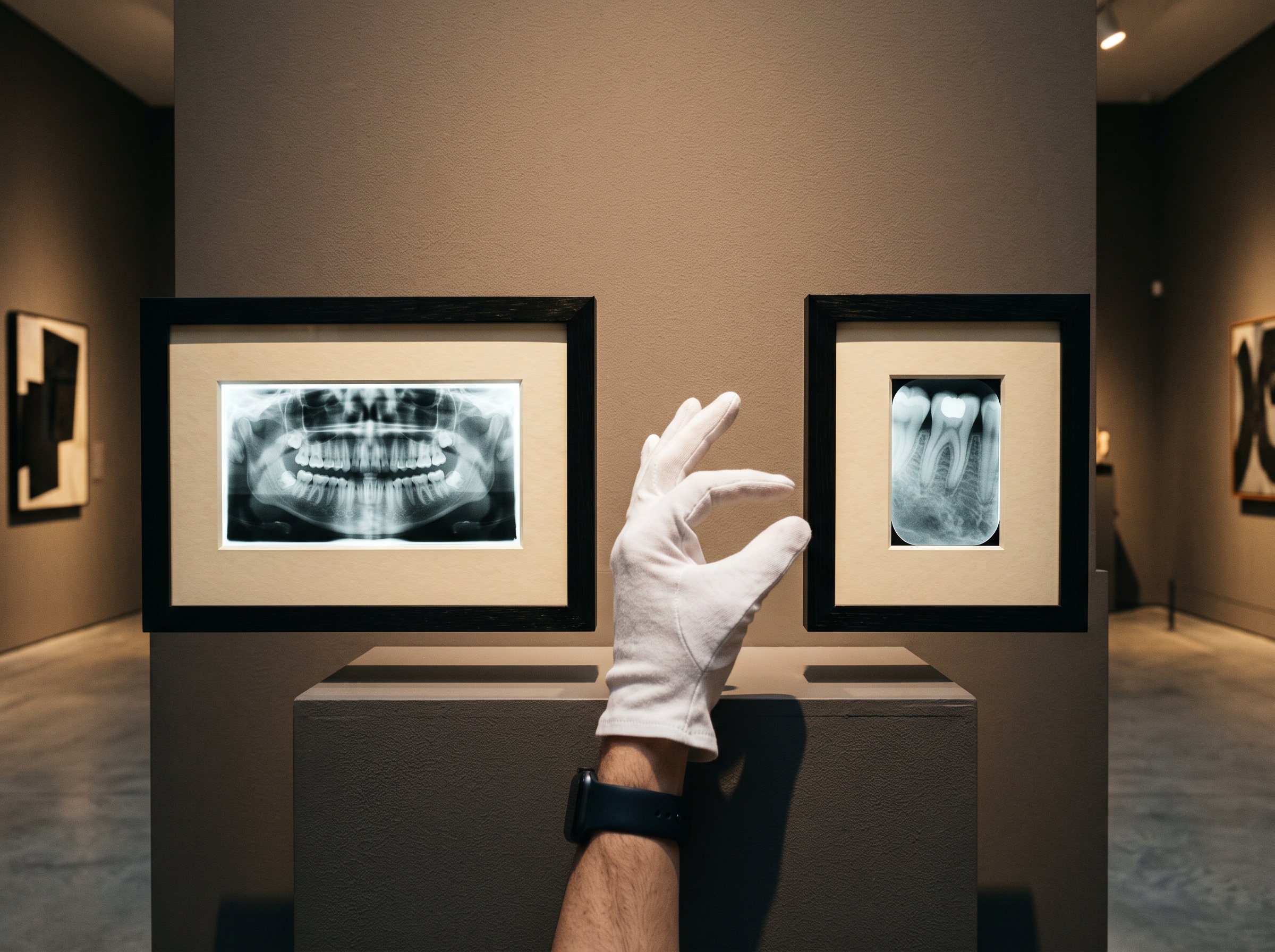
The Radiograph Not Taken
Underlying all of this is the principle of keeping exposure as low as reasonably achievable. The 2026 recommendations are pointed in noting that radiographs should be ordered only when clinically necessary, and that the benefit accrues not only to the patient but to the dental professionals who work amid scattered radiation day after day. Dose is cumulative and quiet; no single justified exposure is dangerous, but a career of unnecessary ones is not nothing. The subtler hazard is that digital sensors are forgiving, and forgiveness invites drift. As we cautioned in The Dose You Cannot See, a modern detector will render a perfectly usable image from an overexposure, so the dose can creep upward unnoticed because the picture never looks wrong. Restraint, then, operates on two fronts: the images one chooses not to take, and the honest exposure of the ones taken. Both require a discipline that the equipment itself will never enforce.
The Justified Image Earns Its Place
There is a generous corollary to all this economy. When an image is genuinely warranted, it should be made well enough to earn the exposure it cost – which means the argument for restraint is also an argument for quality. A radiograph taken sparingly must repay that scarcity with information, and a well-made one is remarkably generous. As we explored in The Whole Picture, a single film carries far more than the tooth that prompted it: bone levels, sinus floors, calcifications, and the quiet incidental findings that reward a careful reader. The discipline of taking fewer images raises the stakes of each one and, properly understood, raises the craft of making it. Fewer exposures, better composed and fully read, serve the patient more faithfully than a reflexive full series glanced at and filed.
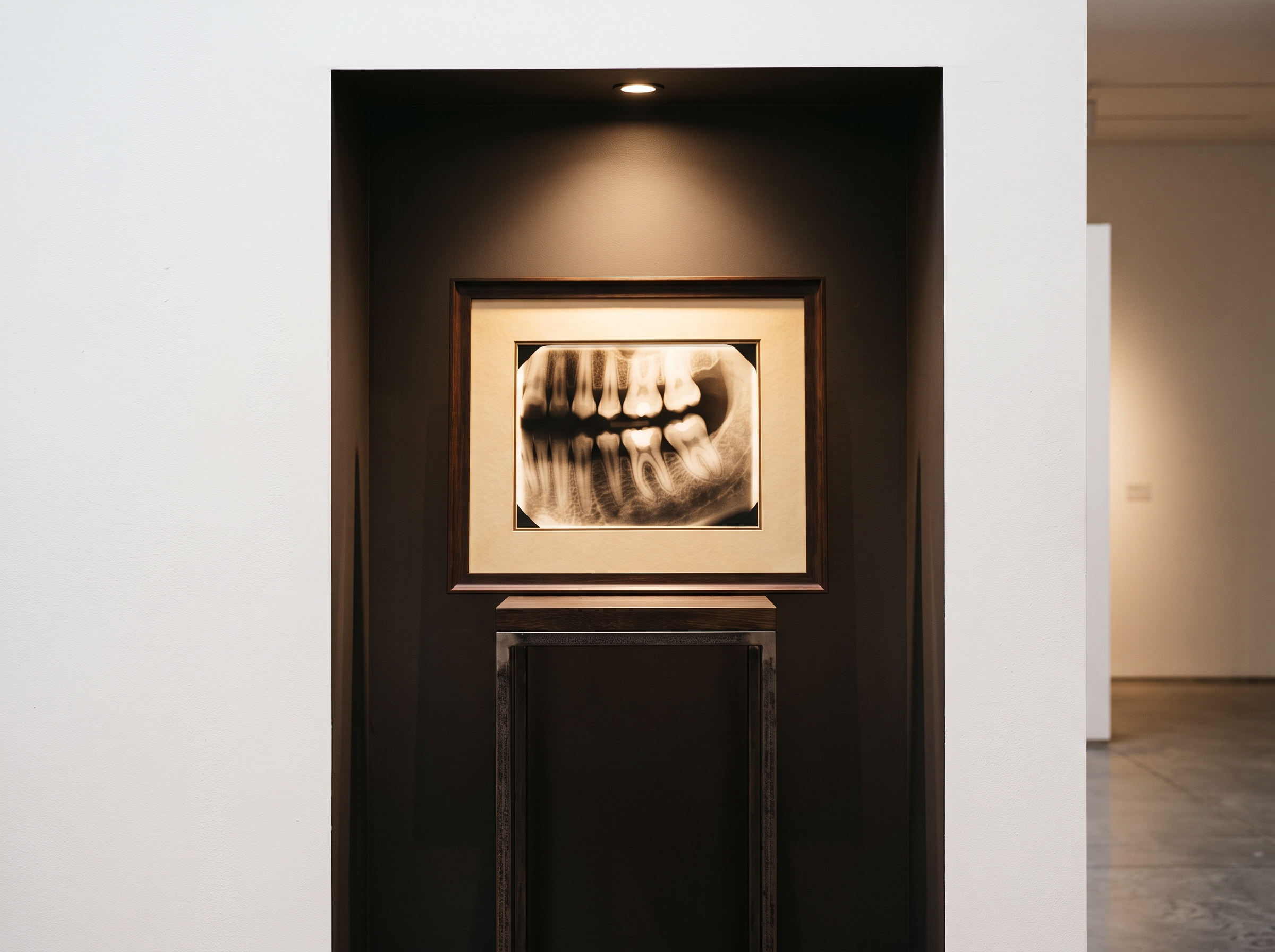
The New Frontier: CBCT Under the Same Discipline
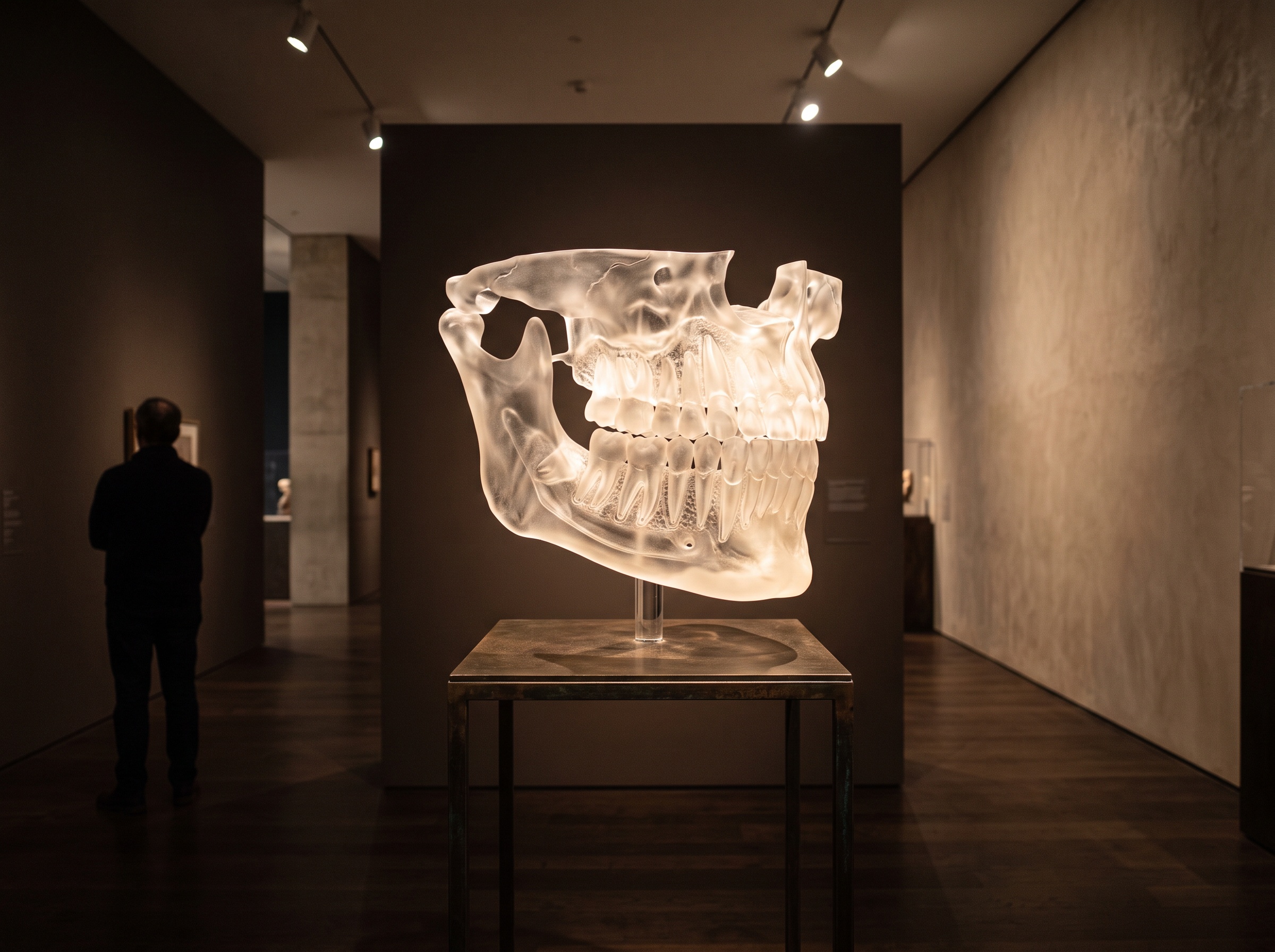
What makes these recommendations genuinely of their moment is that they are the first to bring cone-beam computed tomography under the same principle of selective use. CBCT is the most powerful instrument in the collection – a true three-dimensional view that resolves questions a flat radiograph can only hint at, from impaction geometry to the anatomy of a root canal system. As we described in AI on CBCT, the volume it produces is dense with structure a two-dimensional image simply cannot hold. But that reach comes at a higher dose than a periapical, and power of that order demands a proportionally stronger justification. The guidance resists letting the three-dimensional scan become the reflexive default merely because it shows more; it asks that CBCT be reserved for the specific questions that genuinely require volume to answer. The most capable tool in the room is precisely the one whose use most warrants a pause.
Future Developments
The direction of travel is toward imaging that is ever more precisely matched to the individual. Risk-based, personalized selection will only deepen as we grow better at stratifying who genuinely benefits from imaging and when, replacing the last of the calendar-driven habits with judgment informed by real data. Decision-support intelligence is beginning to assist at the point of the order itself – surfacing the relevant history, flagging when a proposed exposure adds little to what is already known, and helping document the clinical rationale that turns an image from routine into justified. Dose-tracking systems will make cumulative exposure visible in a way it has never been, giving the discipline of restraint an honest ledger. The deeper significance is aesthetic as much as clinical. To treat imaging as a craft is to accept that more is not the same as better, that the finest collection is a curated one, and that the mark of mastery is knowing which image to make, which view to choose, and which radiograph to leave, deliberately and confidently, unmade.
Sources & further reading:
Sorry, the comment form is closed at this time.