


September 17, 2026 The X-Ray That Fits in a Bag: NASA’s Portable Imaging Trials and the Craft of Radiography Far From Any Hospital
Consider the injury that arrives at the worst possible address. A crew member on a long transit to Mars turns awkwardly and something in the wrist gives; a molar cracks on a freeze-dried meal months from the nearest dentist. On Earth these are ordinary problems with ordinary answers, and the answer almost always begins with a radiograph. In deep space there is no radiology department down the hall, no return flight to fall back on, and a conversation with the ground that may lag by many minutes each way. The care has to happen where the crew is – which means the imaging has to happen there too. A broken bone or a dental injury a hundred million miles from help is, before it is anything else, a problem of how to make a good picture of the inside of a body with almost nothing to make it with.
This is the problem that scientists at NASA’s Glenn Research Center in Cleveland set themselves, and in September 2025 the agency described where the work had reached: testing portable, handheld X-ray systems intended to travel on future extended missions to the Moon, Mars, and beyond. The logic is plain once stated. Crews will need non-invasive imaging to diagnose conditions on their own, and a machine light and simple enough to carry aboard a spacecraft is the only kind that can go. The interesting part, for anyone who thinks about imaging as a craft, is that the constraints NASA is wrestling with are not exotic at all. They are the same constraints that quietly shape every portable radiograph taken on Earth – only pushed to their absolute limit.
Choosing From Two Hundred Machines
The engineers did not begin by inventing anything. They began by looking – reviewing more than 200 commercial systems and weighing each against a short, unforgiving list of qualities: size, weight, image quality, ease-of-use, cost, and safety. From that field they selected three systems for deeper testing: MinXray, Remedi, and Fujifilm. What is worth pausing on is that these six criteria pull against one another. Better image quality tends to want more power, more mass, more shielding; portability wants less of all three. Ease-of-use for an untrained operator can be bought only by hiding complexity that a specialist would rather keep visible. Every portable imaging decision is a negotiation among these axes, and the operatory knows the negotiation intimately – it is precisely the tension explored in the physics of image quality in a handheld dental X-ray versus the wall-mounted unit. NASA is simply running that same trade-off with the dial turned to maximum.

The Phantom and the Dental Clinic
You cannot refine a new imaging system by pointing it at astronauts, so the validation runs on stand-ins. Cuyahoga Community College contributed anatomical phantoms – lifelike models of the human body – from its radiography laboratory, and, tellingly for our purposes, from its dental hygiene clinical facility as well. Faculty and students consulted with the NASA researchers on the unglamorous fundamentals that decide whether an image is diagnostic: patient positioning, image acquisition, and image quality. A phantom is a quietly elegant idea – a body that feels no dose, endlessly patient, against which a machine’s honesty can be measured before any real person is exposed. Alongside the bench work, University Hospitals is running a study on real patients, comparing the portable systems against hospital-grade equipment on usability, image clarity, and diagnostic accuracy. That comparison is the crux: a portable machine has earned its place only when its picture can be trusted the way the big machine’s picture is trusted.
What Space Teaches the Operatory
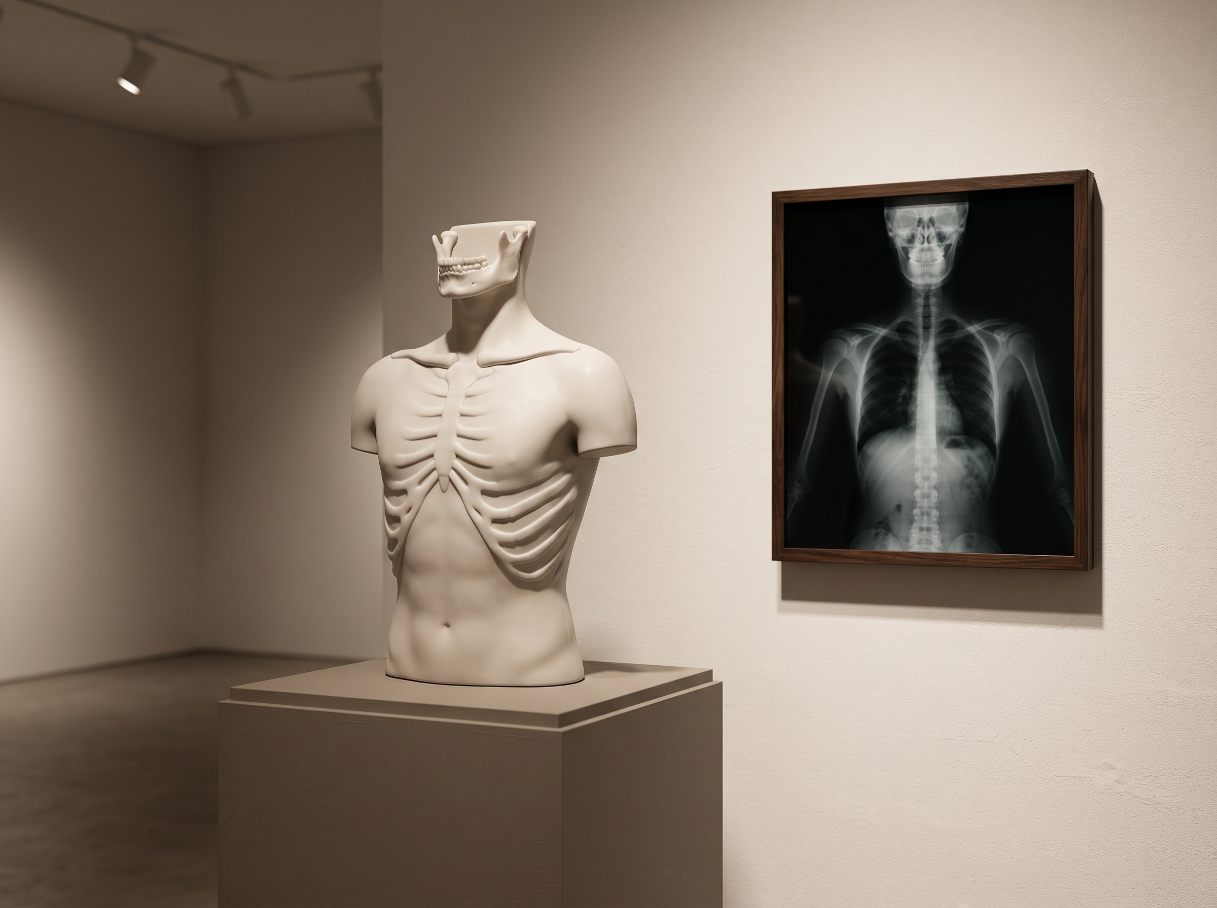
The dental parallel is not a stretch that NASA’s own framing supplies it: dental injuries sit on the short list of things a mini-X-ray must be able to see. But the deeper kinship is in who holds the device. An astronaut is not a radiographer, and the system must produce a diagnostic image in hands that may never have taken one before. That is the handheld intraoral unit’s defining challenge on Earth, too – its results ride on operator technique, on angulation and steadiness and distance, far more than a fixed installation’s do. The lesson runs in both directions. A machine engineered to be forgiving enough for a crew member in microgravity is a machine engineered to be forgiving in a busy clinic, or a mobile setting, or a home visit. And the whole exercise is a meditation on how much diagnostic weight a single image can carry, a theme close to how much a dental radiograph reveals beyond the teeth alone – because in space, as at the chairside, you may get one look and must make it count.

One Beam, Two Kinds of Seeing
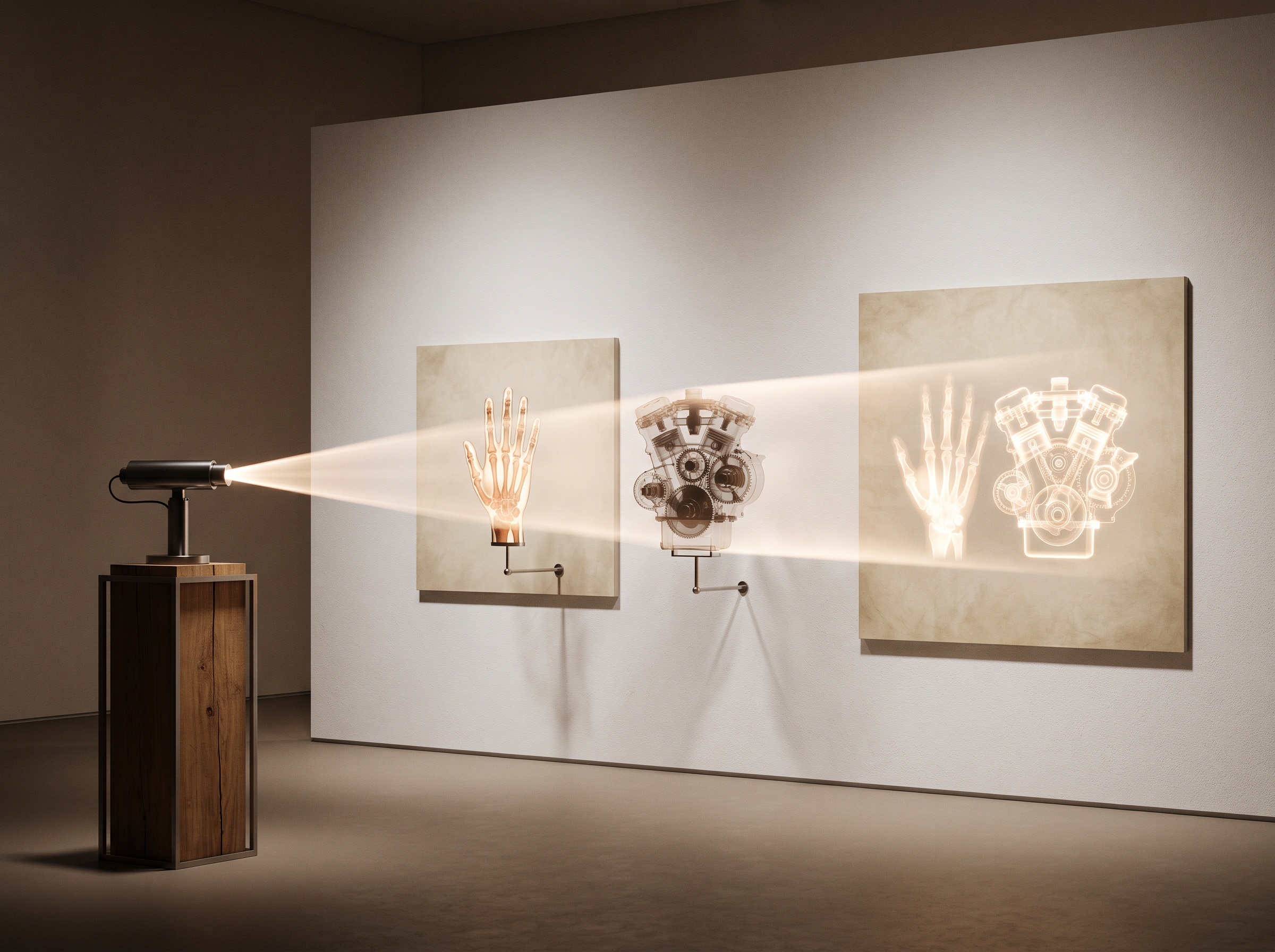
There is a detail in NASA’s account that reframes radiography itself. The portable systems are meant for both clinical and non-clinical diagnostics – the same instrument that checks an astronaut’s body could locate a tear in a spacesuit or inspect onboard electronics without disassembling the gear. That dual purpose is a reminder that an X-ray is not fundamentally a medical device; it is a way of seeing inside things that do not open. The physician and the engineer are, in that light, practicing one craft. Everything downstream of the beam – how the detector catches the photons and turns them into a picture – is shared engineering, the same lineage of sensor technology examined in our comparison of CCD, CMOS, and phosphor-plate detectors. Make that capture lighter, lower-power, and more rugged for a spacecraft, and you have made it better for the operatory by the same stroke.
Future Developments
Imaging built for the harshest, most resource-starved environment we can name has a way of flowing back to the clinic. The pressures NASA is applying – shed mass, cut power and dose, survive rough handling, and become simple enough for any trained-or-untrained hand – are exactly the pressures that, met, make portable dental and medical radiography better on Earth: lighter detectors, gentler exposures, and interfaces that ask less of the operator without asking less of the image. It is easy to read a story about mini-X-rays bound for Mars as a novelty from a distant program. Read more carefully, it is a study in the discipline of imaging far from help – and that discipline, pursued to its edge, refines the craft of imaging everywhere. The frontier, as ever at PatientGallery, teaches the studio; the picture made where nothing else was available turns out to be the picture worth learning from.
Sources & further reading:
- NASA Glenn Tests Mini-X-Ray Technology to Advance Space Health Care (NASA, September 2025)
- Miniature X-ray Systems – NASA Glenn Exploration Medical Technologies
Sorry, the comment form is closed at this time.