


September 4, 2026 Three Ways to Catch a Photon: CCD, CMOS, and Phosphor Plates Compared
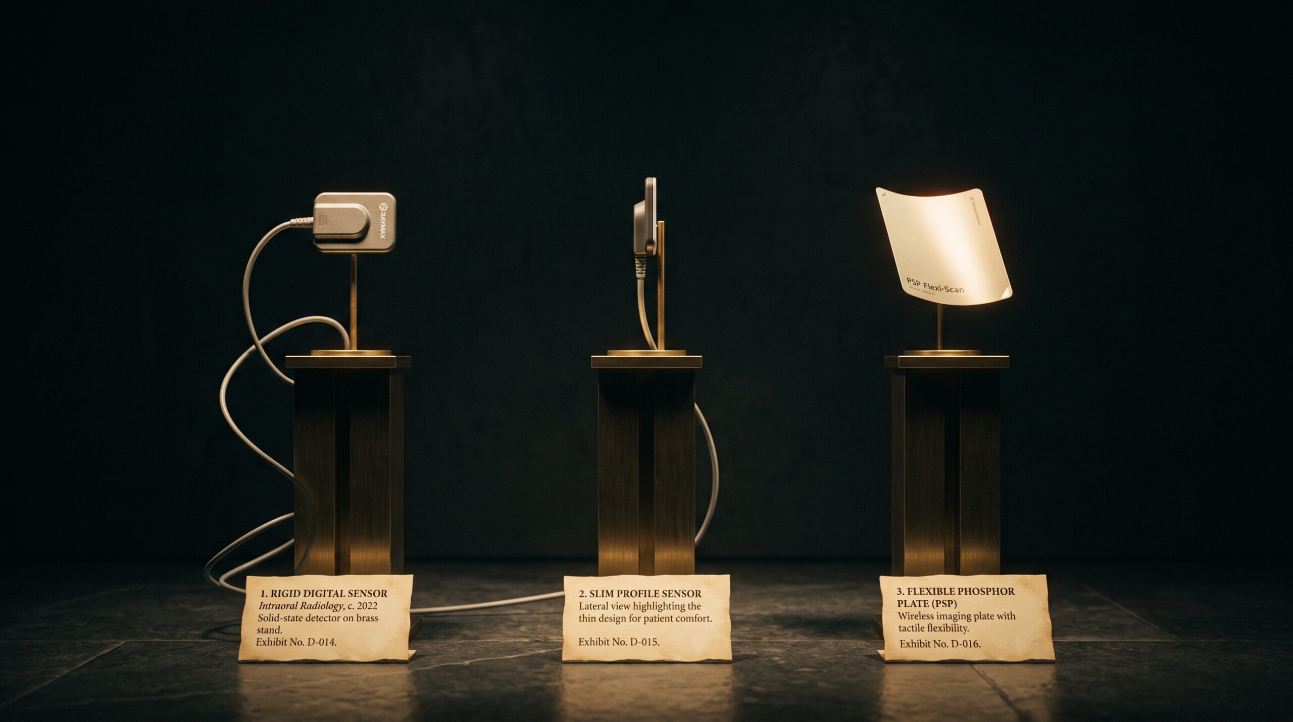
Long before an image reaches the screen, before any software sharpens or measures it, a single physical decision has already been made inside the mouth: how to catch the x-ray. A photon that has passed through enamel and bone carries the entire diagnosis, and it must be turned into a number that a computer can hold. There are three ways this is commonly done in intraoral radiography – the corded solid-state sensor built on a CCD, its close cousin built on CMOS, and the wire-free photostimulable phosphor plate. They are often spoken of as interchangeable roads to the same digital picture. They are not. Each catches the photon by a different logic, and each logic leaves its fingerprints on what the radiograph can show, what it costs the patient in dose, and how it feels to work with.
Two Families, Three Members
The three detectors sort into two families by when they answer. A solid-state sensor – whether CCD or CMOS – is a rigid electronic slab, tethered by a cable, that reports the moment the exposure ends: the image appears on the monitor in a second or two, still warm. A photostimulable phosphor plate belongs to a different family entirely. It is a thin, flexible sheet coated in a storage phosphor, and it answers nothing at the chairside. Instead it remembers. The x-ray energy is trapped in the phosphor as a latent image, held in suspended electrons, until the plate is carried to a scanner where a laser reads it out. One family speaks immediately; the other keeps its secret and confesses later. Nearly every argument about which detector to buy descends, in the end, from that single difference in timing.
The Silicon Slab: CCD and CMOS
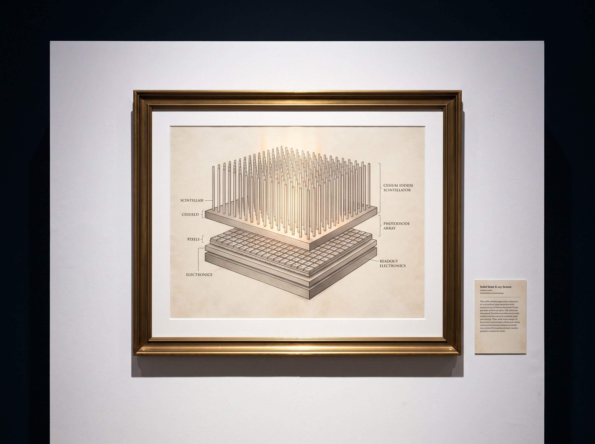
The two solid-state sensors share an architecture. Most are indirect converters: a scintillator layer – frequently columnar cesium iodide – first turns the x-ray into visible light, and a grid of silicon photodiodes beneath it turns that light into charge. We looked closely at that luminous first layer in the essay on the columnar scintillator and how it decides what a sensor can see; the pixel grid below it is the second half of the bargain, and it fixes the sensor’s resolution at manufacture, one square well per picture element. Where CCD and CMOS part ways is in how they empty those wells. A CCD shuttles charge across the chip bucket-brigade fashion to a single corner to be read, an elegant but power-hungry march. A CMOS chip gives each pixel its own tiny amplifier and reads them where they sit, which is cheaper, sips far less power, and lets the sensor be driven straight off the USB cable. For years CCDs held a slight edge in uniformity while CMOS carried a reputation for noise, but modern CMOS has closed that gap so thoroughly that it now dominates new intraoral sensors – the quiet, low-power reader has simply won on economics without conceding image quality.
The Phosphor Memory: PSP Plates
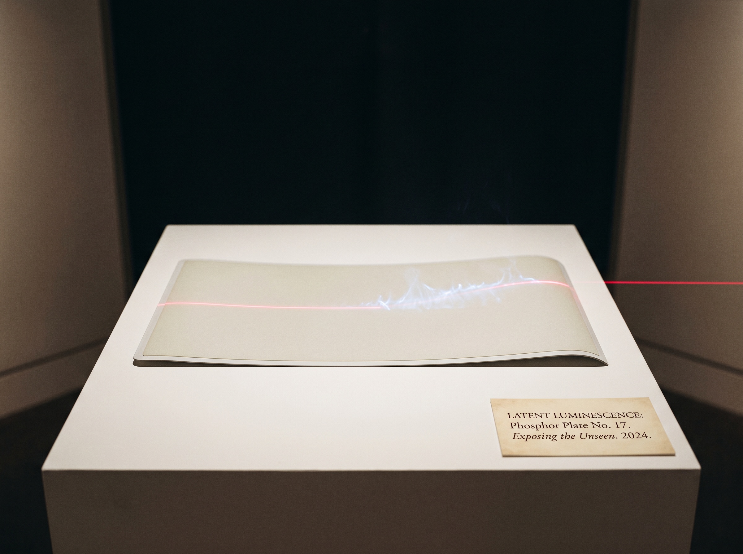
The photostimulable phosphor plate solves the same problem by storing rather than reporting. During exposure, x-rays lift electrons in the phosphor into metastable traps, where they wait – a latent image encoded not in developed silver, as in film, but in held energy. The plate is then fed into a reader, where a fine laser beam sweeps across it. The laser’s energy releases the trapped electrons, which fall back and emit a faint blue glow in exact proportion to the original exposure at each point; a photomultiplier collects that glow and builds the image line by line. The plate is afterward flooded with light to erase it and returned to service. The payoff for this patience is physical: a PSP plate is thin, flexible, and free of any cable, so it seats where a rigid sensor cannot and sits far more kindly against the palate and floor of the mouth. Its resolution is set by the fineness of the laser scan and the spread of light within the phosphor rather than by a fixed pixel grid. The cost is the workflow – a trip to the scanner, a plate that can be scratched, bent, or fogged, and the handling hazards we catalogued among the streaks, fog, and ghosts that haunt phosphor plates.
Resolution, Latitude, and the Honest Trade
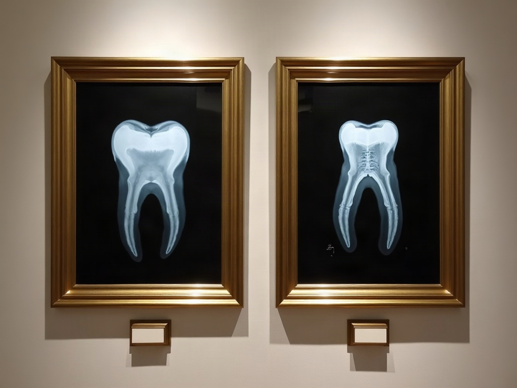
Set the three side by side and the differences resolve into a few honest trades. Solid-state sensors generally deliver the finer spatial resolution – the crisp rendering of a thin periapical film line or an early carious lesion – because their pixel pitch is small and fixed; this is the domain we explored in the piece on pixel pitch, line pairs, and the true limits of sharpness. Phosphor plates, by contrast, tend to offer wider exposure latitude: a broad, forgiving range over which a usable image survives both under- and over-exposure, where a solid-state sensor’s response is narrower and less tolerant of a bad angle or a slipped setting. That forgiveness is a double-edged gift. It rescues an awkward exposure, but it also hides the operator’s errors and invites the slow, invisible climb of dose creep, in which a forgiving sensor quietly accepts ever more radiation because nothing on screen protests. On dose proper, both digital families need far less exposure than film ever did, and the differences between them are smaller than the marketing implies; the more useful figure of merit is how efficiently each turns a given dose into signal – the detective quantum efficiency we treated as the true measure of a dental sensor, beyond resolution – rather than a headline resolution number in line pairs per millimeter.
Choosing the Detector to the Task
The right answer is not a winner but a fit. A practice that lives on speed and single-tooth precision – endodontics chasing a canal, a restorative eye reading a margin – is well served by solid-state CMOS: instant feedback, superb fine detail, no plate to ferry. A practice that images children, gaggers, or difficult anatomy, or that needs full-mouth series without a cable fighting the tissue, will bless the thin, flexible phosphor plate and its gentler seat, accepting the scanner detour as the price of comfort and coverage. Many thoughtful offices simply keep both, reaching for the rigid sensor by default and the plate when the mouth or the patient refuses it. What matters is to choose with eyes open: to know that the sensor is not a neutral window but an instrument with a temperament, and that its temperament is written into every image it makes before the clinician ever looks.
Future Developments
The frontier is moving away from the scintillator altogether. Direct-conversion detectors, which turn the x-ray straight into electrical charge in a photoconductor and skip the light-scattering middle step, promise sharper images at lower dose by eliminating the blur that a scintillator’s glow inevitably spreads – and the same photon-counting logic now reshaping medical CT is beginning its long walk toward the dental chair, where a detector might one day weigh each photon’s energy rather than merely tallying it. Flexible large-area CMOS and thinner, tougher, faster-reading phosphors are narrowing the ergonomic gap from both sides at once. And as the raw capture grows cleaner, more of the image’s fate passes to the algorithms that read it – the reconstruction and enhancement layers we keep returning to – which only sharpens the oldest truth in this craft: that the most sophisticated interpretation can never recover what the detector, in its first silent instant, declined to catch.
Sorry, the comment form is closed at this time.