


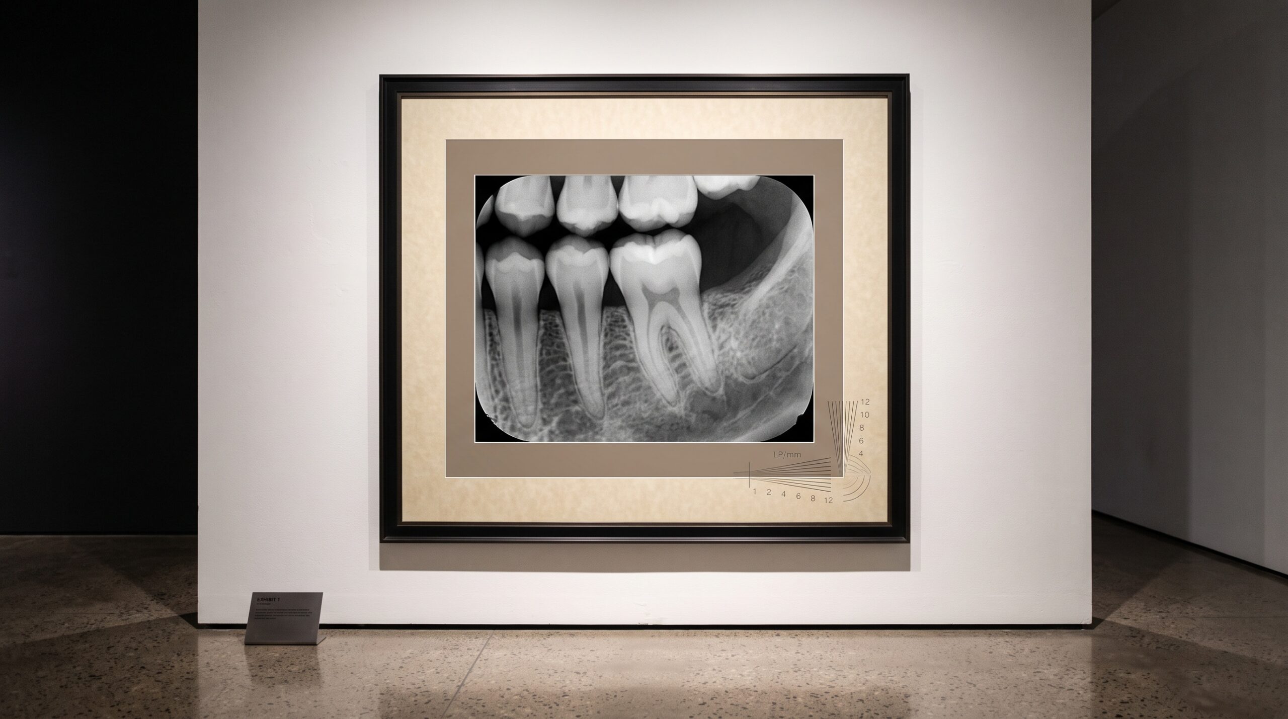
August 3, 2026 How Sharp Can a Dental Image Truly Be? Pixel Pitch, Line Pairs, and the Limits of Resolution
Stand close to any radiograph long enough and a question surfaces that no brightness or contrast slider can answer: how much finer detail is actually there, waiting to be seen, and how much has already been lost before the image ever reached the screen? Sharpness feels like an aesthetic judgment, something we sense the way we sense a well-lit portrait. In imaging it is nothing of the kind. Every dental image arrives with a hard, measurable ceiling on the smallest structure it can resolve — and understanding that ceiling is what separates reading an image from merely admiring it.
Resolution Is a Number, Not a Feeling
The classical currency of sharpness in radiography is the line pair per millimeter (lp/mm): one black bar beside one white bar, packed as tightly as a system can still tell them apart. Traditional dental film could resolve well beyond 20 lp/mm — a stunning figure that modern digital sensors have never fully matched. Contemporary intraoral sensors typically deliver somewhere in the range of 15 to 25 lp/mm depending on generation and technology, while panoramic and cone-beam systems, spreading their detail across a far larger field, resolve considerably less. The point is not to crown a winner but to recognize that limiting resolution is a specification, as concrete as a lens’s focal length, and that it governs whether the earliest hairline of a fracture or the faint widening of a periodontal ligament space can register at all.
Pixel Pitch: The Floor Beneath Every Detail
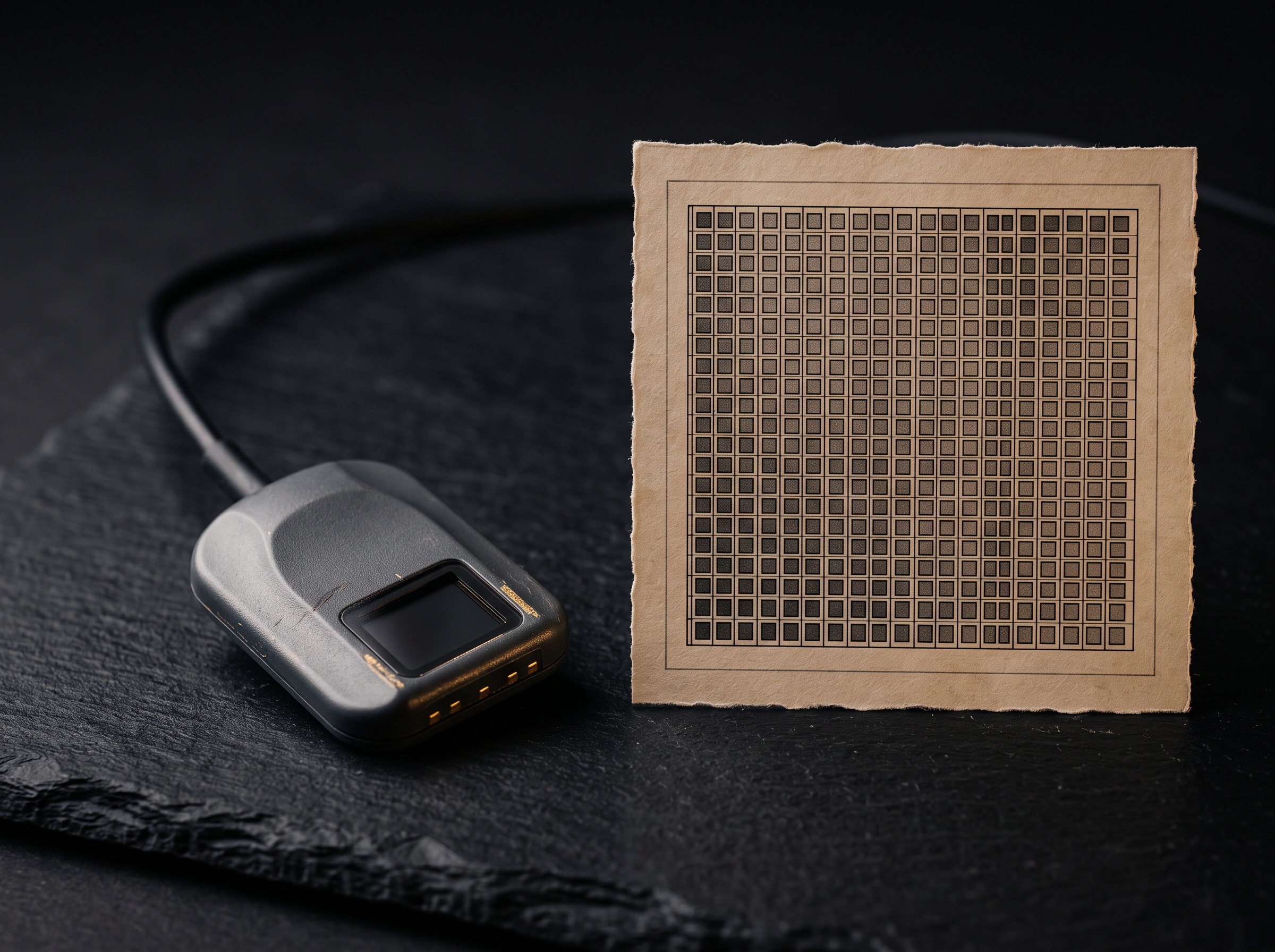
For a digital sensor, the first and most unforgiving limit is pixel pitch — the physical spacing between the centers of adjacent photosites, measured in microns. A sensor cannot record a detail finer than its sampling allows, and the mathematics here is exacting. The Nyquist theorem tells us that to distinguish one full line pair we need at least two pixels, so the theoretical ceiling of a sensor equals half of its sampling frequency. A photosite pitch of roughly 20 microns implies a Nyquist limit near 25 lp/mm; widen that pitch and the ceiling drops in direct proportion. This is why marketing figures about total megapixels are nearly meaningless in dentistry. What matters is not how many pixels blanket the sensor but how small and how closely spaced each one is over the tissue we care about.
The Modulation Transfer Function: Sharpness as a Curve
A single number for resolution flatters the truth. Real imaging systems do not resolve perfectly right up to a cliff and then fail; they fade. The modulation transfer function (MTF) is the honest portrait of that fade — a curve describing how faithfully a system preserves contrast as detail grows finer. At coarse spatial frequencies, a bold restoration margin or a large carious lesion, the MTF sits near unity and contrast passes through almost untouched. As the structures shrink toward trabecular texture and enamel micro-anatomy, the curve slopes downward: the blacks lighten, the whites darken, and the distinction between them dissolves. The frequency at which contrast collapses to roughly a tenth of its original value is often cited as the practical limiting resolution. Read the MTF and you are reading the system’s entire character, not a single boast.
Where the Sharpness Actually Goes
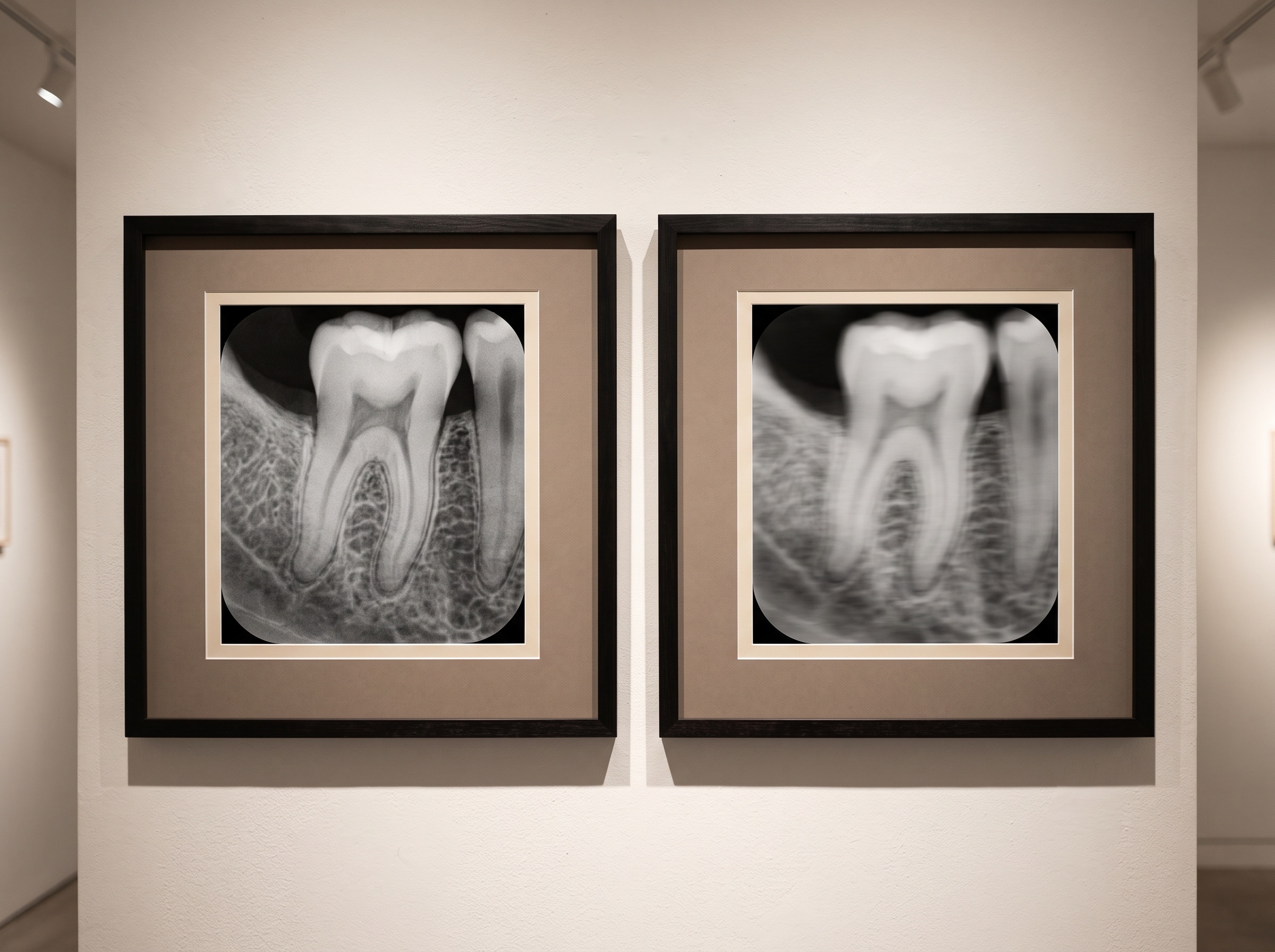
Even a superb sensor rarely reaches its own ceiling, because resolution is degraded at every stage of the imaging chain. Geometric unsharpness arises from the finite size of the x-ray source: a focal spot that is not a true point casts a soft penumbra around every edge, and the effect worsens as the object drifts from the receptor or toward the source. Motion unsharpness — a patient’s swallow, a trembling tongue, a fractional shift of the sensor — smears fine structure into oblivion in a way no post-processing can recover. And the detector itself contributes: in phosphor-based systems the scatter of light within the plate blurs the latent image, while in every system electronic noise masquerades as, and competes with, genuine fine detail. The recorded sharpness is always the product of these losses multiplied together, never the best single component alone.
Sharpness Is Not the Same as Diagnostic Truth
Here the craft turns subtle. More resolution is not automatically more diagnosis. A high-frequency image renders exquisite texture but, all else equal, distributes the available signal across more, smaller elements — and if dose is held constant, each element carries more noise. The clinician’s real quarry is not the finest line pair but the smallest meaningful change against its background, a balance formally captured by the detective quantum efficiency and, more intuitively, by whether a lesion stands clear of the grain. This is why aggressive digital sharpening can betray us: it amplifies edges and noise alike, manufacturing an impression of detail while burying the low-contrast findings — early demineralization, a faint periapical rarefaction — that a calmer image would have surrendered. True image quality is a negotiation between resolution, contrast, and noise, never the pursuit of any one in isolation.
Future Developments
The ceiling is rising, and it is rising along more than one axis. Photon-counting detectors, already reshaping medical CT, count individual x-ray quanta and sort them by energy, promising sharper images with less electronic blur and lower dose — a technology poised to migrate toward dental cone-beam and intraoral use. Learned super-resolution, in which neural networks trained on paired low- and high-resolution images infer plausible fine structure, is advancing quickly, though it raises a genuinely new question for our field: when an algorithm proposes detail the sensor never physically recorded, where does documentation end and interpretation begin? Meanwhile, quieter refinements — smaller focal spots, finer photosite lattices, and MTF-aware processing that sharpens only where the signal warrants it — continue to close the historic gap with film. The enduring lesson is that sharpness is a measurable, negotiable property, not a gift. To photograph the mouth as both science and art is to know precisely how fine your instrument can see, and to compose every image with honest respect for that limit.
Sources & further reading:
Related Reading
- The Seductive Edge: How Sharpening, Contrast, and Noise Reduction Can Deceive on a Digital Dental Radiograph
- The Plane of Sharpness: Depth of Field, Diffraction, and Focus Stacking in Clinical Dental Photography
- The Blur Built Into Every X-Ray: Focal Spot Size, Penumbra, and the Geometry of a Sharp Radiograph
Sorry, the comment form is closed at this time.