


February 6, 2026 The Plane of Sharpness: Depth of Field, Diffraction, and Focus Stacking in Clinical Dental Photography
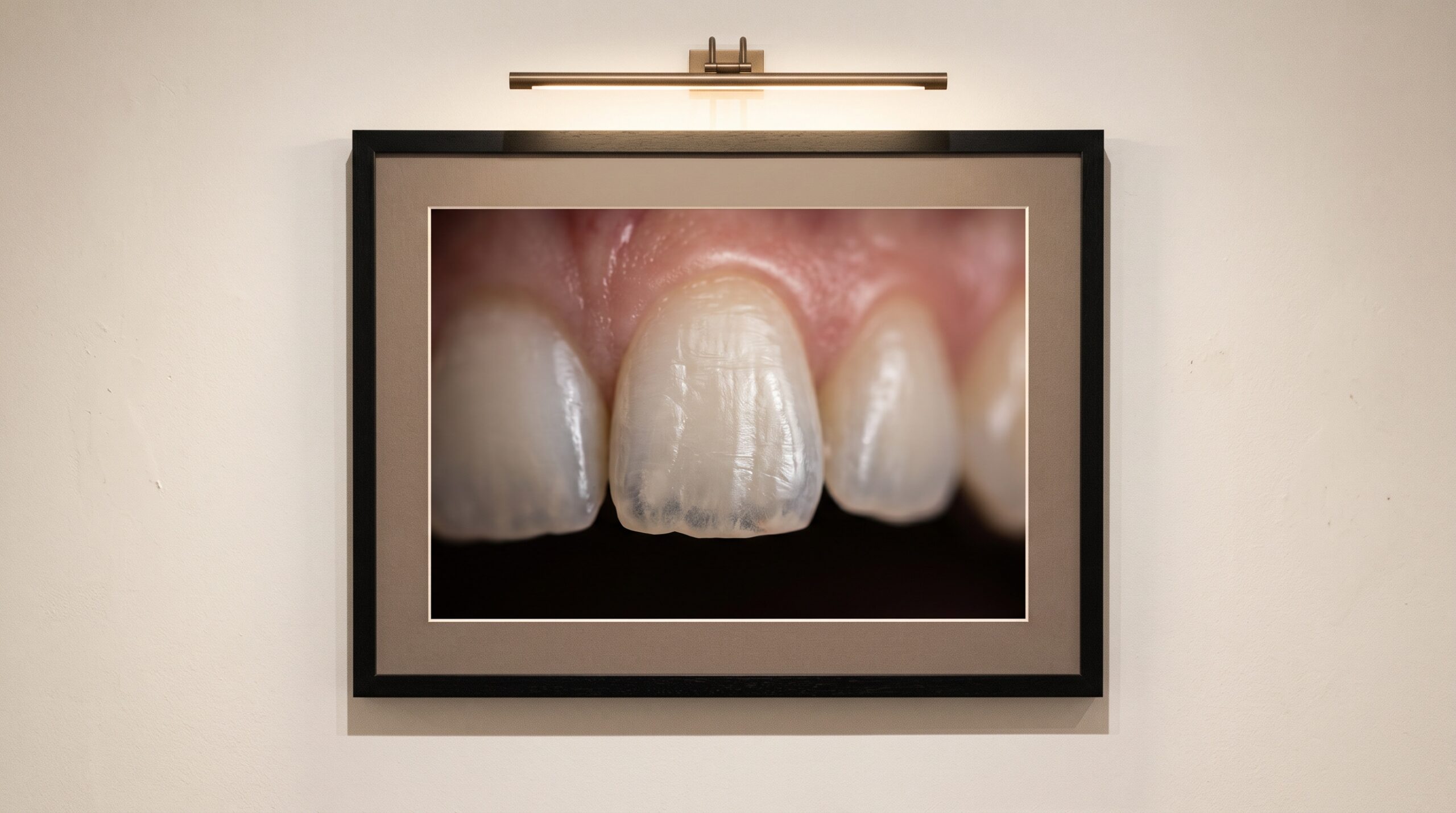
Stand a patient’s arch on end in your imagination. The labial surface of a central incisor and the occlusal table of the second molar behind it are separated by a startling distance — two, three centimetres of curving, receding enamel. Now ask a camera, held close enough to fill the frame with a single quadrant, to render all of it sharp at once. This is the quiet, unglamorous problem beneath every clinical photograph, and it is a problem of physics before it is ever a problem of technique. At the magnifications dentistry requires, the region of acceptable sharpness — the depth of field — is not a comfortable zone but a razor-thin sliver, frequently shallower than a single tooth is deep.
Understanding that sliver, and learning where it can be widened and where it must instead be assembled, is the difference between a photograph that documents and one that merely gestures at the mouth. Depth of field is the first craft decision in clinical imaging, and for a century it obeyed a single unforgiving lever: the aperture.
Why the Sliver Is So Thin
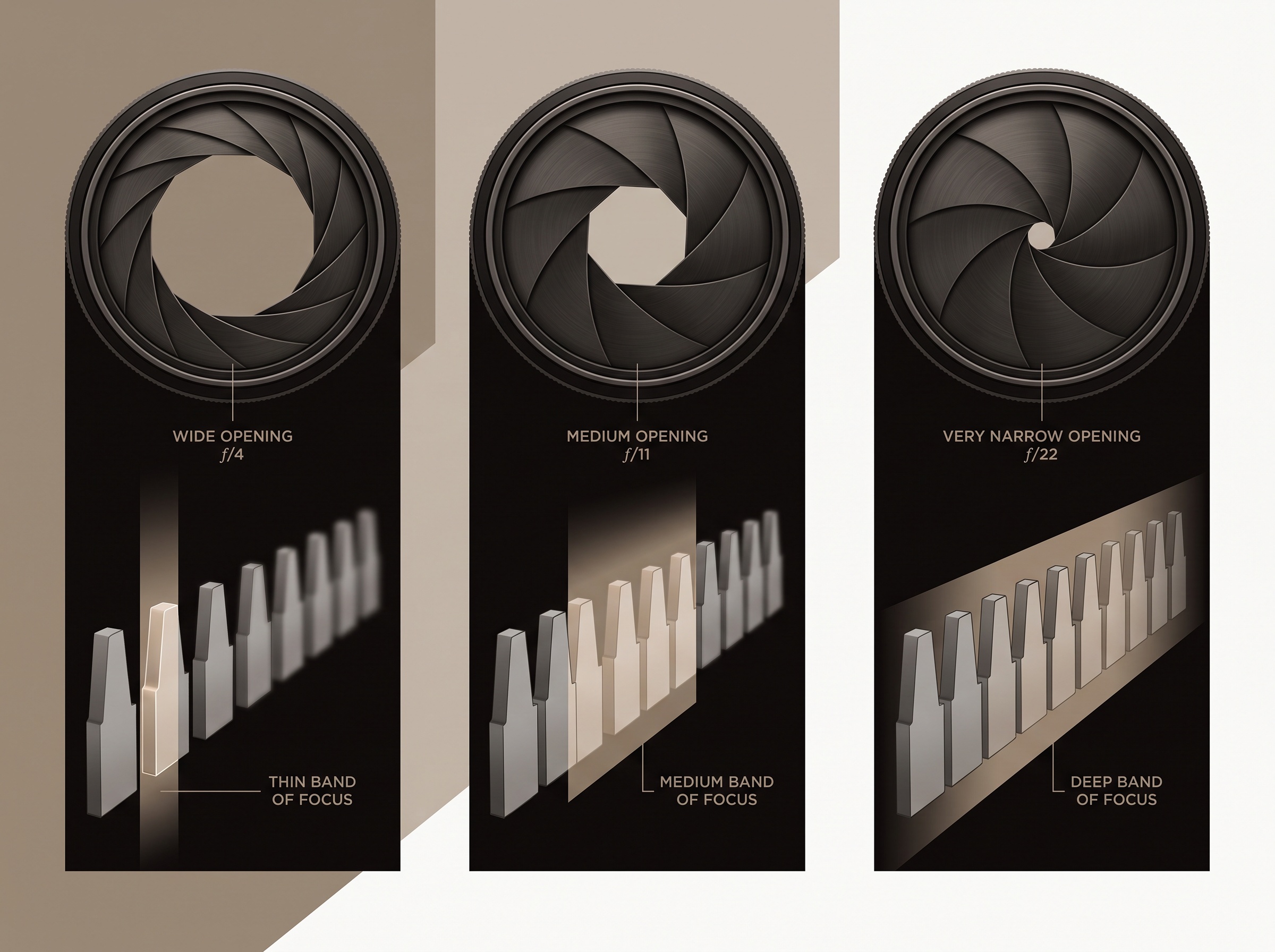
Depth of field is governed by three things, and dental photography pushes all three toward their least forgiving extreme. The first is aperture — the physical opening in the lens, named by its f-number. A wide aperture such as f/4 gathers light generously but renders only a hair’s breadth in focus; a narrow aperture such as f/22 holds far more of the scene sharp. The second is the acceptable circle of confusion, the largest blur spot the eye will still accept as a point — a definition of “sharp” that tightens the more critically the image will be examined. The third, and the cruel one, is magnification.
Depth of field collapses as magnification rises, and it collapses fast — with the square of the reproduction ratio at close range. A landscape at f/8 may hold kilometres in focus; the same lens and aperture at 1:1 macro may hold barely a millimetre. This is why intraoral photography feels like a different discipline from every other kind: the moment you move close enough to see the texture of enamel, the plane of sharpness shrinks to something you can lose by breathing. It is not the clinician’s unsteadiness that blurs the molars behind a sharp incisor. It is geometry.
The Aperture Answer — and Its Price
The century-old solution is simply to stop the lens down. Close the aperture to f/22, even f/32, and the depth of field stretches until the whole arch can fall within it. Much of the clinical literature reflects this instinct: an aperture between roughly f/22 and f/32 is the classic recommendation for intraoral work, precisely because it keeps detail sharp from the front teeth to the back of the oral cavity. For the standard full-arch or quadrant record, this remains sound, dependable advice, and it is why dental flashes are built to deliver so much light — a narrow aperture is a light-hungry aperture.
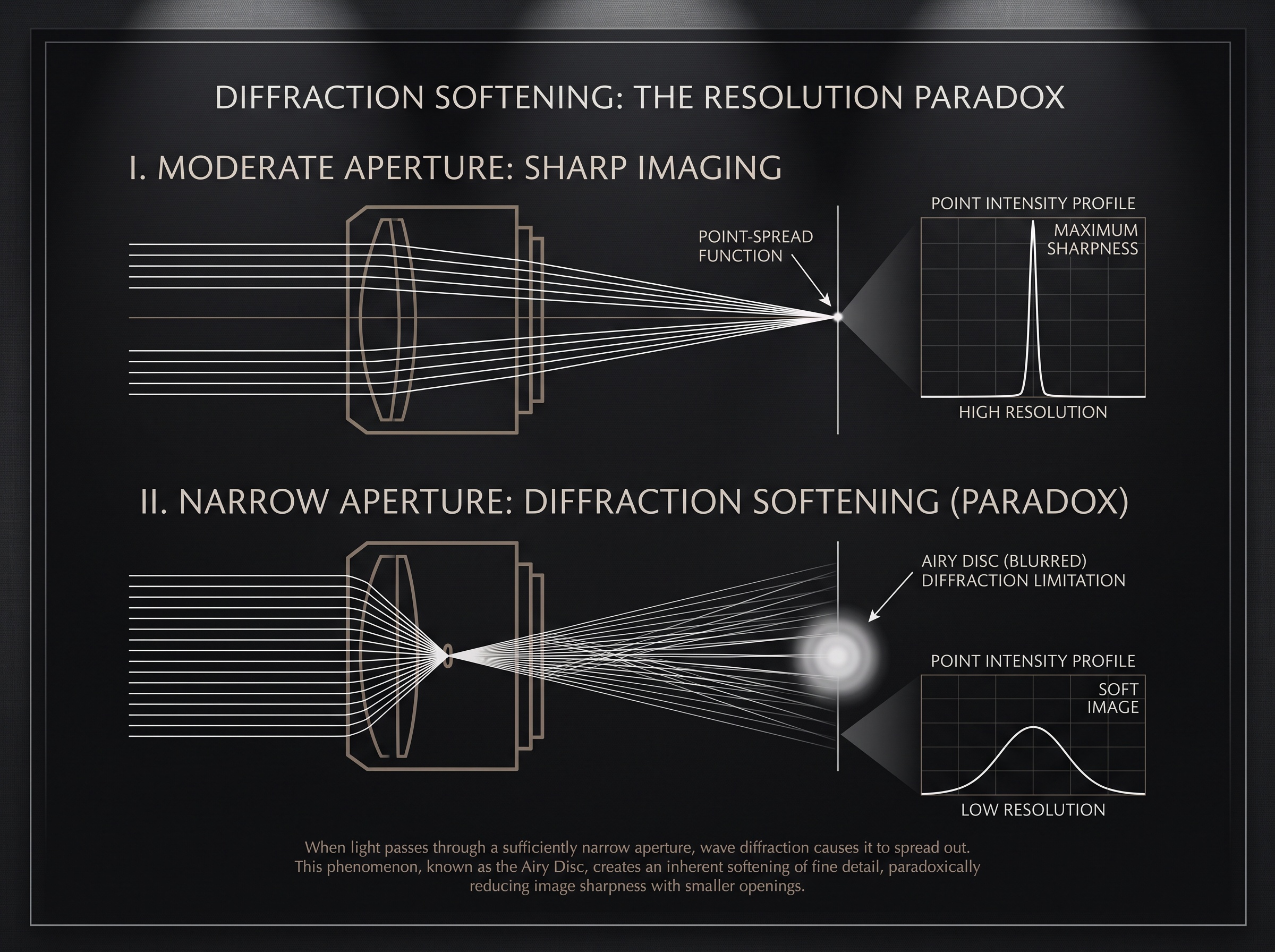
But the small aperture exacts a price that the specification-sheet instinct overlooks, and it is a price written into the wave-nature of light itself: diffraction. As the opening narrows, light bends around its edge and every point in the image spreads into a small disc. Past a certain f-number the whole frame begins to soften, uniformly, everywhere. So a paradox emerges that catches many careful clinicians off guard — at f/22 more of the scene is nominally in focus, yet the image can be less crisp overall than the same scene at f/11, because diffraction has quietly stolen the fine resolution the extra depth was meant to serve. Comparative studies of aperture in dental photography make exactly this point: beyond the diffraction threshold you are trading real sharpness for the appearance of depth.
The Sweet Spot and Its Limits
Every lens has a range where it performs at its sharpest — typically a few stops down from wide open, often around f/8 to f/11 for macro work — where aberrations are tamed but diffraction has not yet taken hold. This is the sweet spot, and it presents the clinician with the central dilemma of the craft in a single sentence: the aperture that renders a tooth most beautifully is not the aperture that keeps the whole arch in focus.
For a portrait of one anterior tooth, a shade-match study, a close record of a restorative margin, the choice is easy — favour the sweet spot and let the background fall away. But for the record that must be uniformly sharp end to end, the clinician has traditionally been forced to compromise: stop down past the sweet spot, accept some diffraction softening, and take the whole arch at f/22 because a slightly soft-but-complete image beats a razor-sharp-but-partial one. For decades that compromise was simply the cost of doing business. It no longer has to be.
Focus Stacking: Assembling the Impossible Frame
Focus stacking dissolves the dilemma by refusing to ask a single exposure to do the impossible. Instead of one photograph at a compromised aperture, the clinician captures a series — each focused on a slightly different plane, the point of sharpness marching steadily from the labial surface of the front tooth back to the distal molar — all taken at the lens’s sharp, diffraction-free sweet spot. Software then examines the stack pixel by pixel, keeps only the sharpest rendering of each region, and fuses them into one image in which every plane is crisp.
The logic is elegant: there is no reason to stop down to f/22 and suffer diffraction to buy depth of field that the stack will supply anyway. You take back the sweet spot’s sharpness and gain unlimited depth, at the cost only of a few extra frames and a patient — and a subject — that hold still. That last clause is the catch. Stacking demands stillness across the sequence, which is why it has found its natural home in extracted-tooth documentation, dental materials science, and controlled esthetic studies rather than in the wet, moving mouth. But as capture speeds rise and in-camera stacking matures, its territory is expanding toward the chairside.
Depth of Field as a Deliberate Voice
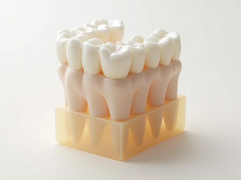
The finest clinical photographers treat depth of field not as a setting to be maximised but as a language to be spoken. A shallow plane, wide open at the sweet spot, isolates a single tooth against a soft fall-off — the visual grammar of the esthetic close-up, drawing the eye to translucency and texture. A deep, stacked field renders an entire arch with uncanny, uniform clarity — the grammar of the comprehensive record and the medico-legal document. Neither is more correct; each answers a different question the image is being asked to pose.
To command that choice is to understand that sharpness in dentistry is never free. It is bought from light with the aperture, taxed by diffraction, shrunk by magnification, and — when the physics of a single frame runs out — assembled from many. The clinician who knows which currency to spend, and when, is no longer at the mercy of the razor-thin plane. They are composing with it.
Future Developments
The trajectory is toward making the impossible frame effortless. Computational photography already brings focus stacking inside the camera body, and the same depth-mapping that powers it points toward a future where a single chairside capture yields a fully-focused image reconstructed after the fact — the aperture dilemma solved in software rather than optics. Light-field sensors, which record the direction as well as the intensity of each ray, promise images whose plane of focus can be chosen after the shutter closes. As these mature, the century-old trade between depth and sharpness may quietly dissolve, and the clinician will be freed to spend their full attention where it always belonged — not on wrestling the physics of the lens, but on seeing, and preserving, the truth of the mouth in front of them.
Sources & further reading:
- Dental Photography 101 (Spear Education)
- Comparison of Different Aperture Values in Dental Photography (Essential Dental / EssentDent)
- Application of macro photography in dental materials science (Journal of Dentistry, ScienceDirect, 2020)
Sorry, the comment form is closed at this time.