


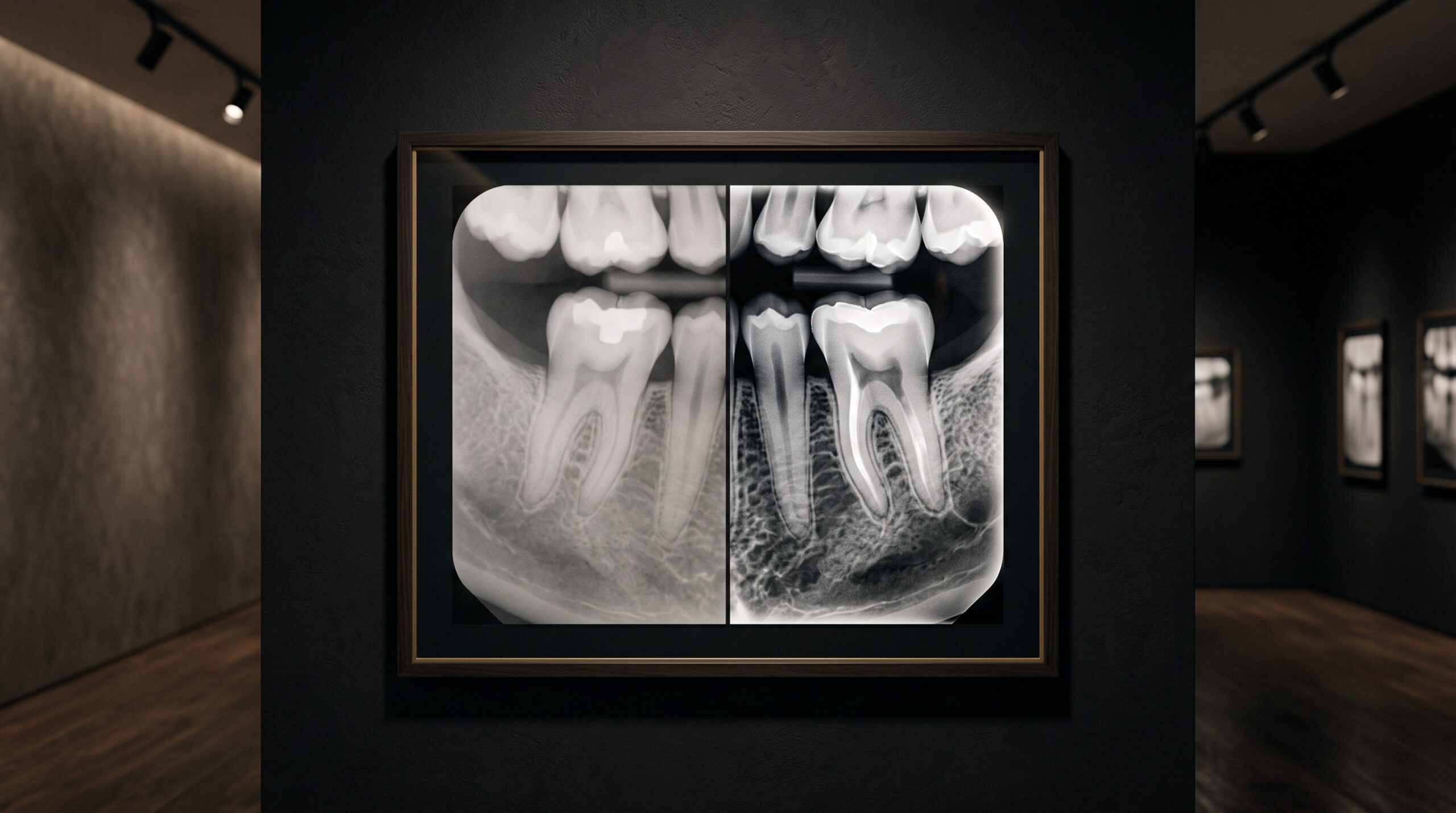
August 13, 2026 The Seductive Edge: How Sharpening, Contrast, and Noise Reduction Can Deceive on a Digital Dental Radiograph
There is a quiet fiction at the heart of every modern radiograph, and it is worth naming plainly: the image a clinician reads is never the image the sensor captured. Between the photons striking the detector and the crisp grey study glowing on the monitor lies a pipeline of arithmetic — gain correction, tone mapping, edge enhancement, noise suppression — that reshapes the raw data into something the eye finds legible and pleasing. None of this is dishonest by intention. Each step exists to serve the reader, to pull a faint lesion out of the murk or calm a storm of grain into something you can actually look at. But every one of those kindnesses is also a lever, and a lever pushed too far can manufacture detail that was never there, or dissolve detail that was. The art of reading a digital image well begins with respect for how thoroughly it has already been interpreted before you ever see it.
The Raw Image No One Ever Sees
Strip a digital dental sensor back to its output and you would not recognise it. The unprocessed signal is flat, low in contrast, freckled with the individual quirks of every pixel and the random speckle of a finite number of X-ray photons. It is faithful, but it is nearly unreadable. So the manufacturer wraps it in a processing chain, tuned to a house aesthetic, that turns that honest sludge into the confident, high-contrast picture the market expects. This is why the same tooth, exposed identically, looks meaningfully different across two vendors’ software: you are not comparing detectors so much as comparing opinions about how a radiograph should look. The processing is not a cosmetic afterthought layered onto the diagnosis — it is the image. Understanding that the picture is a rendering, not a measurement, is the first and most important act of honest interpretation.
Unsharp Masking and the Manufactured Edge
The most seductive tool in the chain is sharpening, and its workhorse is a technique with a wonderfully paradoxical name: unsharp masking. The algorithm blurs a copy of the image, subtracts that blur from the original to isolate the edges, and then adds those edges back in amplified. Boundaries snap into focus; a marginal ridge or the outline of a restoration leaps off the screen. But the amplification does not stop at the true edge. It overshoots, laying a bright rim just inside every dark-to-light transition and a dark trough just beyond it — the tell-tale halo of over-sharpening. On a radiograph those halos are not harmless decoration. A bright overshoot hugging the cervical margin of a crown can mimic the radiolucent line of recurrent decay; a dark undershoot around an implant thread can impersonate the peri-implant bone loss a clinician is trained to fear. The detail looks real because sharpening did not invent the underlying structure — it merely exaggerated the transition until the eye read emphasis as substance. This is the shadow side of the pursuit of acuity described in the physical limits of sharpness in a dental image: true resolution is set by physics, but apparent sharpness can be conjured by software, and the two are not the same thing.
Contrast, Gamma, and the Enhanced Lesion
If sharpening manipulates edges, contrast processing manipulates the grey scale itself — the very currency of radiographic diagnosis, where disease so often announces itself as a subtle difference in density. A tone curve maps the captured shades onto the ones displayed, and steepening that curve stretches a narrow band of greys across a wider range of brightness. Do it judiciously and an early carious lesion, invisible in the flat raw data, becomes plainly visible. Do it aggressively and the same steep curve will exaggerate an area of perfectly normal variation into something that looks like demineralisation, or, conversely, flatten a real lesion into the surrounding bone until it disappears. Gamma adjustment, histogram equalisation, and the automatic “optimisation” that many systems apply by default all reshape this mapping, often invisibly and differently from one image to the next. Two radiographs of the same tooth, processed with two curves, can tell two different diagnostic stories. The discipline this demands is the same one that governs reading a bitewing honestly: knowing what the image is capable of showing you, and what it is capable of inventing.

Noise Reduction and the Vanishing Detail
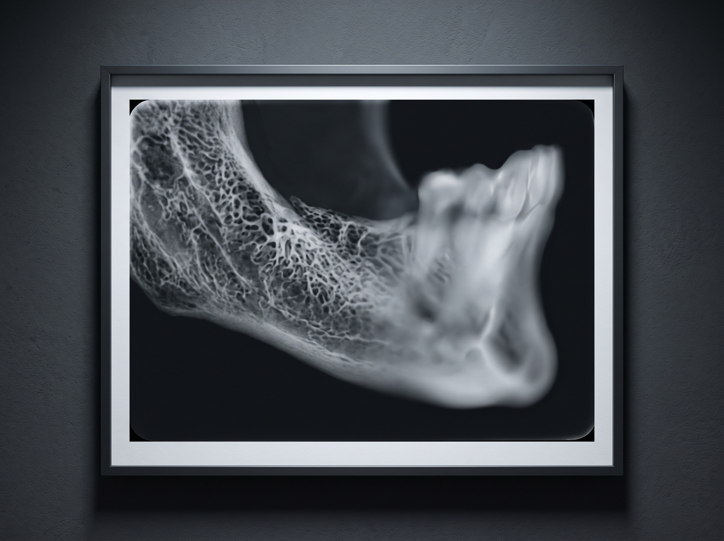
At the opposite pole sits the gentlest-seeming manipulation of all: noise reduction. A digital radiograph, especially a low-dose one, carries an irreducible grain — quantum mottle, the statistical speckle of counting a finite number of photons. Smoothing filters average neighbouring pixels together to calm that grain, and the result looks cleaner, more confident, more diagnostic. The catch is that the algorithm cannot always tell noise from signal. The fine, lace-like trabecular pattern of bone, the hairline of a root fracture, the faint texture that distinguishes healthy marrow from early change — these live at the same fine scale as the grain the filter is trying to erase. Push denoising too far and the image acquires a waxy, plastic smoothness in which real anatomy has quietly gone missing. Nothing looks wrong; the study appears pristine. That is precisely the danger. A radiograph’s ability to render true detail is bounded by the physics captured in its detective quantum efficiency, and no filter can add information the exposure never recorded — it can only rearrange, and sometimes remove, what is there.
When Processing Becomes Diagnosis
Each of these tools is defensible in isolation and indispensable in practice; the hazard is that their effects are silent, cumulative, and inconsistent. A false radiolucency conjured by a sharpening halo can send a healthy tooth toward unnecessary treatment. A genuine early lesion smoothed into oblivion by over-eager denoising can be missed until it is no longer early. And because the same clinician may view images from different sensors, different software versions, and different default settings, the visual language shifts underfoot without warning. The defence is not to distrust the image but to know its dialect: to recognise the flanking overshoot of a sharpening halo for what it is, to be wary of a study that looks implausibly clean, to reproduce a suspicious finding on a second exposure or a different modality before acting on it, and to keep processing settings consistent so that today’s image can honestly be compared with last year’s. These are cousins of the projection and reconstruction distortions catalogued in the artefacts that distort a CBCT scan — the difference is that these ones are introduced not by the beam or the anatomy, but by the software acting in good faith on the clinician’s behalf.
Future Developments
The frontier now moving fastest is also the one that most sharpens this concern. Machine-learning models are increasingly folded into the processing chain, denoising low-dose exposures and reconstructing detail with a fluency no classical filter approaches — promising cleaner images at a fraction of the radiation. But a learned model does not average pixels; it predicts what a clean image should look like based on the thousands it was trained on. When such a model fills a noisy or missing region, it is not measuring the patient in front of it so much as painting in the most statistically plausible anatomy. Most of the time it is right, and the result is genuinely better. Occasionally it will render a convincing structure that the photons never described, and unlike a sharpening halo, this fabrication carries no obvious tell. The task ahead is not to refuse these tools — they are too useful — but to demand transparency from them: an honest record of how much of a study was measured and how much was inferred, and a way to see the plainer, uglier, more truthful image beneath the beautiful one. The radiograph has always been a rendering. The work of the coming years is making sure we never forget how much of it we drew ourselves.
Sources & further reading:
Sorry, the comment form is closed at this time.