


August 30, 2026 Two Hundred Patents Deep: What a Two-Decade Portfolio Reveals About How the Dental Image Learned to See
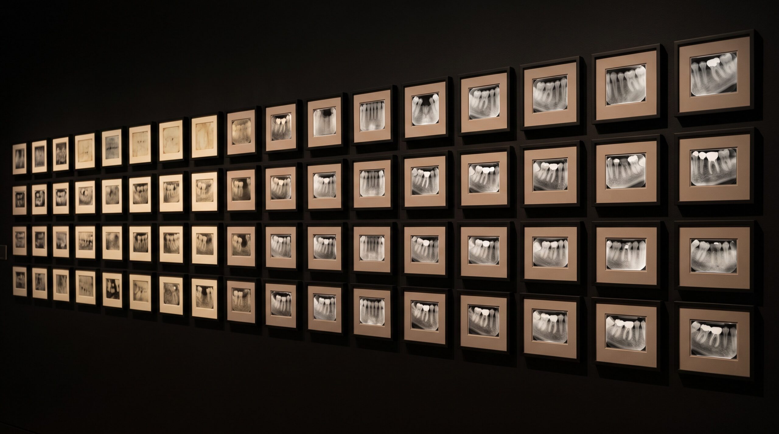
A dental-imaging manufacturer recently did something quietly revealing: it counted itself. Over a twenty-year span, one vendor tallied 330 patent applications and 217 granted patents in dental diagnostic imaging alone. As corporate announcements go it is easy to skim past – another firm marking its own homework. But read less as a boast and more as an archive, that portfolio is something more interesting: a map of how the dental image was actually engineered. Not one breakthrough, but a couple of hundred small, patient inventions, each shaving away a little noise or a little dose or a little uncertainty. The picture we take for granted on the operatory monitor is the sum of that accumulation, and it rewards a closer look at where all those increments went.
The Image Begins in the Material
Before geometry, before software, there is the question of what catches the X-ray in the first place – and this is where a great many of imaging’s incremental gains are hidden. An indirect detector converts X-rays to light in a scintillator and then that light to charge in a sensor, and the microscopic structure of that scintillator governs how much of the original signal survives the conversion. Grow the scintillator as a dense forest of fine, needle-like columns rather than a uniform slab, and each column channels its light straight down instead of letting it spread sideways into neighboring pixels; the result is a sharper, less noisy image for the same exposure. That single materials insight, which we explored in detail in the story of columnar cesium iodide, is the kind of quiet refinement a patent portfolio is full of – not visible in any one image, but present in all of them.

Alongside the scintillator sit two decades of refinement in the sensor itself: smaller pixels for finer detail, better electronics to read charge with less added noise, and readout schemes that widen the range between the faintest and brightest recoverable signal. None of these is a headline. Together they are the difference between an image that merely shows a tooth and one that resolves the thin dark line of a hairline fracture.
Geometry, Patented Curve by Curve
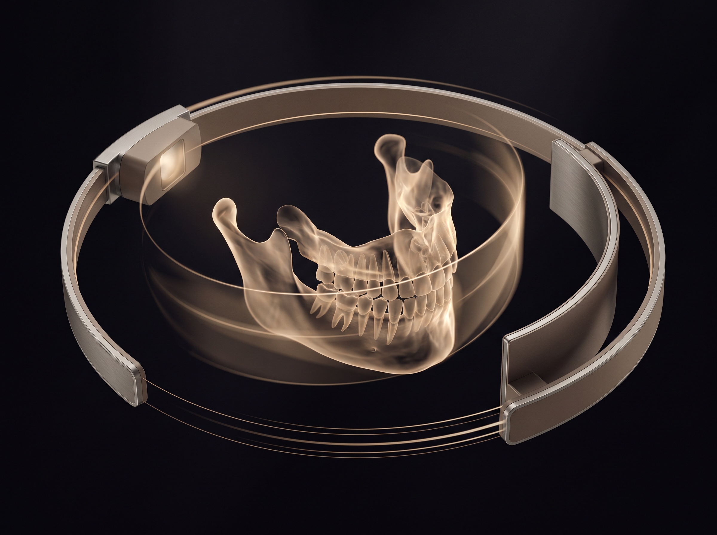
A panoramic radiograph is a feat of choreography more than optics. The source and detector sweep around the head in a coordinated arc, and the machine keeps only a curved ribbon of space – the focal trough – in sharp focus while everything nearer or farther smears into blur. Shaping that trough to hug the real, varied anatomy of human arches, and holding structures inside it as patients differ in size and position, is a genuine engineering problem, and it is exactly the sort of thing that generates filing after filing. The visible payoff over the years is images with fewer patients pushed out of the focal zone, less overlap at the contacts, and geometry that adapts rather than forcing every jaw into one idealized shape.
The same is true of the physics that shapes contrast within a single exposure – collimation that trims the beam to only what must be imaged, and the management of the scattered radiation that would otherwise fog the picture. Those choices, which we unpacked in the anatomy of scatter and collimation, are not settled science frozen in a textbook; they are living design variables that each generation of hardware tunes a little further.
The Picture Finished in Software
The most consequential shift of the last two decades may be that the image is no longer finished at the moment of exposure. In cone-beam computed tomography, a few hundred flat projections are captured as the machine circles the head, and a reconstruction algorithm assembles them into a three-dimensional volume. The early workhorse method was fast but unforgiving, prone to streaks and to the bright starbursts that a metal crown or implant throws across the surrounding bone. Iterative reconstruction changed the character of that output: rather than computing the volume in a single pass, it repeatedly refines an estimate against the measured data, suppressing noise and taming metal artifact until anatomy that was previously buried becomes legible.
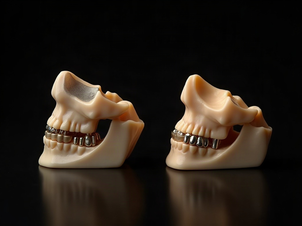
This is a profound reframing of what a scan is. The measured X-ray data is fixed the instant the exposure ends, but the diagnostic image extracted from it keeps improving as the mathematics improves. A volume acquired today and one acquired years ago may rest on the same physics, yet the newer reconstruction pulls a cleaner, quieter picture from the same photons. It is one reason a modern dataset travels so gracefully into downstream uses – the clarified volume is what later gets aligned to an optical surface in multimodal fusion, and everything built on top inherits that quality.
The Discipline That Runs Through All of It
What keeps this from being a simple story of “more” is that every advance is weighed against the dose it costs. A clearer image is trivial to obtain if one is willing to irradiate more; the actual achievement is holding clarity while pushing dose down. So the portfolio is threaded with work aimed at the opposite of excess – smaller fields of view that expose only the region of interest, pulsed and shaped exposures, and reconstruction that extracts a diagnostic result from fewer photons. The through-line of two decades is not raw power but efficiency: more diagnostic information per microsievert.

That is the discipline a patent count, oddly, makes visible. Hundreds of filings are not hundreds of revolutions. They are the granular, unglamorous labor of moving a well-understood craft forward a millimeter at a time – a better crystal here, a smarter curve there, an algorithm that forgives a restless patient or a metal restoration. The dental image did not arrive; it was assembled, increment by increment, by people solving small problems well.
Future Developments
The next stretch of that ledger is already legible in outline. Detector development is moving toward direct-conversion and photon-counting designs that measure the energy of individual X-rays rather than merely their sum, promising cleaner contrast and material discrimination at lower dose. Reconstruction is folding in learned models that can suppress noise and artifact more aggressively than hand-built algorithms, though they carry their own obligation to be validated so they clarify rather than invent. And the boundary between capture and interpretation continues to soften, as the same systems that make the picture begin to help read it. Whatever specific inventions fill the next twenty years of filings, the pattern will hold: the dental image will keep learning to see a little more, at a little less cost, one patient increment at a time – and the finished picture on the screen will go on concealing, as it always has, the enormous quiet craft that made it clear.
Sources & further reading:
Sorry, the comment form is closed at this time.