



August 15, 2026 The Needle Forest: How Columnar Cesium Iodide and the Scintillator Decide What a Sensor Can See
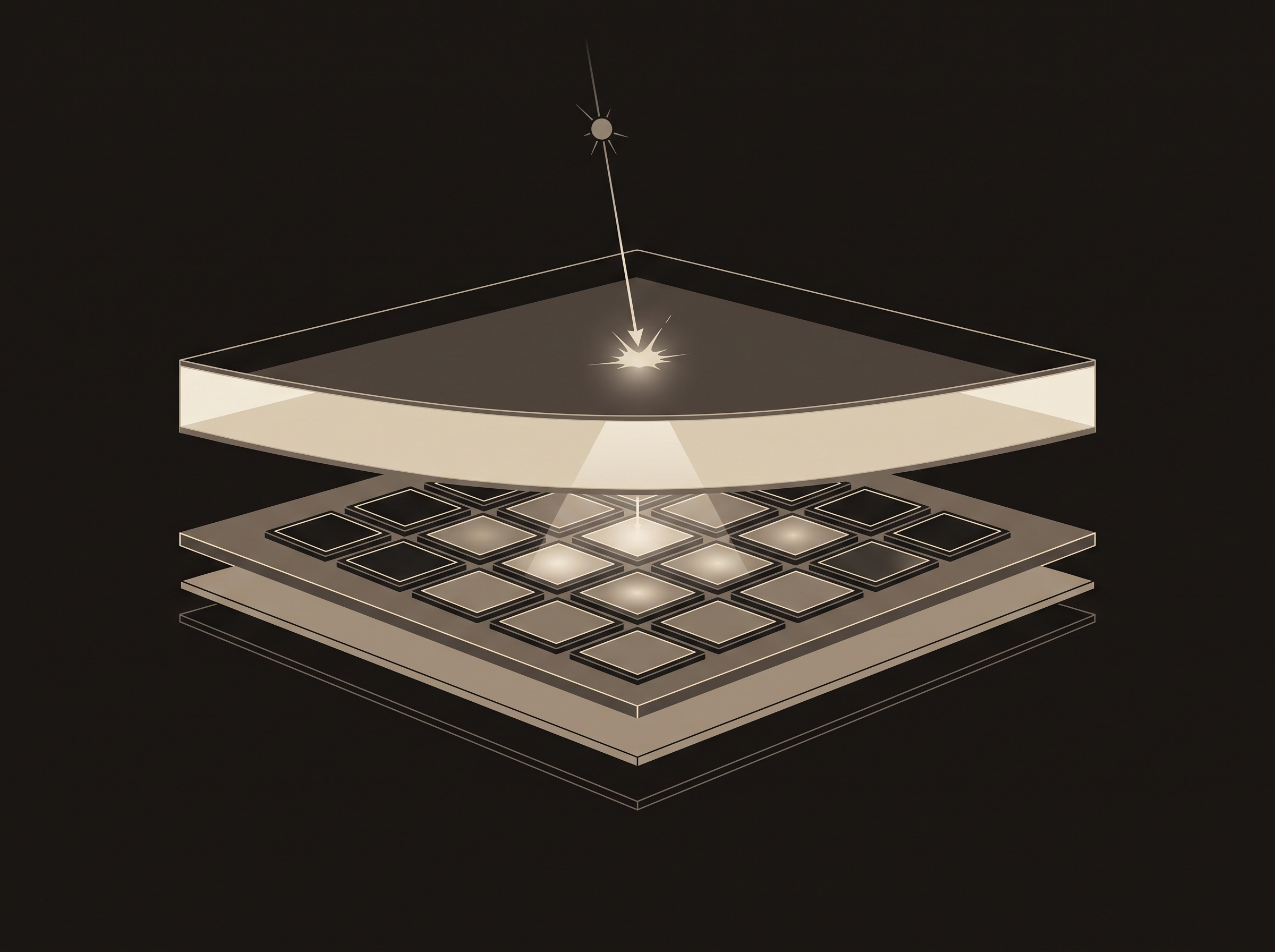
There is a quiet deception at the heart of nearly every digital dental sensor, and it is worth admiring rather than resenting. The pixel array — the grid of silicon that we think of as the detector — is, in most sensors, blind to X-rays. It cannot see them and does not try to. Instead, a thin crystalline screen is laid over the silicon, and it is that screen which meets the radiation, absorbs it, and answers with a faint flash of ordinary visible light. The array simply watches the screen glow. This is indirect conversion, and the screen has a name that belongs more to a physics bench than a dental operatory: the scintillator. Long before any circuit sharpens or displays the image, the choice of scintillator material has already set the ceiling on how sharp, how quiet, and how efficient that sensor can ever be.
Two Ways to Catch a Photon
Broadly, a digital detector can do one of two things with an incoming X-ray. It can convert the photon straight into electric charge inside a photoconductor such as amorphous selenium — direct conversion, the approach that reaches its purest form in the individual-photon counting explored in the true measure of a dental sensor. Or it can take the two-step, indirect route: convert the X-ray first into visible light in a scintillator, and then let a bed of photodiodes convert that light into charge. The indirect path dominates dental imaging because scintillators are robust, sensitive, and manufacturable at scale. But it introduces an intermediary, and that intermediary has a material personality all its own.
The Grain and the Needle
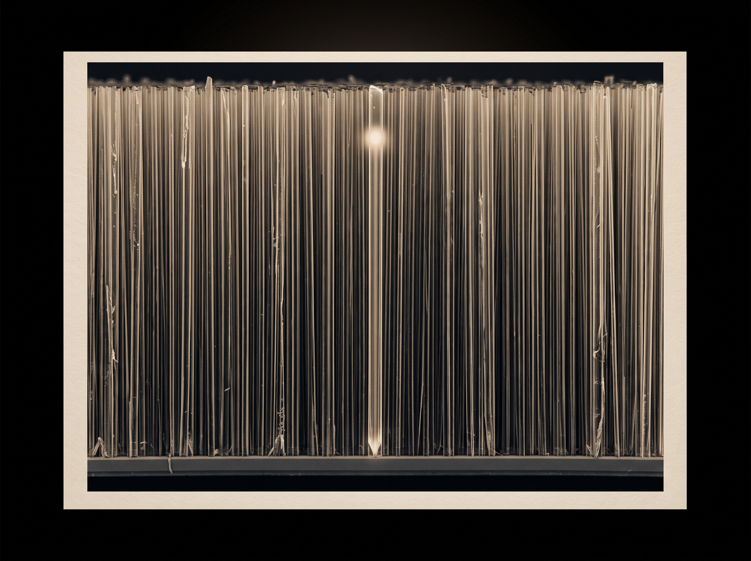
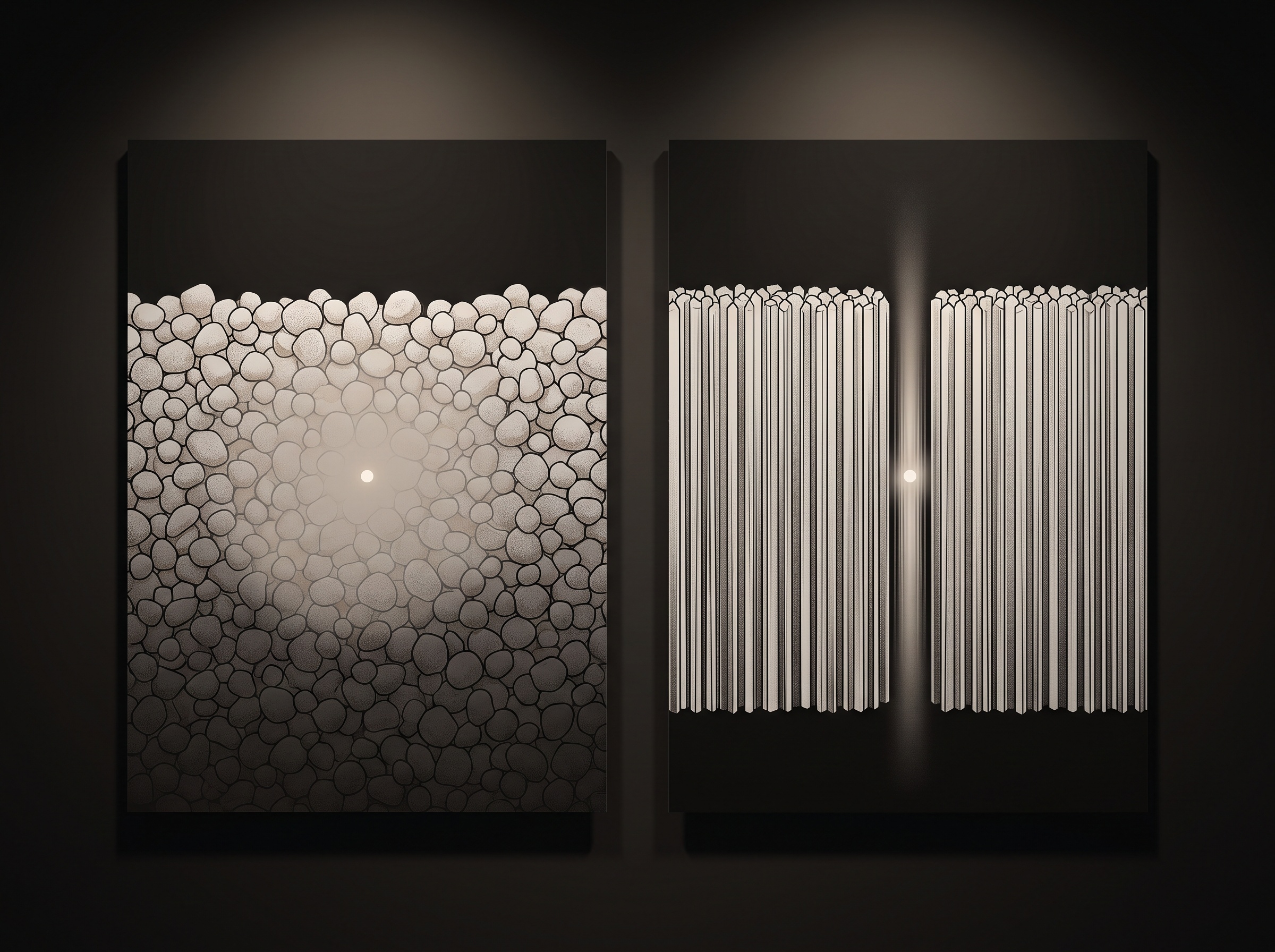
Two materials have long divided the field. The first is gadolinium oxysulfide — Gd2O2S, known affectionately as Gadox or GOS — a granular phosphor settled onto the array as a bed of tiny crystalline grains. It is inexpensive, stable, and perfectly serviceable. The second is cesium iodide doped with thallium, CsI:Tl, and it has a trick the granular phosphor cannot match. Grown under the right conditions, cesium iodide does not settle as a powder; it crystallises into a dense forest of slender vertical needles, each one standing perpendicular to the array beneath. Those needles behave like a bundle of microscopic fibre-optics, and that single structural fact is the whole story.
Why Structure Beats Chemistry
When an X-ray is absorbed deep inside a scintillator, the flash of light it produces radiates in every direction at once. In a granular material, that light is free to scatter sideways as it works its way toward the photodiodes, so a single point of absorption arrives at the array smeared across a small disc rather than a point. That lateral spread is blur, plainly and directly — it is one of the ways the scintillator itself quietly caps the resolution described in how sharp a dental image can truly be. Columnar cesium iodide fights this by channelling. Each needle guides its light down its own length, reflecting it off the needle walls and delivering it to the array in a tight column beneath where it was born. The light stays where it started, and the image stays sharp.
The Bargain of Thickness
Every scintillator lives inside a single unforgiving trade-off, and it is a trade-off of thickness. A thicker layer absorbs more of the incoming X-rays. That is unambiguously good: photons that pass straight through the screen unabsorbed are photons wasted, dose delivered to the patient for nothing, and their absence shows up as quantum mottle — the grainy statistical noise that haunts an under-caught image. Catch more of them and the picture grows quieter and the dose more honest. But in a granular phosphor, making the layer thicker also gives the scattered light more room to spread before it lands, so sharpness decays as efficiency improves. The two goods are chained in opposition. This is precisely where columnar cesium iodide earns its cost: because the needles keep the light collimated regardless of how far it must travel, the layer can be made thick enough to catch a large fraction of the beam without paying the usual penalty in blur. It partly unchains the two, letting a sensor be both efficient and sharp — a rare thing in imaging physics.

Light Yield, Colour, and the Photodiode’s Taste
Absorption is only half of a scintillator’s job; the other half is generosity. A good scintillator returns many visible photons for each unit of X-ray energy it swallows — its light yield, or conversion efficiency — because a brighter flash rises further above the electronic noise of the readout. Cesium iodide is notably generous, emitting on the order of tens of thousands of light photons per absorbed X-ray, and, no less importantly, it emits them in a green band that sits almost exactly where amorphous-silicon photodiodes are most sensitive. That spectral courtesy — the scintillator speaking in precisely the colour the array hears best — is as much a part of the material’s fitness as its stopping power or its structure. A phosphor that glowed brilliantly in a colour the silicon could barely register would squander its own light.
The Ghost the Material Leaves Behind
No material is without a flaw, and cesium iodide’s is memory. The same doped crystal that glows so readily is also prone to afterglow, or lag — a faint continued emission that lingers after the exposure has ended. In a single still radiograph this is invisible, but where images are captured in rapid succession, that residual glow can carry a whisper of one frame into the next, a cousin of the ghosting catalogued among the artifacts that distort a CBCT scan. It is a reminder that the scintillator is not a passive window but an active, imperfect participant, one whose virtues and vices are both written into its crystal lattice.
Future Developments
The material science beneath the pixel is far from settled. Structured cesium iodide continues to be refined toward ever-finer, straighter needles that leak still less light sideways, and manufacturers are experimenting with dual-layer and thicker structured screens that push detection efficiency higher without surrendering resolution. Beyond the two incumbents, a new generation of scintillators — nanostructured phosphors and perovskite crystals engineered for high light yield and low afterglow — is moving from the laboratory toward the clinic, while direct-conversion photon-counting detectors quietly threaten to make the intermediary screen unnecessary altogether. Yet for now, the humble scintillator remains the first and most consequential decision in the imaging chain: a thin, glowing layer of grown crystal that meets the radiation on our behalf and, in the character of its material, decides what the rest of the sensor will ever be allowed to see.
Sources & further reading:
- Scintillators and indirect-conversion flat-panel detectors (radiographic detector physics reference)
Sorry, the comment form is closed at this time.