



September 1, 2026 The Sensor’s Fingerprint: Dark Current, Fixed-Pattern Noise, and the Flat-Field Correction That Makes a Detector Honest
There is a comforting assumption behind every digital radiograph: that the image is a faithful record of the patient, and nothing else. Where the picture is bright, the tissue was radiolucent; where it is dark, dense structure stopped the beam. But a digital sensor is not the neutral pane of glass this assumption imagines. It is a dense grid of millions of tiny light-detecting cells, each one manufactured to a tolerance rather than a perfection – and each with its own small bias, its own sensitivity, its own quiet flaw. Left to speak for themselves, those cells would print their own fingerprint over every tooth they were asked to record. The reason they do not is a piece of unglamorous, invisible craft performed before the clinician ever sees a thing: the detector, in effect, first learns to subtract itself.
Why No Two Pixels Are Equal
Imagine trying to paint a portrait on a canvas whose weave is uneven, whose primer is thicker in some patches than others, with a handful of threads missing entirely. The likeness you produced would carry the canvas’s defects as surely as your brushwork. A digital detector is that imperfect canvas. During fabrication, every photodiode and its associated electronics end up slightly different from its neighbor. Some cells respond a little more strongly to the same dose – they have higher gain. Some sit at a slightly higher baseline even in total darkness – they have a larger offset. A few are simply broken: dead pixels that never respond, or hot pixels that read bright no matter what. None of this is failure; it is the ordinary reality of building a grid of millions of identical-in-theory sensors. The task of calibration is not to fix the canvas but to know it so precisely that its flaws can be cancelled.
The Dark Frame: What a Sensor Sees in the Dark
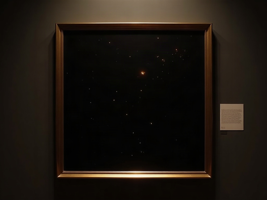
Begin with the strangest exposure in imaging – the one taken with no x-rays at all. Even in complete darkness, a detector is not silent. Warmth alone liberates a slow drizzle of electrons within the silicon, a thermally generated signal called dark current that accumulates in every cell during the exposure. Because each cell leaks at its own rate, the result is not uniform: it is a fixed, structured pattern of faint bias, brighter here, dimmer there, with the occasional hot pixel blazing away. Capture that pattern deliberately – an exposure of the same duration with the beam off – and you have a dark frame: a portrait of everything the sensor contributes before the patient contributes anything.
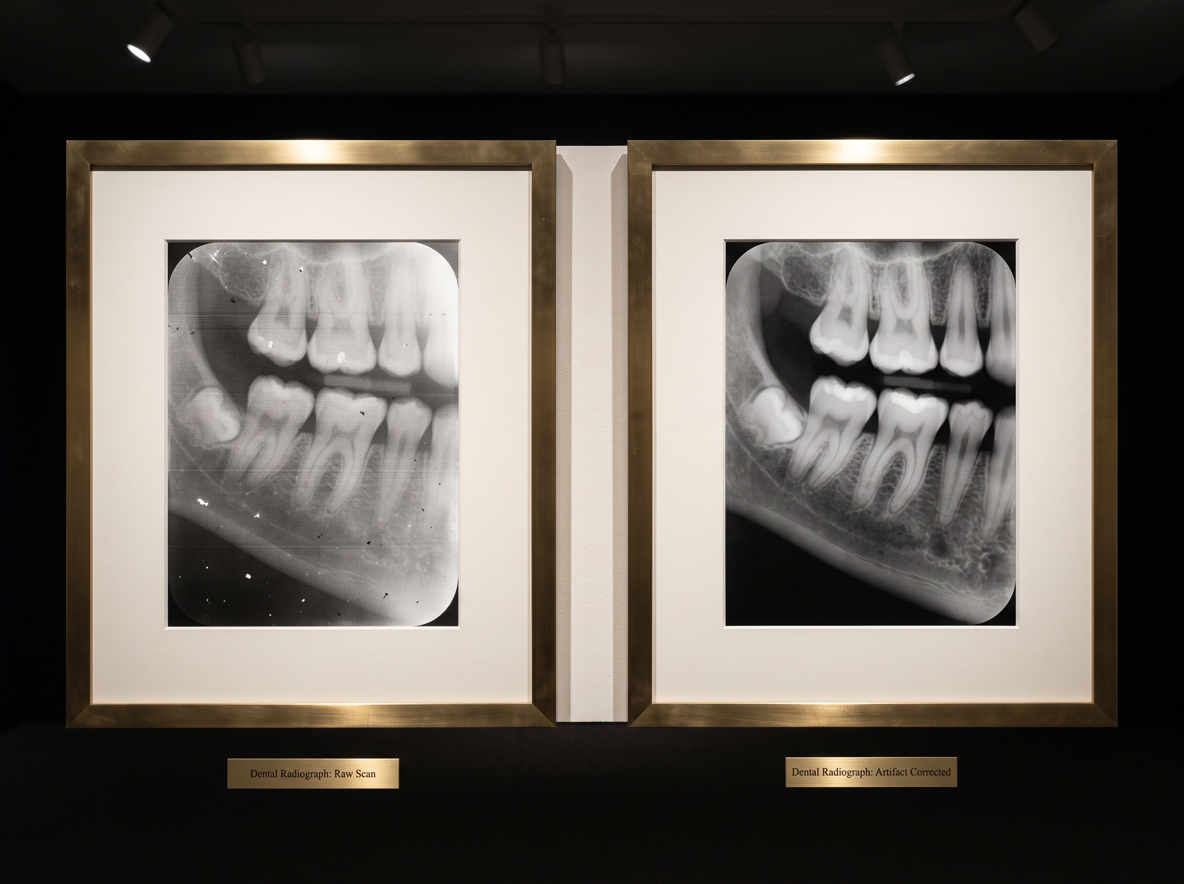
The dark frame is the first thing subtracted from every real image. Whatever fixed bias the sensor was going to add, it added to the dark frame too; take one away from the other and that contribution vanishes. This is the same disciplined skepticism we bring to the exposure index and the slow drift of dose creep – the refusal to trust a number, or a pixel, until you know what it would have read with nothing there.
The Flat Field: Mapping Every Cell’s Appetite
Subtracting the dark frame removes the offset, but it says nothing about gain – the fact that identical doses land differently on different cells. To map that, the detector is given the opposite exposure: an even flood of radiation with nothing in its path, a uniform field that should render as a perfectly flat, featureless grey. It never does. The captured flat field reveals the whole landscape of non-uniformity at once – the per-cell differences in sensitivity, the gentle brightness gradient of the anode heel effect sloping across the frame, and the subtle structural signature of the scintillator layer that first catches the x-rays before they ever reach the silicon.
That scintillator is itself a patterned thing, as we explored in the forest of cesium-iodide needles that decides what a sensor can see; its unevenness is baked into the flat field along with everything else. The correction then does something elegant: it divides each pixel of the real image by that pixel’s value in the flat field. A cell that read 10 percent hot to the calibration flood is assumed to read 10 percent hot to the patient too, and is scaled back down. In one stroke, the whole map of appetites is levelled.
Fixed-Pattern Noise Versus the Noise You Cannot Remove
What dark-frame and flat-field correction eliminate together has a name: fixed-pattern noise. The word noise is slightly misleading, because this noise does not flicker. It is the same every time – a structural, repeatable signature stamped on image after image by the same offending cells. Precisely because it is fixed, it can be measured once and subtracted forever. That is what makes it curable.
It is worth being clear about what correction does not remove. Every individual exposure also carries random noise – the grainy quantum mottle of a finite number of x-ray photons arriving by chance, different in every frame. No calibration can subtract that, because there is no fixed pattern to learn; it is the irreducible statistical texture of the dose itself. Confusing the two is a costly error. The seductive temptation is to lean on aggressive processing to smooth away random grain, and, as we have cautioned in the essay on how sharpening and noise reduction can deceive, that path invents detail as readily as it removes it. Flat-field correction is the honest counterpart: it removes only what genuinely does not belong to the patient.
The Map of the Broken Cells
Then there are the pixels beyond correction – the truly dead and the incurably hot, cells whose reading means nothing at all. These cannot be scaled into honesty; they must be disowned. The manufacturer characterises the detector and records a defective-pixel map, a register of every cell not to be trusted. In the finished image, each of those locations is quietly filled in by interpolation from its healthy neighbours – an educated reconstruction rather than a measurement. It is a small act of forgery in the service of truth, and an acceptable one, provided the defects are few and scattered. When they cluster into lines or blooming patches, interpolation can no longer paper over them, and the detector is telling you it is reaching the end of its working life.
A Calibration That Ages
None of this is done once and forgotten. A detector’s fingerprint drifts. Dark current climbs with temperature, so a sensor calibrated in a cool morning operatory is subtly miscalibrated by a warm afternoon. Gain relationships shift as the panel ages and its scintillator wearies. The best systems recalibrate on a schedule – refreshing their dark frames and flat fields periodically – so that the corrections keep pace with the hardware they describe. A calibration is not a permanent fact about a detector; it is a photograph of the detector as it was, and like every photograph it slowly stops resembling its subject.

This is why image-quality faults so often trace back not to a broken sensor but to a stale calibration – a gradient that crept back, a grid texture that resurfaced, a gentle unevenness that a fresh flat field would have erased. The correction is only as honest as it is current.
Future Developments
The direction of travel is toward calibration that thinks for itself. Detectors are beginning to monitor their own temperature and defect counts and to flag when their corrections have drifted out of tolerance, rather than waiting for a technician to notice a fault on a patient’s film. Machine-learning approaches promise to model non-uniformity and dark behaviour more subtly than a single subtracted-and-divided frame can, adapting the correction to the exact conditions of each exposure. And as detectors grow more sensitive in the pursuit of lower dose, the fixed-pattern noise they must overcome only becomes more visible against the fainter signal, making this quiet arithmetic more important, not less. There is a lesson in the whole exercise that outlasts any technology: the most trustworthy image is the one whose maker knew, and accounted for, its own flaws. A detector earns its honesty the same way – by rendering nothing, studying the result, and learning to subtract itself before it is ever asked to render a tooth.
Sorry, the comment form is closed at this time.