


August 10, 2026 The Ghosts in the Volume: Beam Hardening, Metal Streak, and the Artifacts That Distort a CBCT Scan
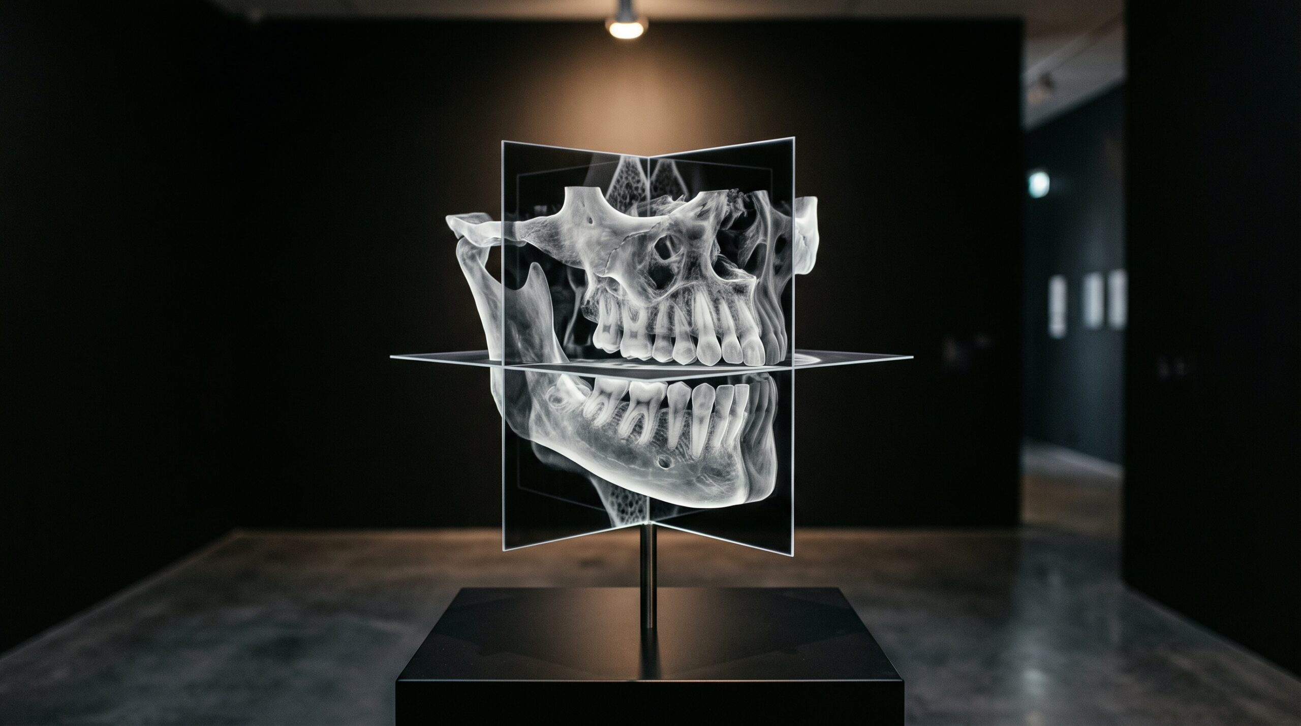
A cone beam scan arrives on the screen with the quiet authority of a photograph. The jaw turns in three dimensions, the roots splay, the canal traces its path through the bone, and it is easy to forget that none of it was ever seen. A CBCT volume is not a picture of anatomy; it is a mathematical reconstruction, assembled from several hundred flat projections taken as the source and detector swept around the head. Every reconstruction is an inference, and every inference carries ghosts — the artifacts that appear in the image but were never in the patient. The skilled reader does not pretend they are absent. The skilled reader learns their faces, names them on sight, and reads past them to the anatomy underneath.
Why the Volume Lies
The root of most CBCT artifacts lies in a single inconvenient fact of physics: the X-ray beam is not a single pure color. It is polychromatic, a spectrum of photon energies, and the reconstruction algorithm assumes — for the sake of tractable mathematics — that it is not. That gap between the honest physics and the convenient assumption is where the ghosts are born. Add the cone-beam geometry, a comparatively low radiation dose, a flat-panel detector with its own quirks, and a living patient who cannot hold perfectly still, and the surprise is not that artifacts appear but that the image is as faithful as it is.
Beam Hardening: The Cupping and the Bands
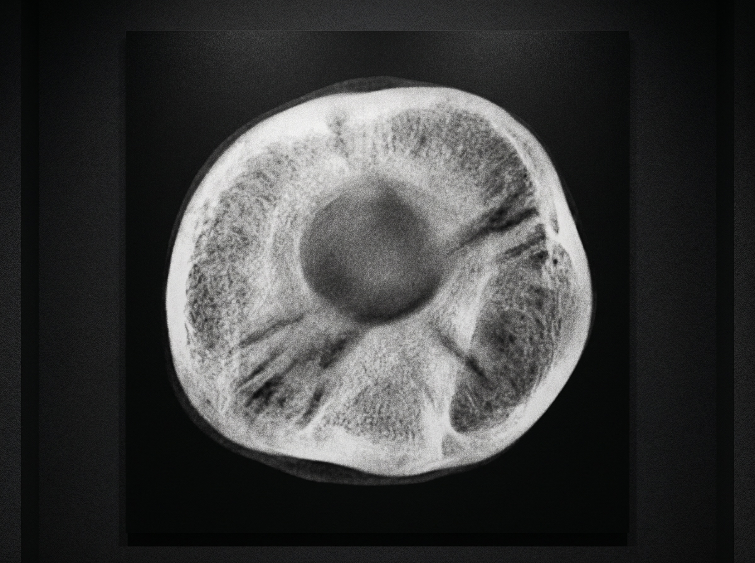
As that polychromatic beam passes through tissue, the lower-energy photons are absorbed first. The beam that emerges is therefore ‘harder’ — skewed toward higher energies — than the beam that went in, and the algorithm, expecting a consistent energy, mismeasures how much material the ray actually crossed. Two signatures result. The first is cupping: the center of a uniform dense structure is reconstructed as darker than its edges, as though the bone were hollowed out. The second is dark banding — broad shadowy streaks that stretch between two dense objects, most notoriously between the roots of adjacent teeth or across the dense cortical plates. A dark band drifting near a root apex can masquerade as a lesion or a fracture; recognizing it as beam hardening, tethered to the geometry of the dense structures around it, is what keeps it from becoming a misdiagnosis.
Metal: The Loudest Ghost of All
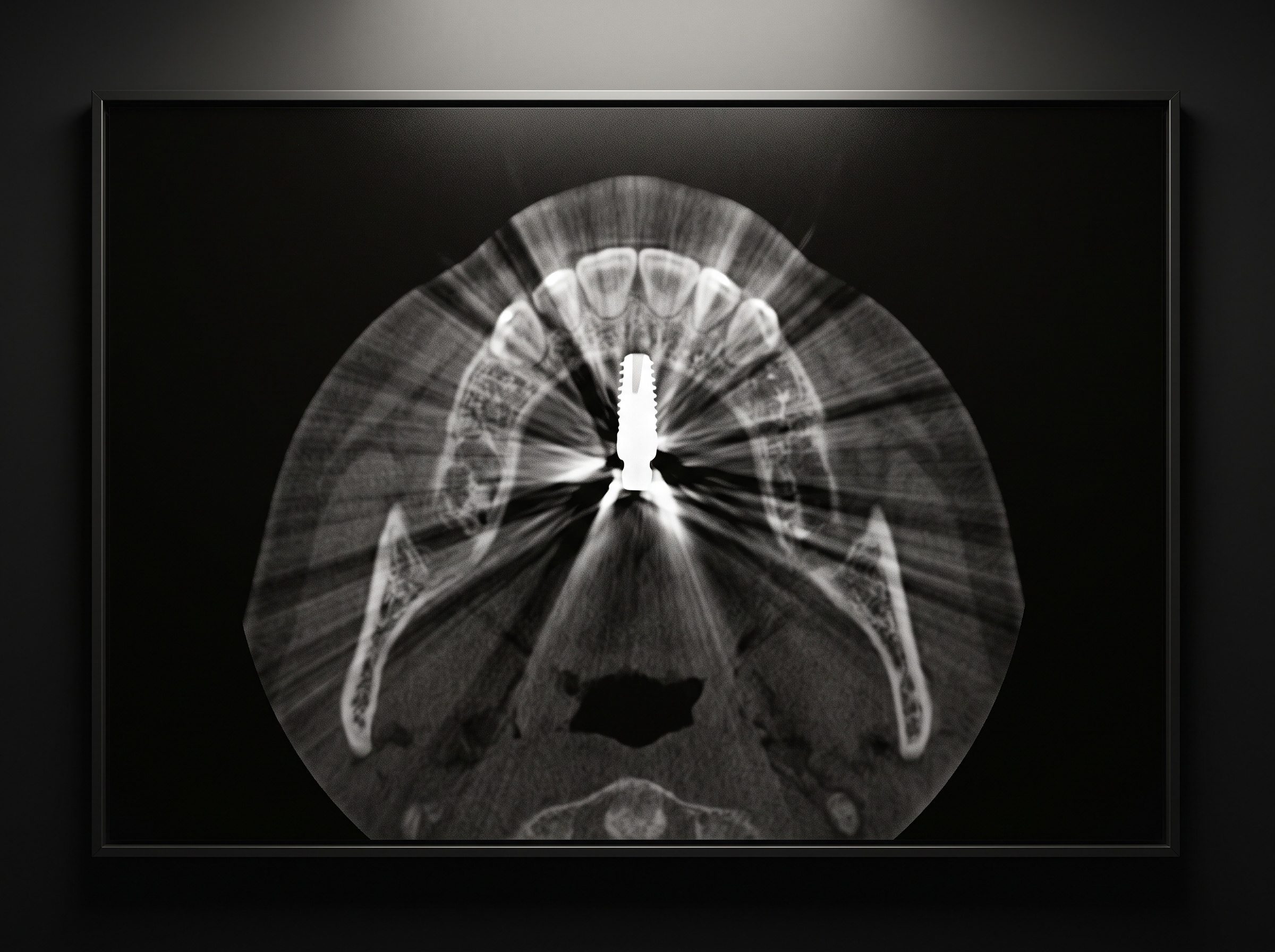
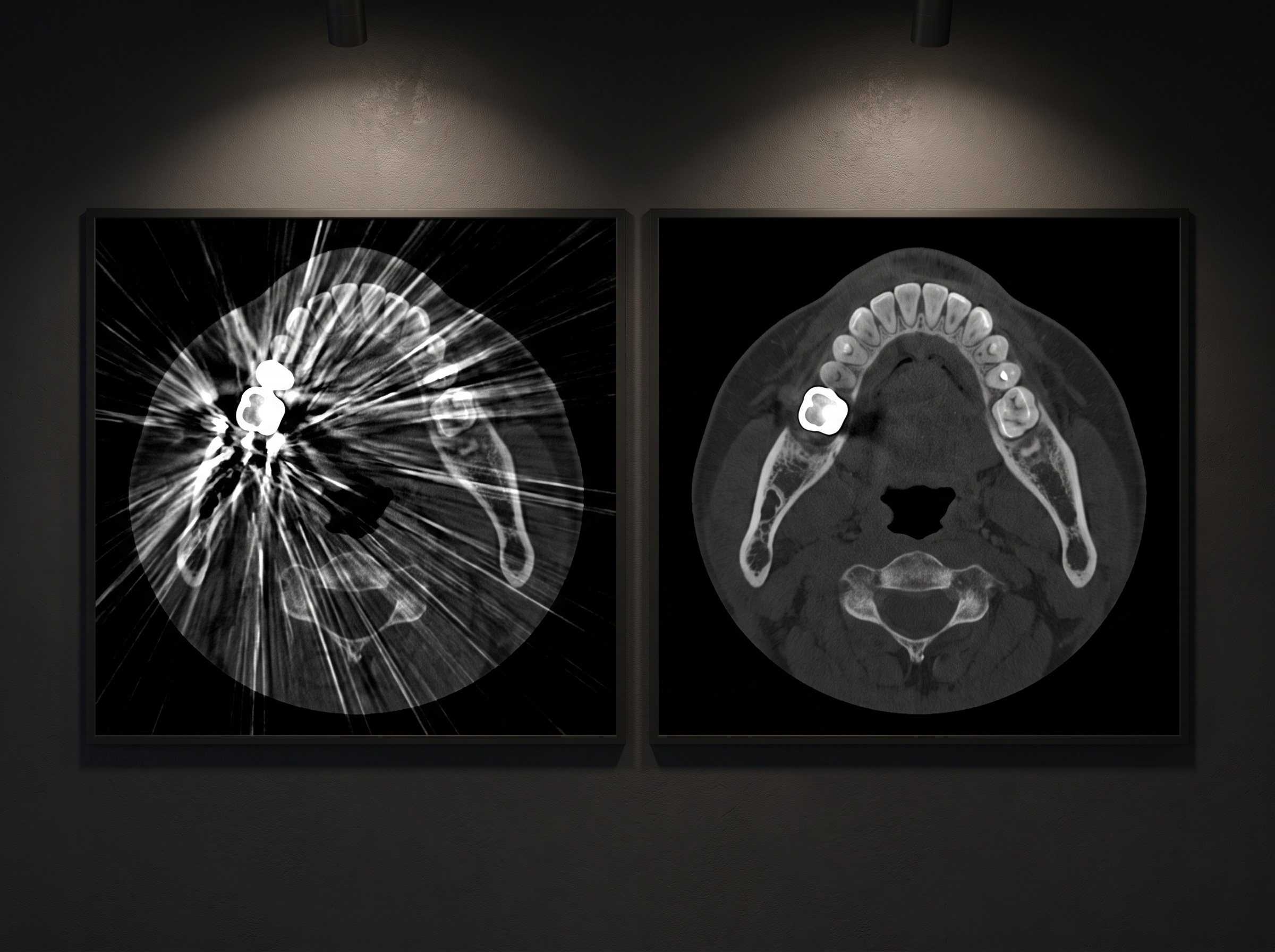
Nothing disturbs a dental CBCT like metal. Amalgam, gold, titanium implants, stainless posts, orthodontic hardware — each is far denser than any tissue, and each provokes the most dramatic artifact in the catalogue. The mechanism is really beam hardening pushed to its extreme, compounded by outright signal starvation: so little of the beam survives the metal that the detector records almost nothing along those rays, and the reconstruction, given no honest data, invents. The result is a starburst of brilliant white streaks and pitch-dark shadows radiating across the slice, often obliterating the very region the clinician most needs to see — the bone around an implant, the margin of a restoration, the canal beside a post. These are not subtle. The danger is not that they will be missed but that they will erase genuine findings hiding in their glare, and that a dark streak between two restorations will be read as a fracture that is not there.
Scatter and Noise: The Grain Beneath Everything
Cone-beam geometry irradiates a whole volume at once rather than a thin fan, and that broad exposure generates abundant scattered radiation — photons that ricochet off their original path and strike the detector where they do not belong. Scatter lowers contrast, adds a milky haze, and feeds the general graininess that softens fine trabecular detail. It is the reason CBCT, for all its geometric precision, cannot rival medical CT at distinguishing subtle differences in soft tissue. Lower dose protocols, chosen wisely to protect the patient, buy that protection partly in noise. The craft is knowing which questions the grain still permits you to answer confidently — bone volume, root morphology, canal position — and which it does not.
Motion and the Rings
A CBCT acquisition is not an instant; it is a rotation lasting several seconds, and the volume is only coherent if every projection describes the same, still anatomy. Let the patient swallow, breathe deeply, or drift a millimetre, and the reconstruction smears: edges double, cortical lines blur into ribbons, and fine detail dissolves. Motion is the most preventable artifact of all — a firm head support, a clear instruction, and a calm few seconds defeat it — and also the most quietly corrosive when ignored, because a gently blurred volume can still look plausible. Distinct from motion are the ring artifacts: concentric circles centered on the axis of rotation, the fingerprint of miscalibrated or defective detector elements repeating the same error at every angle. Rings point not at the patient but at the machine, and their appearance is a prompt to calibrate.
Reading Around the Ghosts
Naming an artifact is most of the battle, because a named artifact loses its power to deceive. A dark band that connects two dense structures, a starburst locked to a piece of metal, a haze that thickens with lower dose, concentric rings on the axis — each announces its own origin once the eye is trained. Beyond recognition, the reader has real tools. Scrolling through adjacent slices reveals whether a suspicious shadow persists in three dimensions like true anatomy or flickers like the projection-bound ghost it is. Adjusting the window and level lifts genuine detail out from under a streak. Metal-artifact-reduction algorithms, now standard on most units, re-estimate the starved data and recover much of what the streaks conceal — though never quite all of it, and sometimes at the cost of introducing new, subtler distortions of their own. And when the artifact truly buries the answer, the honest move is to reach for a supplementary view: a well-angled periapical, a targeted small-field rescan, a different modality altogether.
Future Developments
The physics that breeds these ghosts is being met, at last, with equal cleverness. Iterative reconstruction — refining the volume through repeated passes rather than a single back-projection — already suppresses noise and metal streak far better than the classical algorithms, and dual-energy and photon-counting detectors promise to attack beam hardening at its polychromatic root by measuring the spectrum instead of assuming it away. Most striking is the arrival of deep-learning reconstruction, in which networks trained on vast libraries of scans learn to distinguish true anatomy from artifact and to reconstruct clean volumes from lower-dose, sparser data. The trajectory is clear: quieter images, at lower dose, with the loudest ghosts increasingly hushed. Yet the reader’s eye will not be retired. An algorithm that erases an artifact can also, on occasion, erase a finding or invent one, and the clinician who knows what a real ghost looks like remains the last, indispensable safeguard between a beautiful reconstruction and a true one.
Sources & further reading:
Sorry, the comment form is closed at this time.