


October 8, 2026 Painting Back the Edge of the Scan: How Diffusion AI Erases CBCT Truncation Artifacts
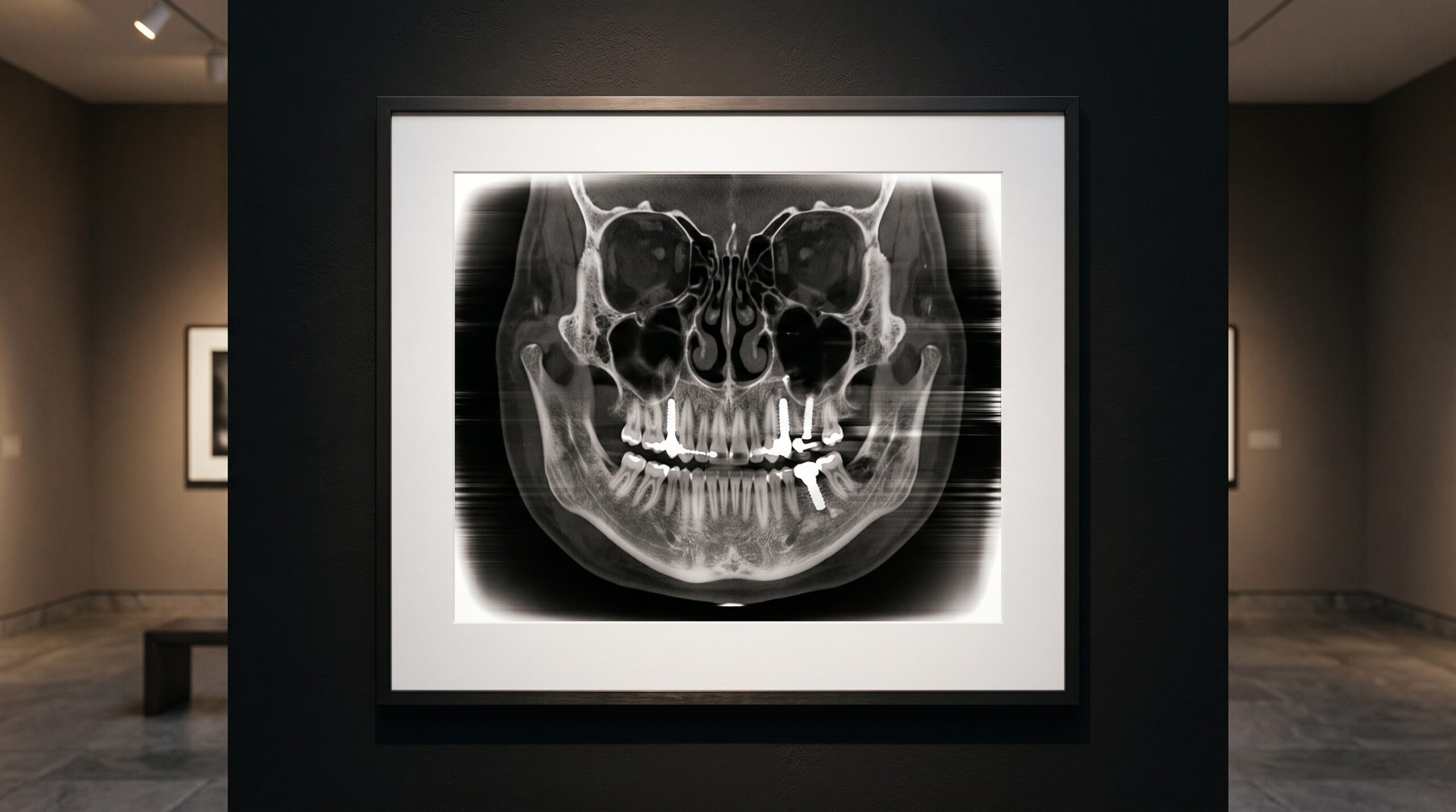
Look closely at the margin of a cone-beam CT volume and you will sometimes find the place where the image quietly stops telling the truth. The center may be exquisite — crisp trabecular bone, a root canal traced in fine gray — but toward the edges a bright halo blooms, densities drift, and faint streaks rake inward from the border. These are truncation artifacts, and for years they have been treated as an unavoidable tax on CBCT’s compact geometry. A new study suggests that tax may finally be negotiable, and the instrument of the bargain is a kind of generative artificial intelligence usually associated with making pictures, not restoring measurements.
The edge where the picture breaks down
Cone-beam CT earned its place in dentistry and beyond by doing a great deal with a little: high spatial resolution and three-dimensional anatomy at a fraction of the radiation dose of conventional multi-detector CT. But the compactness that makes it affordable and low-dose carries a structural flaw. The flat-panel detector is finite, and patients are not conveniently sized to fit within it. When the shoulders, the full breadth of the skull, or any dense structure extends beyond the detector’s field of view, the X-rays passing through those regions are never recorded at all. The projection data, in the language of the physics, are truncated — and a reconstruction built on incomplete data breaks down exactly where the data ran out.
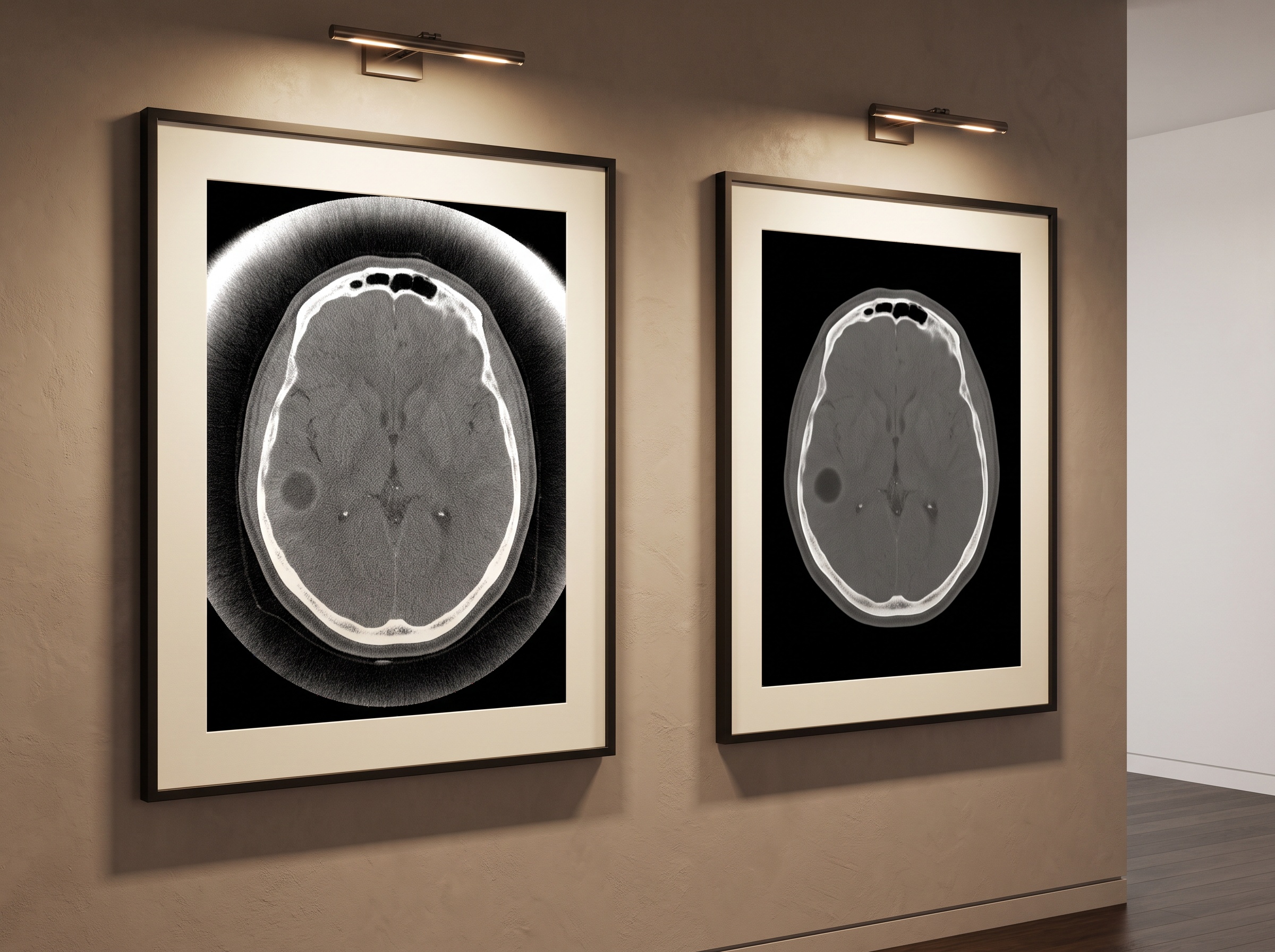
The visible signature is familiar to anyone who reads these scans: bright cupping along the periphery, distorted intensities near the boundary, and shading that propagates inward far enough to shift the apparent density of real anatomy. Subtle, low-contrast findings near the edge can vanish into that artifact background entirely, which is precisely where a quiet lesion is least forgiving of being missed.

Why filtered backprojection cannot forgive a missing measurement
To understand why a gap in the data is so destructive, it helps to recall how the image is built. Most CBCT systems reconstruct with filtered backprojection, an elegant algorithm that rests on one unforgiving assumption: that every X-ray line through the object has actually been measured. Feed it a complete dataset and it returns a faithful volume. Feed it projections with their edges missing and it does not fail gracefully — it honors the missing data as if they were zero, and the mathematics retaliates. Intensity gradients pile up at the boundary of the field of view and spill inward as the cupping and streaking we see. The artifact is not a flaw in the hardware so much as a mismatch between what the algorithm was promised and what it was given.
This is the same family of problem that motivates much of the recent work on smarter reconstruction, from the dose-saving mathematics behind deep-learning reconstruction of the low-dose dental scan to the careful benchmarking of diagnostic reference levels in CBCT dose. In every case the question is the same: how do we recover a trustworthy image when the measured signal is, by design, imperfect?
The old fix was a polite guess
The conventional remedy for truncation is mechanical and modest. The widely used Reconstruction Toolkit, among others, relies on padding: the truncated projection is extended at its edge with synthetic values, often mirrored or smoothly decayed, so the reconstruction algorithm at least receives a nominally complete dataset. Padding tames the worst of the cupping, and for many scans it is good enough. But it is, at heart, a guess. Those padded values carry no real information about the anatomy that lay outside the detector; they are a plausible-looking filler. Where the truncated region is dense or structurally complex, the guess shows its seams and residual artifacts remain.
A model that completes the measurement, not the picture
A research team from the Beijing Institute of Technology and Beijing WeMed Medical Technology, writing in BMC Medical Imaging, took a different line of attack. Instead of patching the corrupted image after reconstruction, they went upstream to the raw projection data and asked a model to complete the measurement itself. Their tool is a conditional generative diffusion model — the same class of AI that has reshaped image synthesis elsewhere, repurposed here for a strictly scientific end.
A diffusion model learns by reversing a gradual noising process: shown data corrupted by progressively more noise, it learns to denoise each step, and so learns the deep statistical structure of the images it was trained on. Left unconstrained, such a model will happily invent. The crucial word in this work is conditional. The generation is anchored to the specific truncated projection in hand, so the model does not dream up arbitrary anatomy — it fills in the missing detector regions with values that are realistic, anatomically plausible, and consistent with what was actually measured. The completed projection is then reconstructed by the ordinary filtered-backprojection pipeline, which finally receives the complete dataset it always demanded.

The numbers behind the restoration
The gains reported are not subtle. Measured at the projection level against the established padding method, the diffusion-based completion improved peak signal-to-noise ratio by roughly 12 decibels and structural similarity by 0.15 — a 12 dB PSNR gain representing a very large reduction in error, and an SSIM improvement of that size indicating substantially better preservation of real structure. Carried through to the final three-dimensional volumes, the method delivered an average 9 dB PSNR gain and a 0.10 rise in SSIM over the reference approach, with visibly better image uniformity and, most meaningfully for the clinic, improved visibility of low-contrast structures — the faint soft-tissue and density distinctions that decide whether a finding is seen or missed.
The team also tested the harder case of region-of-interest reconstruction and found the diffusion approach held a statistically significant advantage over earlier generative adversarial network methods while preserving diagnostically relevant features. That comparison matters for more than bragging rights. GANs, long the default for image completion, are notorious for hallucinating convincing detail and for unstable training — and in a diagnostic image, a fabricated structure is not a curiosity but a hazard. A diffusion model’s tendency to stay anchored to the conditioned, measured data, erring toward fidelity rather than invention, is exactly the temperament a clinician should want from a machine that touches a scan before a human reads it.
What it means in the dental operatory
For dentistry the relevance is direct. The skull and shoulders routinely exceed a dental CBCT detector’s field of view, so truncation shading near the image periphery is a familiar complication when assessing structures at the margins of a volume. The appeal of this approach is that it asks nothing new of the hardware. It is a software correction that operates on projection data the scanner already acquires — no detector upgrade, no longer acquisition, no additional radiation dose — and because it feeds a completed dataset into the standard reconstruction chain, it is compatible with the pipelines already in commercial use. A cleaner volume from the same scan is a rare kind of improvement: one with no dose penalty attached. It is also a reminder that the richest returns in imaging increasingly come from software and data, the same shift that makes a unified record like the fused virtual patient possible at all.
The caution that accompanies any deep-learning method in medicine applies here too. The study was retrospective, conducted with ethics approval from Xuanwu Hospital, Capital Medical University, and published as an early-release accepted manuscript still subject to final edits. Broad adoption will depend on validation across diverse patient populations, scanner geometries, and anatomical sites, and on confidence that the model’s completions stay reliable for anatomy far outside its training distribution. A model that fills a gap must never be allowed to fill it with something that was not there.
Future Developments
What makes this work quietly radical is its restraint: it does not use generative AI to make a prettier image, but to solve a genuine physics problem — restoring missing measurements with enough fidelity that the century-old reconstruction mathematics simply works as it was always meant to. That framing points to where the field is heading. Expect correction to migrate earlier and earlier in the imaging chain, from post-processing toward the moment of acquisition, and expect the diffusion architecture — faithful, stable, anchored to data — to displace more of the older generative methods wherever a fabricated detail would be dangerous. If these results hold at scale, the bright, cupped edges that have haunted cone-beam volumes for years may come to look like a historical artifact in both senses of the word: a flaw we once tolerated, preserved only in the older images we kept. The art of the dental scan has always been a negotiation between dose, detector, and mathematics; here the machine simply gives the mathematics back the complete picture it was always owed.
Sources & further reading:
Related Reading
- The Ghosts in the Volume: Beam Hardening, Metal Streak, and the Artifacts That Distort a CBCT Scan
- The Seductive Edge: How Sharpening, Contrast, and Noise Reduction Can Deceive on a Digital Dental Radiograph
- The Dose You Don’t Take: Deep-Learning Reconstruction and the Craft of the Low-Dose Dental Scan
Sorry, the comment form is closed at this time.