


September 30, 2026 The Dose You Don’t Take: Deep-Learning Reconstruction and the Craft of the Low-Dose Dental Scan
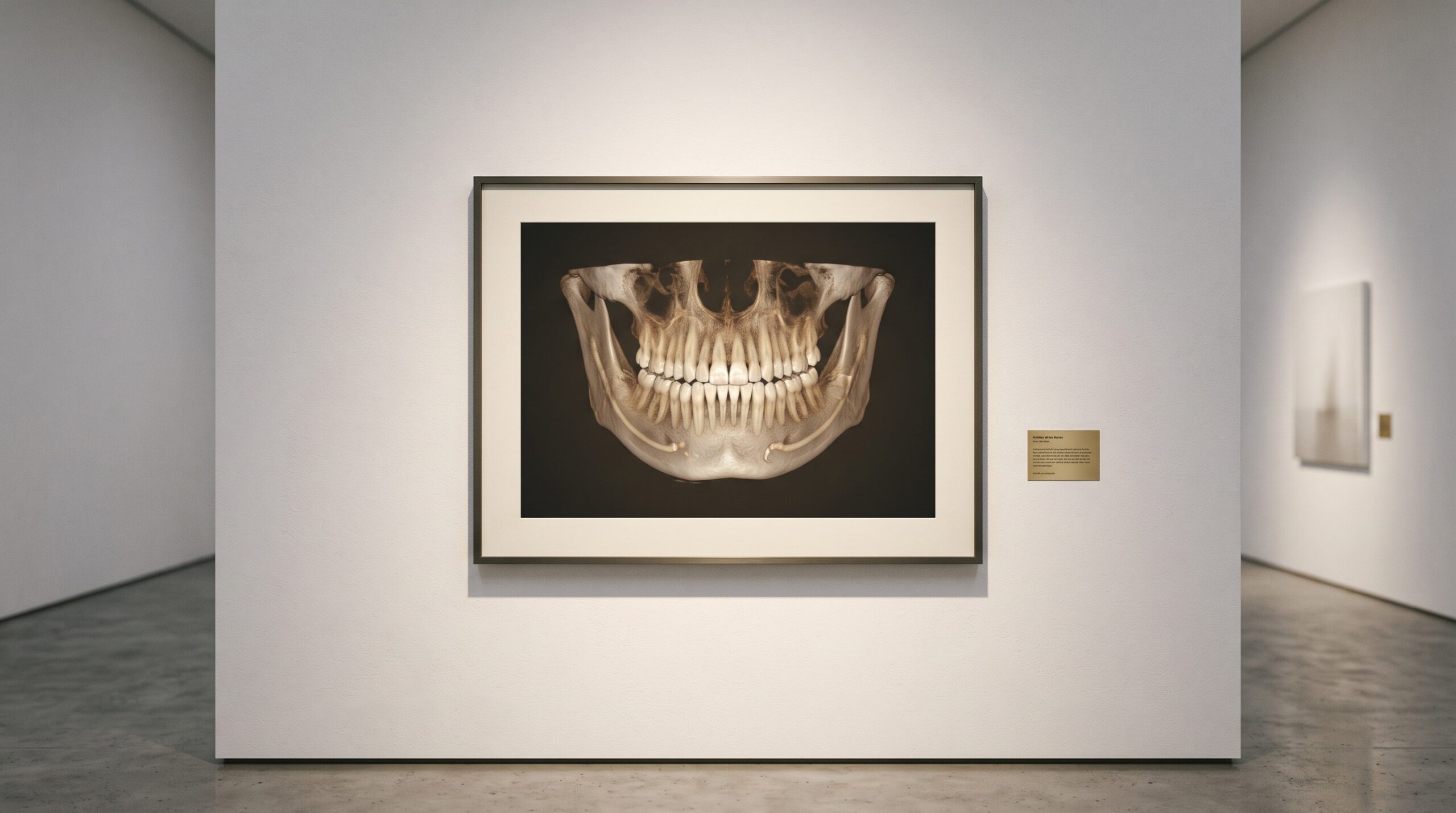
Every cone-beam scan is a negotiation. Turn the dose up and the image arrives clean, its grey values settled and quiet; turn it down — as every guideline and every conscientious clinician now urges — and the picture pays for that restraint in noise. Photons are the currency of a radiograph, and a low-dose acquisition simply buys fewer of them. The grain that results is not a flaw in the machine but the honest texture of a frugal exposure. For decades the craft of imaging lived inside that trade: accept the noise, or accept the dose. A new class of deep-learning reconstruction is quietly rewriting the terms.
The Noise Floor
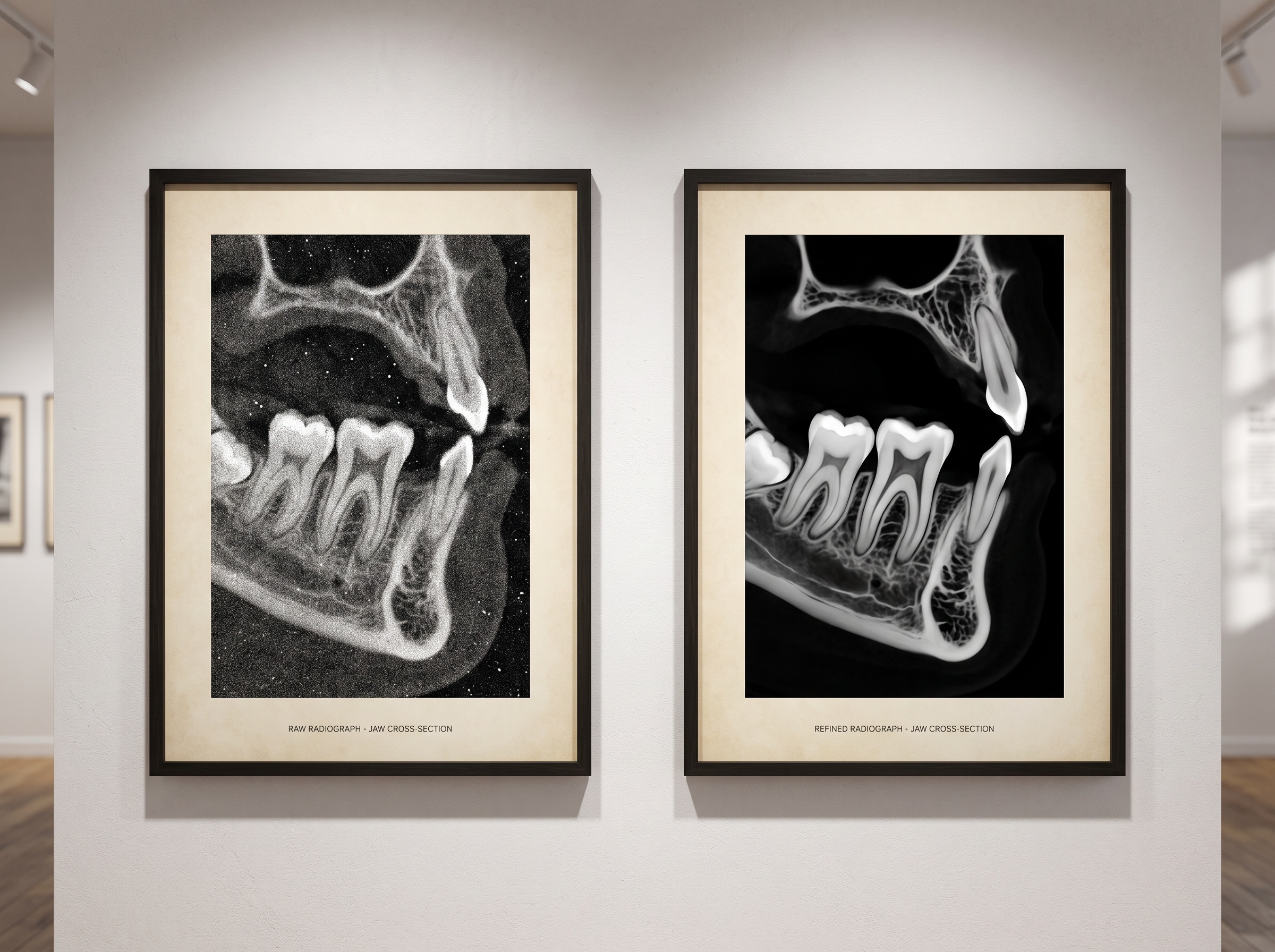
Lower the milliampere-seconds and you starve the detector. Fewer X-ray photons reach each element of the sensor, and the statistical fluctuation between neighbouring measurements — quantum mottle — rises relative to the signal you care about. The result is a scan speckled with grain, its fine trabecular architecture dissolving into static, its low-contrast boundaries softening into ambiguity. This is the noise floor, and conventional filtered back-projection cannot lift a diagnosis out of it without also blurring away the very edges a clinician reads. The classical remedies — iterative reconstruction, edge-preserving regularization — pull the image partway back, smoothing noise while trying to spare boundaries. They help, but they reach a limit: push them harder and the image turns waxy, its detail sacrificed to its own cleanliness.
Two Places to Clean an Image
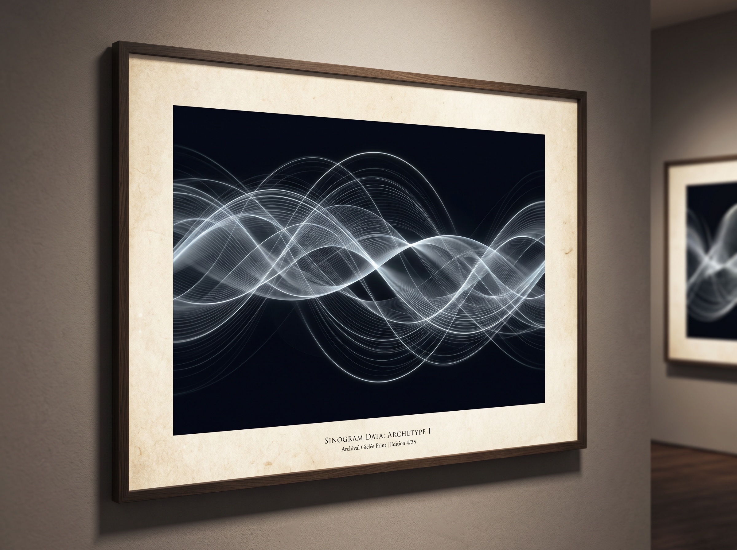
A learned reconstruction can intervene in two very different rooms. The first is the projection domain — the sinogram, the raw sweep of measurements the detector records as the source arcs around the head, before any recognisable picture exists. Cleaning noise here, in the data itself, keeps the subsequent reconstruction physically consistent. The second is the image domain, where a network takes the already-reconstructed slice and denoises it as a radiologist’s eye would wish, learning what a clean scan of teeth and bone ought to look like and steering the noisy one toward it. The most ambitious systems work in both rooms at once, and the field’s reviews now sort the literature roughly this way: filtering and iterative refinement, vendor-agnostic denoising software layered onto existing scanners, and bespoke networks trained to reconstruct a high-quality volume directly from a low-dose acquisition.
The Metal Problem
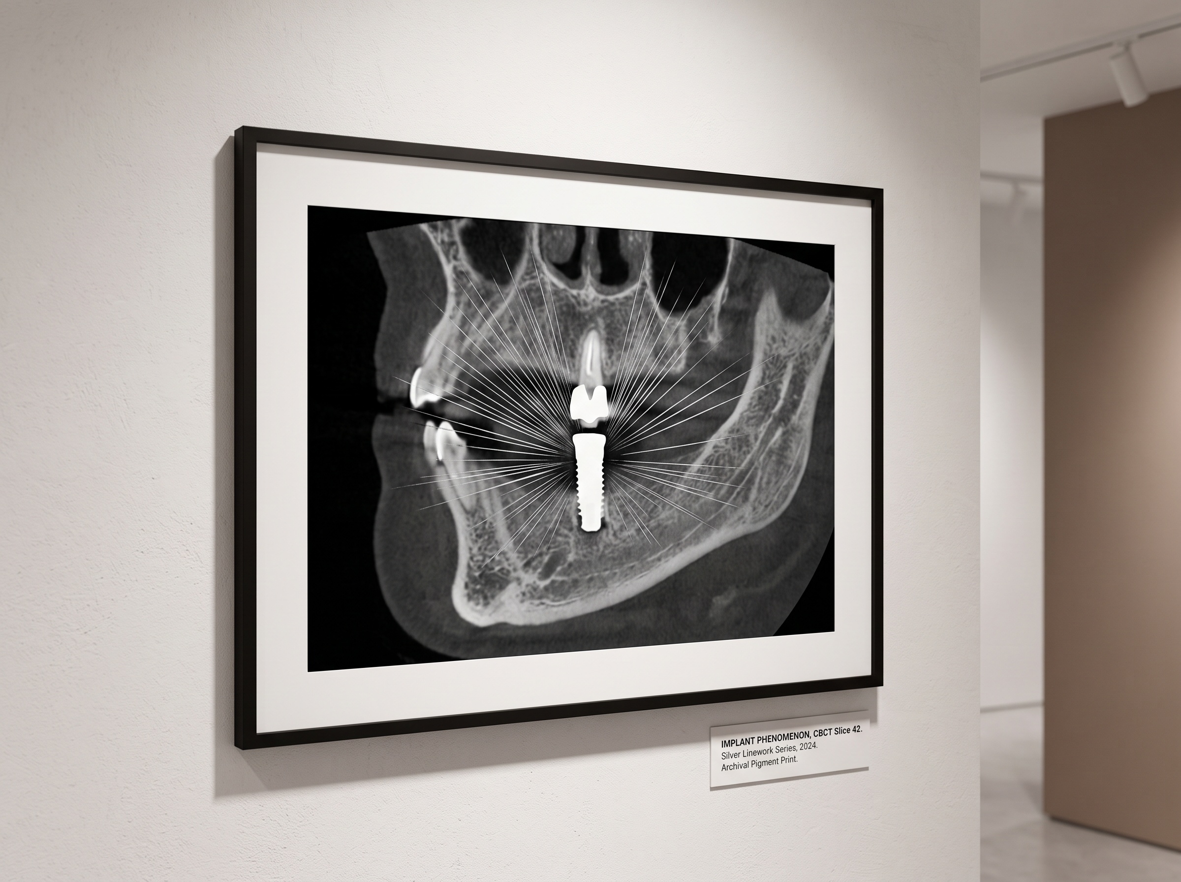
The hardest subject in the dental mouth is the one most often in it: metal. A crown, a post, an implant, an amalgam — each is a high-attenuation body that swallows photons whole and throws bright streaks and dark bands across the surrounding slice. In a low-dose scan the problem compounds, because the missing projection data and the limited field of view give a reconstruction algorithm less to work with precisely where it must work hardest. Recent work published in 2025 in the Philosophical Transactions of the Royal Society A takes this case head-on, developing deep-learning artefact reduction for low-dose dental cone-beam CT in the presence of high-attenuation materials — the setting where sparsity-based regularizers, so effective in metal-free tissue, tend to fail against the complex, structured artefacts that metal creates. It is a telling choice of battlefield: the technique is only as trustworthy as its performance around the restorations it will actually meet.
Teaching the Network What Clean Looks Like
A network cannot denoise what it has never seen cleaned. The craft, then, moves upstream into the training data — into the pairing of a noisy input with the quiet target it should become. Some groups assemble paired datasets across domains, teaching an ultra-low-dose scan to resemble its full-dose twin; a 2025 SPIE Medical Imaging study did exactly this, using paired data drawn from different domains to drive noise reduction for ultra-low-dose dental CBCT. Others reach for a more exacting teacher altogether. A 2025 study in the Journal of Dentistry trained a super-resolution network for dental CBCT against a micro-CT reference — the laboratory instrument whose resolution dwarfs any clinical scanner — and added an edge-loss term so the model would sharpen boundaries rather than merely smooth them, reporting gains in image-quality metrics and in the accuracy of the resulting three-dimensional reconstruction. The clinical scan learns, in effect, to aspire toward the specimen on the bench.
The Image the Algorithm Partly Imagines
Here the craft turns philosophical. A denoised, super-resolved, artefact-corrected slice is no longer a direct transcription of the photons that struck the detector; it is, in part, the network’s considered guess about what those photons would have shown had there been more of them. That guess is usually right, and often beautiful. But a model trained to produce plausible teeth can, at the margins, produce a plausible tooth that was not quite there — smoothing over a hairline fracture, inventing a crisp margin, resolving an ambiguity the raw data never settled. This is why the honest reconstruction papers report not only how clean the image looks but how faithful it remains: structural similarity, edge fidelity, downstream diagnostic accuracy. To read such an image well is to hold two ideas at once — gratitude for the clarity, and a disciplined awareness of where that clarity came from. The gallery has always asked this of us: to admire the print while remembering the hand that made it.
Future Developments
The trajectory is clear enough. As these networks move from the literature into the reconstruction engines of commercial scanners, the dose-versus-clarity negotiation that defined a century of radiography will loosen its grip; the scan a child receives will cost a fraction of today’s exposure and still read cleanly. What must travel alongside the technology is a reciprocal craft of interpretation — validation against reference standards, transparency about where a pixel came from, and the clinician’s trained scepticism kept sharp rather than surrendered to a smooth result. The most advanced dental image will increasingly be a collaboration between a physical measurement and a learned imagination. Our task, as ever in this gallery, is to see both at once: the science that captured the light, and the art that completed it.
Sources & further reading:
- Deep learning-based artefact reduction in low-dose dental cone beam computed tomography with high-attenuation materials (Philosophical Transactions of the Royal Society A, 2025)
- Enhancing Image Quality in Dental-Maxillofacial CBCT: The Impact of Iterative Reconstruction and AI on Noise Reduction — A Systematic Review (PMC, 2025)
- Deep learning super-resolution for dental CBCT using micro-CT reference and edge loss function (Journal of Dentistry / ScienceDirect, 2025)
- Deep learning-based noise reduction for ultra-low-dose dental CBCT images using paired datasets from different domains (SPIE Medical Imaging 2025)
Sorry, the comment form is closed at this time.