16 Mar AI-Enhanced Low-Dose CBCT: Revolutionizing Dental Imaging Safety and Quality
Cone-beam computed tomography (CBCT) has transformed dental diagnosis by providing detailed 3D imaging capabilities. However, radiation exposure concerns have led researchers to develop groundbreaking artificial intelligence solutions that maintain image quality while significantly reducing patient radiation doses.
The Challenge of Radiation Exposure
Traditional CBCT imaging requires substantial radiation doses to produce high-quality diagnostic images. While these doses are generally considered safe, minimizing patient exposure remains a priority in modern dental practice. Recent studies have shown that AI-based image processing can achieve diagnostic quality images with dramatically reduced radiation levels.
Breakthrough in AI Image Enhancement
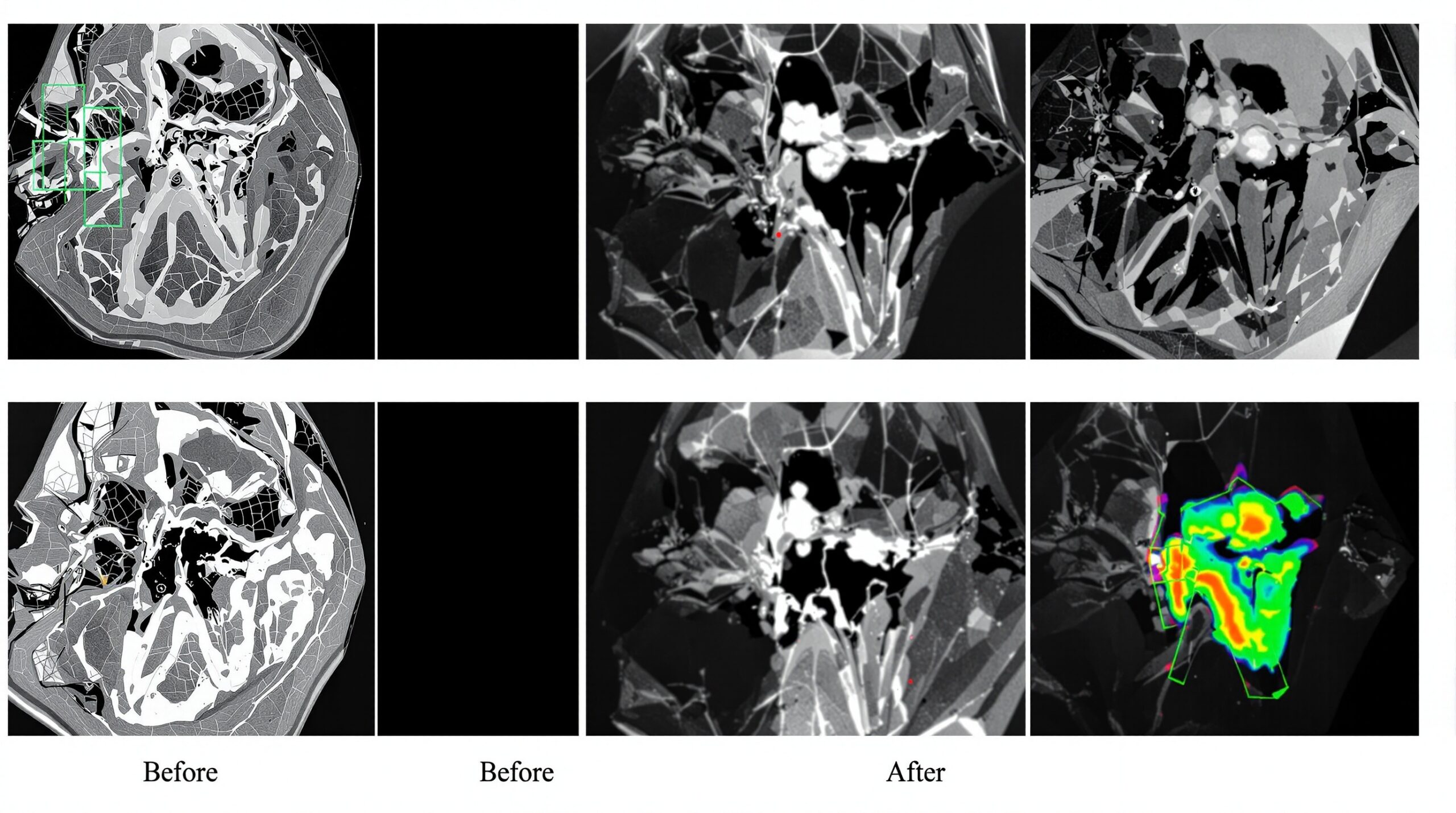
A recent feasibility study published in March 2026 demonstrates that artificial intelligence can process low-dose CBCT images to match the quality of full-dose scans. The research evaluated AI-processed images at various radiation levels, with remarkable results showing that 20% dose images processed through AI enhancement achieved comparable diagnostic quality to standard 100% dose images.
Key Research Findings
- AI-processed 20% dose images showed no statistically significant difference in quality compared to 100% raw dose images
- Five dental specialists independently assessed image quality using standardized criteria
- Anatomical visibility and structural delineation remained excellent even at reduced doses
- Overall diagnostic acceptability was maintained across all enhanced low-dose images
Clinical Applications and Benefits
This technology represents a significant advancement for dental practices, offering multiple benefits for both patients and practitioners. The ability to reduce radiation exposure by up to 80% while maintaining diagnostic quality opens new possibilities for routine imaging protocols.
Patient Safety Improvements
The most immediate benefit is enhanced patient safety through dramatically reduced radiation exposure. This is particularly important for pediatric patients, pregnant women, and individuals requiring multiple imaging sessions. The AI enhancement process allows practitioners to achieve excellent diagnostic results while prioritizing patient well-being.
Enhanced Diagnostic Capabilities
AI processing not only maintains image quality at lower doses but can also enhance specific anatomical features that are crucial for diagnosis. The technology excels at noise reduction, contrast enhancement, and structural delineation, potentially revealing details that might be less visible in traditional imaging.
Future Implications
As AI-enhanced CBCT technology continues to evolve, we can expect further improvements in image quality and additional reductions in required radiation doses. This advancement represents a paradigm shift in dental imaging, where patient safety and diagnostic excellence can coexist without compromise.
The integration of artificial intelligence into dental imaging workflows will likely become standard practice, offering practitioners powerful tools to deliver superior patient care while maintaining the highest safety standards.
Sorry, the comment form is closed at this time.