13 Mar AI-Enhanced Dental Imaging Techniques: 2026 Best Practices for CBCT and Radiographic Interpretation
The integration of artificial intelligence into dental imaging represents one of the most significant advances in clinical dentistry in recent years. As we move through 2026, AI-enhanced diagnostic tools are revolutionizing how practitioners interpret radiographic images, improving accuracy while reducing interpretation time.
Current State of AI in Dental Imaging
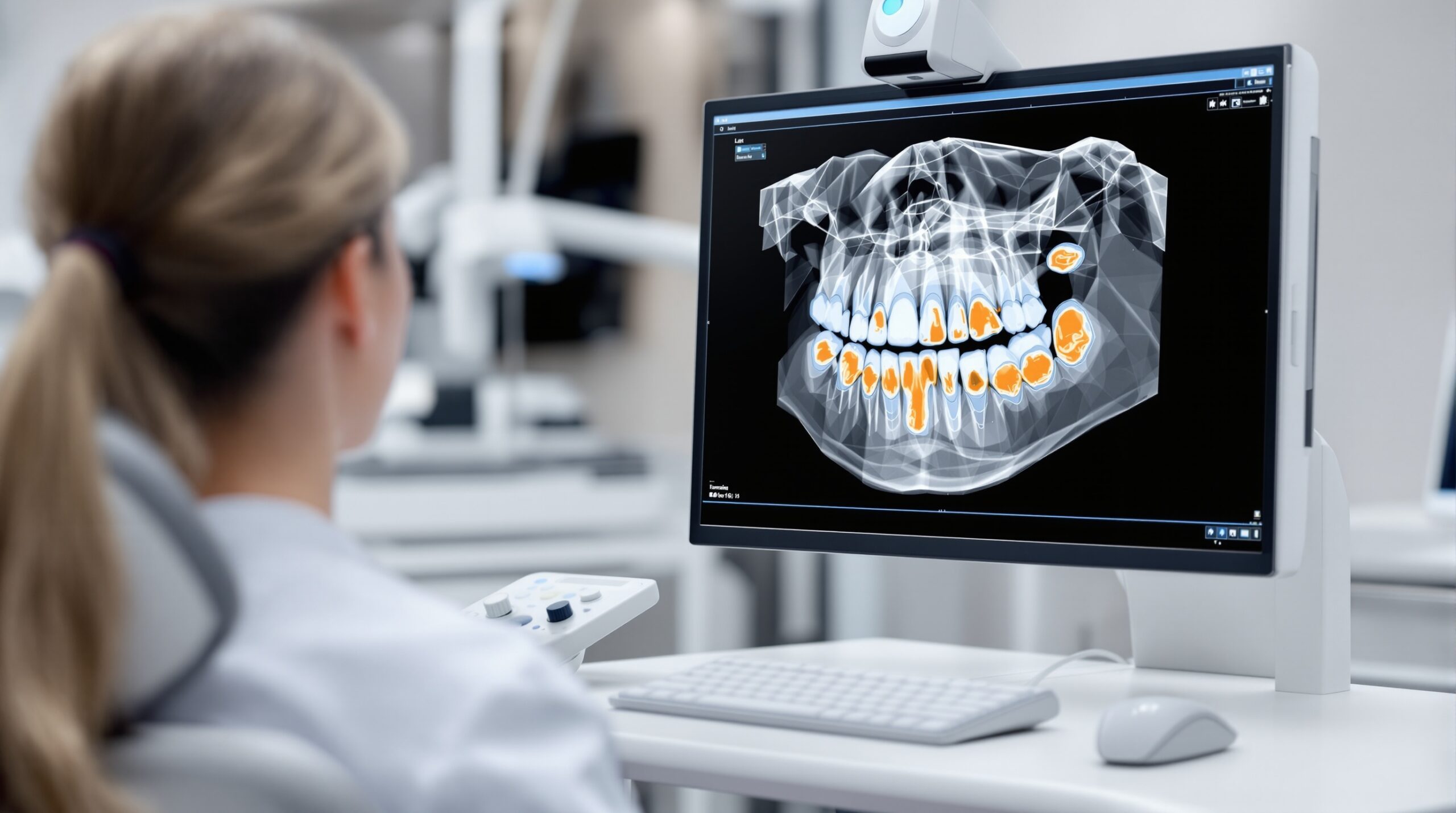
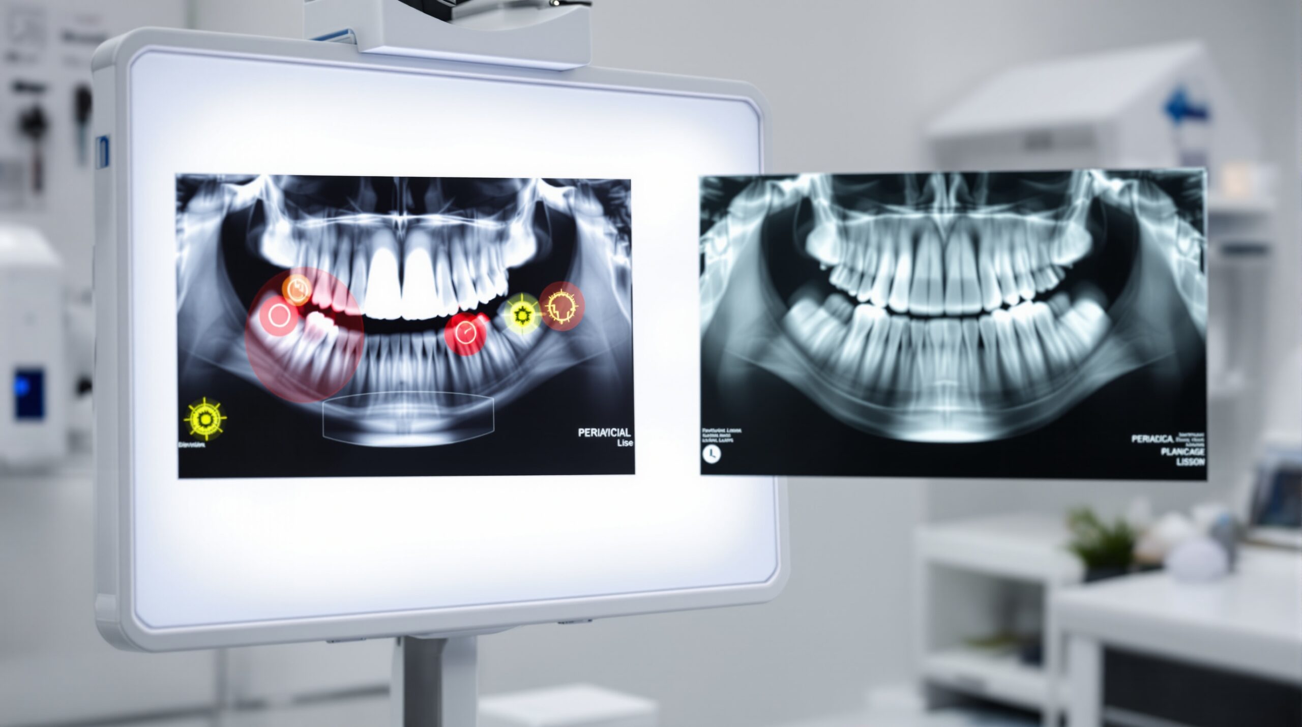
Computer vision models now read bitewings, periapicals, panoramic radiographs, and CBCT volumes to flag cavities, periapical changes, bone loss, and anatomical risks. Current sensitivity and specificity vary by lesion depth and device type but generally range around 80-92% and 75-90%, respectively.
The American Dental Association’s 2026 recommendations emphasize using dental imaging most effectively in moderation, with panoramic radiography for initial assessment before dental implant procedures and CBCT for presurgical planning and placement.
CBCT Technology Advances
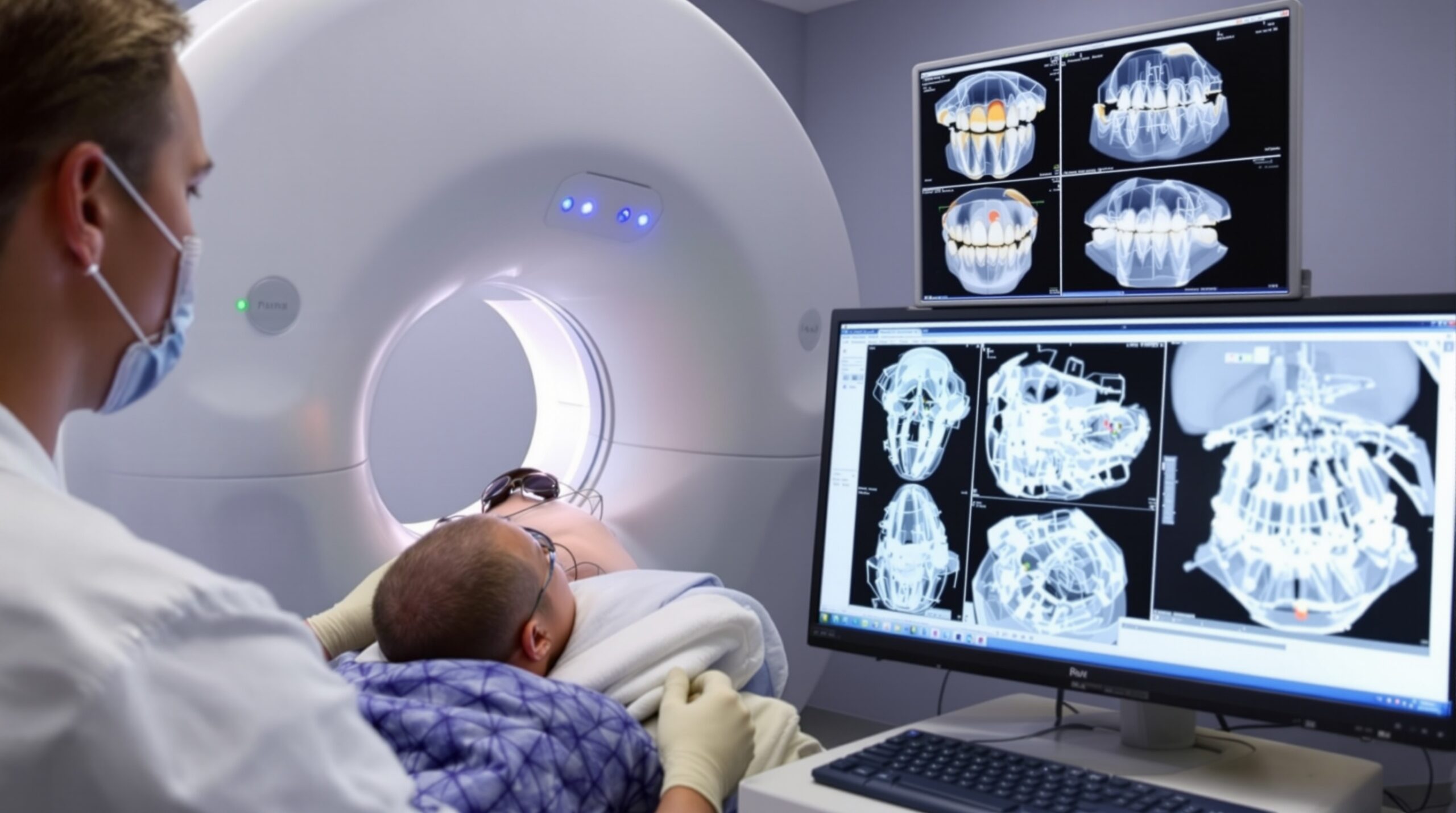
Cone Beam Computed Tomography (CBCT) provides three-dimensional images, enhancing the ability to assess complex dental conditions and plan treatments effectively. Modern CBCT systems integrated with AI algorithms can automatically identify anatomical landmarks, measure bone density, and highlight potential pathology.
Key Benefits of AI-Enhanced CBCT
- Automated measurement tools that calculate bone height and width for implant planning
- Pathology detection algorithms that highlight suspicious areas requiring attention
- Airway analysis capabilities for sleep apnea and orthodontic treatment planning
- Dose optimization through intelligent exposure parameter selection
Digital Radiography Best Practices
Bitewing X-rays remain useful to examine segments of upper and lower arch simultaneously to detect inter-proximal caries. AI enhancement helps identify early-stage lesions that might be missed by human interpretation alone.
Implementation Guidelines
Successful AI integration requires proper workflow design:
- Calibrate systems regularly to maintain diagnostic accuracy
- Train staff on AI output interpretation and limitations
- Maintain human oversight for final diagnostic decisions
- Document AI findings alongside traditional clinical observations
Future Outlook
The dental imaging market continues expanding with advanced techniques providing enhanced diagnostic capabilities. AI tools are becoming increasingly sophisticated, with some systems now capable of predicting treatment outcomes and suggesting optimal intervention timing.
As these technologies mature, practitioners must balance technological advancement with clinical judgment, ensuring that AI serves as a diagnostic aid rather than a replacement for professional expertise.
Conclusion
AI-enhanced dental imaging represents a significant step forward in diagnostic accuracy and clinical efficiency. By following 2026 best practices for CBCT and radiographic interpretation, dental professionals can leverage these tools to provide superior patient care while maintaining the human element essential to quality dentistry.
Sorry, the comment form is closed at this time.