


September 4, 2026 Garbage In, Garbage Out: Image Quality and Why AI Still Needs a Good Radiograph
There is a quiet assumption underneath every claim made for dental artificial intelligence, and it is almost never stated aloud: that the radiograph handed to the model is a faithful record of the tooth. Strip that assumption away and the whole edifice wobbles. A detection network does not examine a patient; it examines a grid of grey values that a sensor produced under a particular exposure, a particular geometry, in a particular half-second during which the patient may or may not have held still. If those grey values misrepresent the anatomy, the model will misrepresent it too – fluently, confidently, and without a flicker of doubt. The oldest maxim in computing turns out to be the most important thing to understand about the newest tools: garbage in, garbage out.
We have spent this series looking at what happens once a good image is in hand – how a network turns a flat field of grey into flagged caries and bone loss in How AI Reads a Bitewing, how it names a three-dimensional volume in AI on CBCT, and how those findings become a signed report in From Detection to Diagnosis. This piece steps back to the thing all of them stand on: the quality of the image itself.
The Model Reads the Sensor, Not the Tooth
It is worth being precise about what a detection model actually receives. Not a patient, not a diagnosis, not even really a tooth – a matrix of pixel intensities, each a proxy for how much radiation reached that point on the detector. Everything the network knows about the mouth arrives through that matrix and nowhere else. The clinician standing in the room has a thousand other channels: the patient’s history, the tap of an explorer, the look of the tissue, the memory of last year’s film. The model has the pixels. When the pixels are honest, that constraint is manageable. When they are not, the model has no fallback, because it never had the other channels to begin with.
This is why image quality is not a housekeeping concern that sits beside the AI question. It is the AI question, moved one step upstream. A tool cleared to aid detection at a stated sensitivity was validated on images of a certain standard. Feed it images below that standard and you are no longer operating the device that was tested – you are operating a different, unvalidated one that happens to share a login screen.
Exposure: When the Tones Collapse
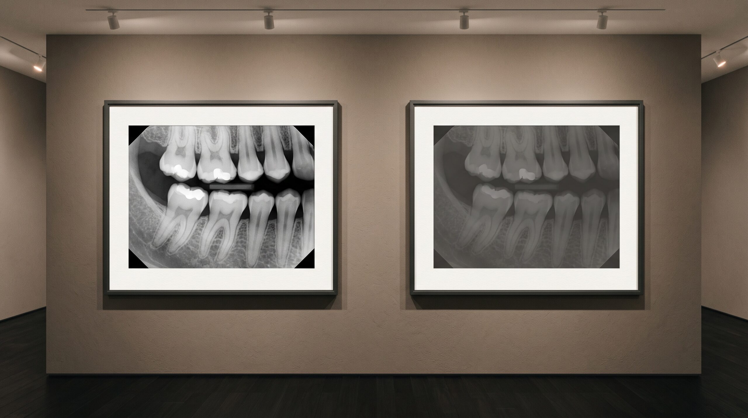
The first axis of quality is exposure, and it fails in both directions. Underexpose a bitewing and the image goes muddy and flat: the enamel-dentine boundary softens, early interproximal lesions vanish into noise, and the model – which learned the appearance of caries as a specific pattern of local contrast – simply has less signal to find. Overexpose and the opposite happens: thin structures burn out, subtle radiolucencies are washed to white, and detail that a properly exposed film would have preserved is gone before the network ever sees it. Digital sensors have a working range within which the mapping from radiation to grey value is informative; drift outside it and the data at the top or bottom of the scale is compressed or clipped, which is to say destroyed.
Humans compensate for a poor exposure to a surprising degree, tilting the monitor, adjusting the window, drawing on experience to read through the murk. A fixed model does none of that. It applies the same learned filters to a degraded image as to a pristine one and returns a result with the same crisp confidence – a result now built on evidence that partly is not there.
Geometry: Cone-Cuts, Overlap, and Foreshortening
The second axis is geometry, and it is the one clinicians most underestimate as an AI problem. A cone-cut – where the beam misses part of the field and leaves a clear unexposed wedge – does not merely look untidy; it removes anatomy the model was never given the chance to read. Whatever sat in that corner is not flagged as missing, it is simply absent, and a network scoring the rest of the image will report on what it sees without noting the hole.

Positioning errors are subtler and just as consequential. Overlapping contacts hide exactly the interproximal surfaces where caries hides. Foreshortening and elongation distort the vertical relationships a bone-loss measurement depends on, so a model quantifying crestal height from cementoenamel junction to bone can be measuring a projection that never matched the patient’s true anatomy. Angulation that a seasoned dentist reads around and mentally corrects for is, to the model, just the geometry of the world. It measures what it is shown.
Artifacts: Motion, Ghosting, and Noise
The third axis is artifact – everything that corrupts the image without changing the anatomy. Motion during exposure smears the fine edges a detector leans on, and because the smear is smooth rather than obviously broken, the model tends to read straight through it rather than flag it. Double exposures, ghosting, sensor noise on an aging plate, software sharpening cranked to introduce halos, foreign objects and processing streaks: each shifts the local pixel statistics away from the clean images the network was trained on. Overjet, whose detection platform we have discussed across this series, notes in its own guidance that motion, exposure errors, and positioning faults are among the recurring imaging problems practices must solve – and its system flags under- and over-exposed captures at chairside precisely because those faults poison everything downstream. That framing is exactly right: the artifact is not a cosmetic flaw, it is corrupted input.
The Domain-Shift Trap
There is a more insidious version of the quality problem that has nothing to do with a single bad film. A model trained largely on images from one sensor, one manufacturer’s processing, one clinic’s exposure habits, learns the texture of those images as much as the anatomy within them. Move it to a different sensor with a different noise profile and pixel pitch, and performance can quietly erode even when every image looks perfectly diagnostic to the eye. This is domain shift, and it is dangerous because it is invisible: nothing on the screen announces that the model is now slightly outside the distribution it was validated on. A 2025 evaluation of a commercial dental AI on panoramic radiographs found that reliability for comprehensive charting remained uncertain across real-world images – a reminder that a headline accuracy figure is a claim about the images the vendor tested, not a promise about yours. It is one more reason to read a tool’s regulatory and validation status precisely, as we argued in From Detection to Diagnosis, rather than trusting the marketing number.
Why a Confident Wrong Answer Is the Worst Outcome
The failure mode that should worry clinicians most is not the model that misses on a bad image. It is the model that doesn’t miss visibly – that takes a cone-cut, underexposed, motion-blurred film and returns a clean, well-formatted, confident set of findings anyway. A blank result invites suspicion. A tidy result invites trust. The whole discipline of the clinician-in-the-loop, which we described as the seam where authorship transfers, assumes the human is reviewing findings drawn from a trustworthy image. When the image is quietly bad and the output is quietly wrong, that safeguard is reviewing a fiction with a straight face. Good input is not a nicety that improves AI performance at the margin; it is the precondition that makes the human check meaningful at all.
Turning the Tools on the Image Itself
The encouraging development is that the same machine learning is now being pointed at the problem it created a need for: judging the image before anything is read from it. Deep-learning models have been trained to score panoramic radiographs for quality directly – one 2025 study reported classifiers reaching roughly 98% accuracy for contrast and density and around 87% for artifact detection, grading exposure, coverage, positioning, and overall diagnostic acceptability automatically. Chairside, Overjet analyzes each capture immediately and flags under- or over-exposed images so staff can retake before the file is ever saved. This is a quiet but important inversion. Instead of trusting the detector and hoping the image was good, the newest workflows put a quality gate first – an automated triage that decides whether an image is worth trusting a diagnosis to, and prompts a retake when it is not.
Future Developments
The trajectory points toward capture that is quality-aware from the first exposure. One can see the shape of it already: a sensor pipeline that grades its own output in real time, refuses a cone-cut before the patient is dismissed, estimates its own confidence conditioned on the measured image quality, and hands the detection model not just a picture but a candid assessment of how much that picture can be trusted. The deeper shift is philosophical. For a century the radiograph was evidence a clinician learned to read around – to compensate mentally for the imperfect film. AI does not compensate; it consumes. That makes the craft of acquisition more central, not less, in an automated practice. The better these tools become at reading a radiograph, the more everything rests on the radiograph being worth reading – and the more the unglamorous work of exposure, positioning, and a clean, artifact-free image becomes the quiet foundation on which all the intelligence stands.
Sources & further reading:
Sorry, the comment form is closed at this time.