


September 2, 2026 AI on CBCT: What 3D Segmentation Actually Finds
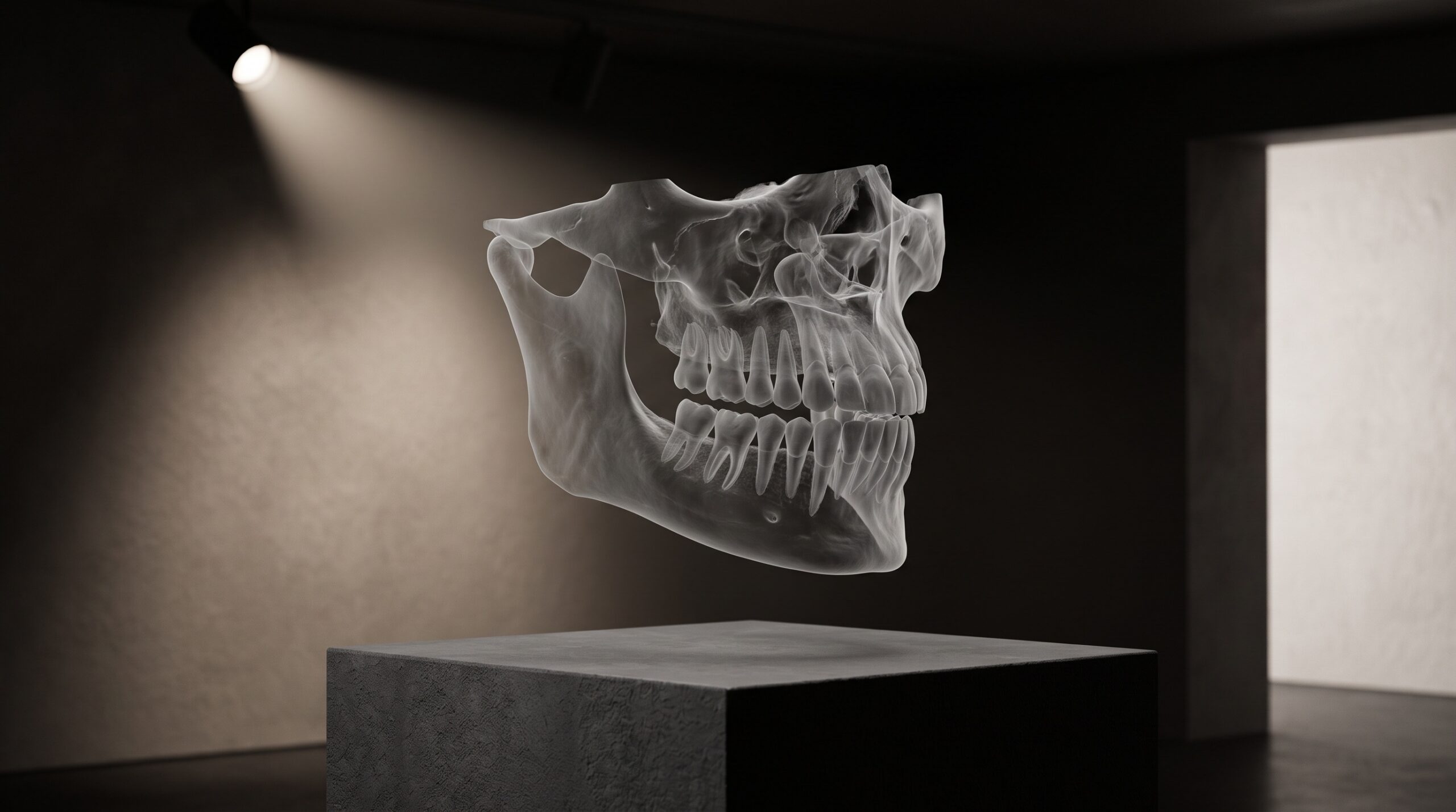
A bitewing is a picture; a CBCT is a volume. That single difference changes everything about how a machine reads it. Where a two-dimensional radiograph is a flat field of shadow to be interpreted, a cone-beam scan is a solid block of the patient – hundreds of thin slices stacked into a lattice of tiny cubes called voxels, each holding a single value of density. Rotate it, cut it, fly through it: the anatomy is all there, but it arrives undifferentiated, a fog of grey in which tooth, bone, nerve, and air are only degrees of brightness. Segmentation is the act of ending that ambiguity – of walking through the volume and giving every voxel a name. It is the quiet, foundational task on which every clever 3D application downstream depends, and understanding it is the difference between trusting a rendering and reading one.
The word itself is worth dwelling on. To segment a CBCT is to partition it: this cluster of voxels is the lower left first molar, that channel is the inferior alveolar nerve, this cavity is the maxillary sinus, everything else is bone or background. Done by hand on a good volume, it is the work of hours – a clinician painting structures slice by slice. Done by a trained neural network, it is the work of a minute or two. What follows is what the machine actually finds when it does.
From Voxels to Structures
A CBCT does not measure teeth. It measures how much of the X-ray beam each point in space absorbed, and stores that as a grid of numbers. Enamel is dense and reads bright; the marrow spaces of trabecular bone read darker; the air of the airway and sinus reads darkest of all; the nerve canal is a low-density tube threading through denser bone. Every structure a clinician cares about is, to the scanner, simply a region where the density falls within a certain range and forms a certain shape. The problem is that those ranges overlap and those shapes touch. Cortical bone and enamel can read at similar densities; two adjacent teeth share a contact where their voxels blur together. Thresholding by brightness alone – the old way – produces a mess.
A three-dimensional convolutional network solves this the way its 2D cousins solve caries detection, but in an extra dimension. Instead of sliding filters across a flat image, it convolves through the volume, learning features that are genuinely spatial: the curvature of a root, the tubular continuity of a canal, the way a sinus wall encloses a pocket of air. Architectures in the U-Net family – and the self-configuring nnU-Net that has become the research workhorse – encode the volume down to its essential features and then decode it back out as a labeled map, assigning every voxel to a class. The output is not a picture with an outline drawn on top; it is a second volume, perfectly registered to the first, in which the anatomy is already named.
The Teeth: Instance Segmentation
Finding “tooth” versus “not tooth” is the easy part. The harder and more useful task is instance segmentation – separating each individual tooth as its own object, correctly numbered, even where crowns touch and roots interweave. This is what lets a system report on the lower left second premolar specifically rather than on a generic bright mass. It is the same distinction between detection and identity we drew for 2D film in How AI Reads a Bitewing, but three-dimensional and, if anything, more demanding: the network must resolve the contact points where neighbours merge and assign each voxel to exactly one tooth.
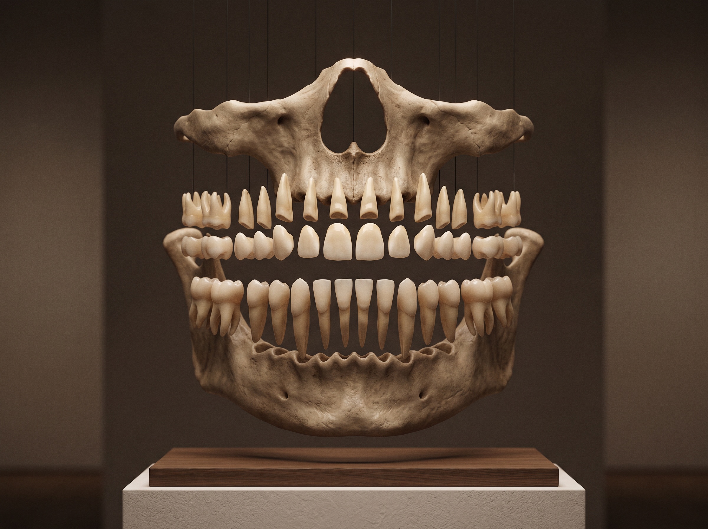
Get it right and the payoff is immediate. Each segmented tooth becomes a measurable object – root length, angulation, proximity to its neighbours, position relative to the bone. This is the substrate for automated orthodontic set-ups, for endodontic assessment of root morphology, and for the digital-twin workflows that align the segmented dentition with an intraoral scan and a facial photograph. Tools such as Relu’s segmentation engine were built precisely to fuse those layers – CBCT bone and teeth, digital impression, facial scan – into one coherent model, and none of it is possible until each tooth is cleanly its own.
The Canal, the Sinus, the Airway
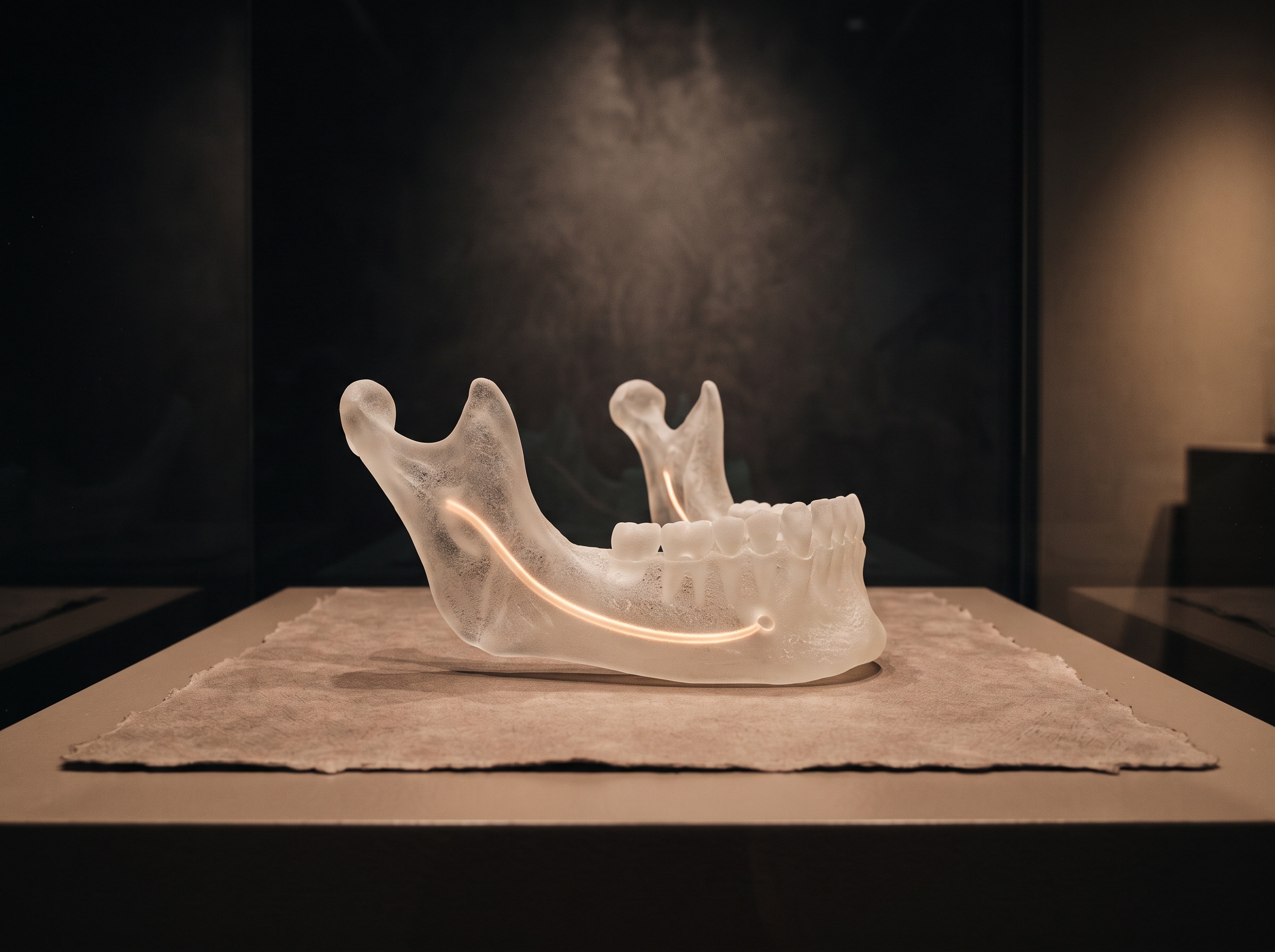
The structure that earns segmentation its keep is the mandibular canal – the bony channel carrying the inferior alveolar nerve along the lower jaw. Nick it during implant placement and the patient leaves with a numb lip that may never fully recover. Tracing it by hand is tedious and error-prone precisely where it matters, deep in the posterior mandible where contrast is poor. AI canal segmentation has become one of the most studied problems in the field; the public ToothFairy challenges, built on expertly annotated CBCT volumes, have pushed automated methods to overlap scores (measured by the Dice coefficient, where 1.0 is perfect agreement) around 0.87 for the canal – close enough to human annotation to serve as a genuine safety layer in implant planning.
The same machinery maps the maxillary sinus – its floor, its septa, the membrane whose thickness matters before a sinus lift – and the pharyngeal airway, whose volume and minimum cross-section feed orthodontic and sleep-related assessment. FDA-cleared platforms have folded these into routine reporting: Overjet’s CBCT Assist, cleared in 2025, automatically labels and quantifies bone levels, airway dimensions, and nerve proximity; Diagnocat offers a first-of-its-kind FDA-cleared 3D diagnostic pipeline; Pearl’s Second Opinion extended its detection lineage into 3D. The details of Diagnocat’s volumetric findings and how the data reaches its cloud are traced in Diagnocat on CBCT Volumes; the point here is the shared foundation beneath all of them – a segmentation map, computed first, on which every measurement rests.
How Good Is the Boundary?
Segmentation quality is not judged by whether a structure was found but by where the boundary was drawn. The field’s standard measure is the Dice coefficient – the overlap between the machine’s labeled region and an expert’s – and the honest reading of a Dice of 0.9 is that the two agree on ninety percent of the disputed volume, not that the result is ninety percent ‘correct’ in any clinical sense. A canal segmentation can score well overall and still stray by a millimetre at the one apical point where an implant will actually approach it. This is why the mature systems render the segmentation as an editable object, not a verdict: the clinician can see the traced canal, judge its plausibility against the raw greyscale, and nudge the boundary where the anatomy disagrees. The overlay is a proposal. The responsibility for the millimetre remains human.
Where It Fails: Metal and Blur
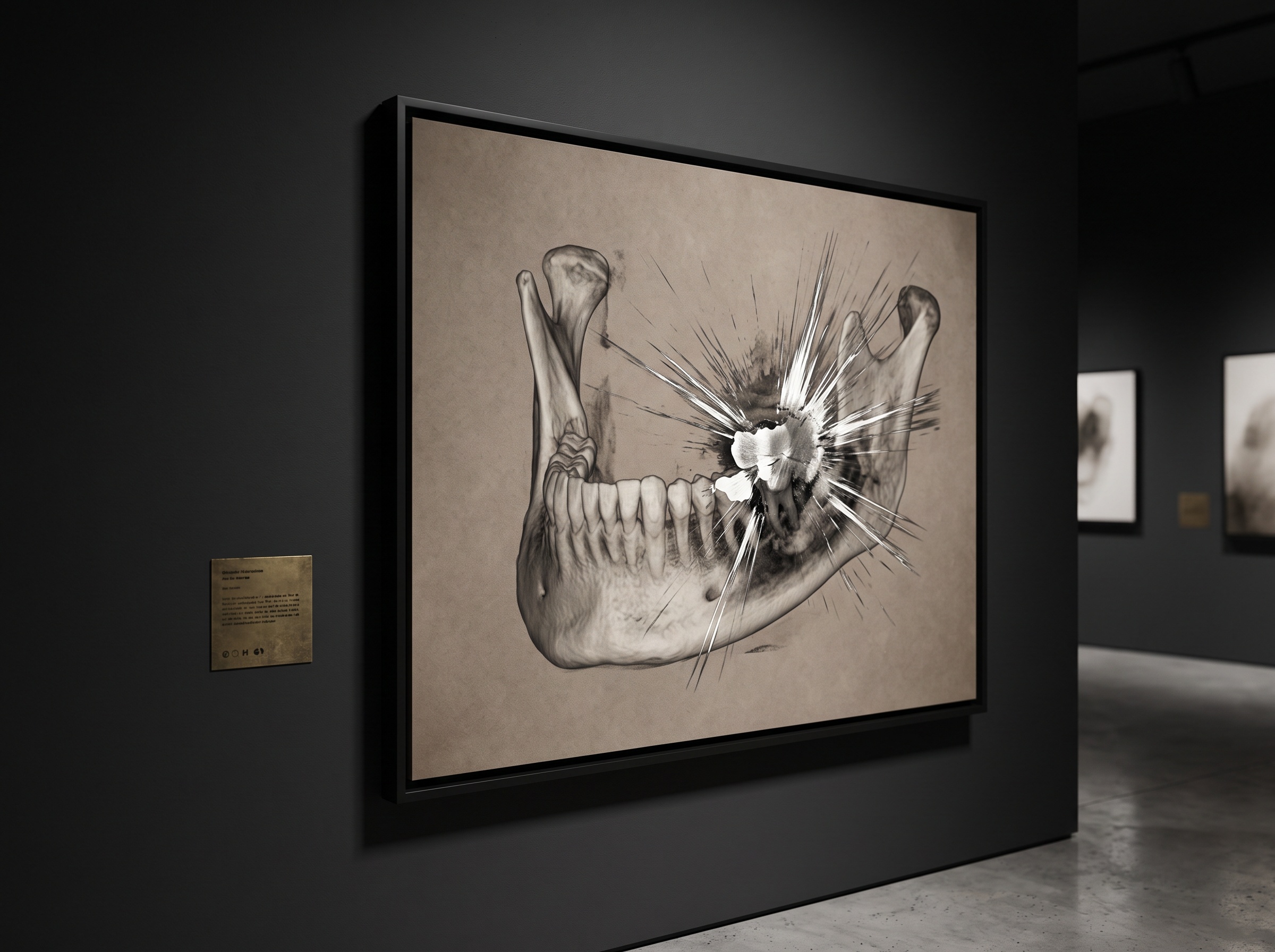
Every promise above degrades in the presence of the two great enemies of CBCT: metal and low contrast. A crown, a post, or an old amalgam scatters the beam into radiating starbursts of false density – beam-hardening artifact – that no network can reliably tell from real anatomy, because the information was corrupted before it was ever recorded. Partial-volume effects blur the thin cortical walls and the fine canal where voxels straddle two tissues at once. A low-dose or poorly-positioned scan starves the network of the contrast it needs to place a boundary at all. The system will still return a confident, beautifully rendered segmentation – and that is exactly the trap. A clean, well-exposed volume is not a nicety here; it is the precondition for the machine finding anything true.
Future Developments
Segmentation is quietly becoming the substrate of everything three-dimensional in dentistry – the layer that turns a scan from something you look at into something you can compute on. The near future is convergence: the segmented CBCT, the intraoral scan, and the facial photograph fused into a single patient model in which the planned implant already knows where the nerve is, the surgical guide prints itself from the labeled bone, and the orthodontic movement is simulated against real root anatomy rather than an idealized average. Further out, the interesting frontier is longitudinal – comparing this year’s segmented volume to last year’s automatically, watching a canal, a lesion, or a graft change over time in three dimensions. But the discipline underneath does not change. A CBCT is only ever as honest as the geometry, dose, and positioning that made it, and segmentation, for all its intelligence, can only name what the volume truly holds. The machine has learned to see in three dimensions. The craft of giving it something worth seeing remains ours.
Sources & further reading:
Sorry, the comment form is closed at this time.