


August 21, 2026 Two Ways of Seeing, One Frame: Fusing the Intraoral Scan and the Radiograph Into a Single Image
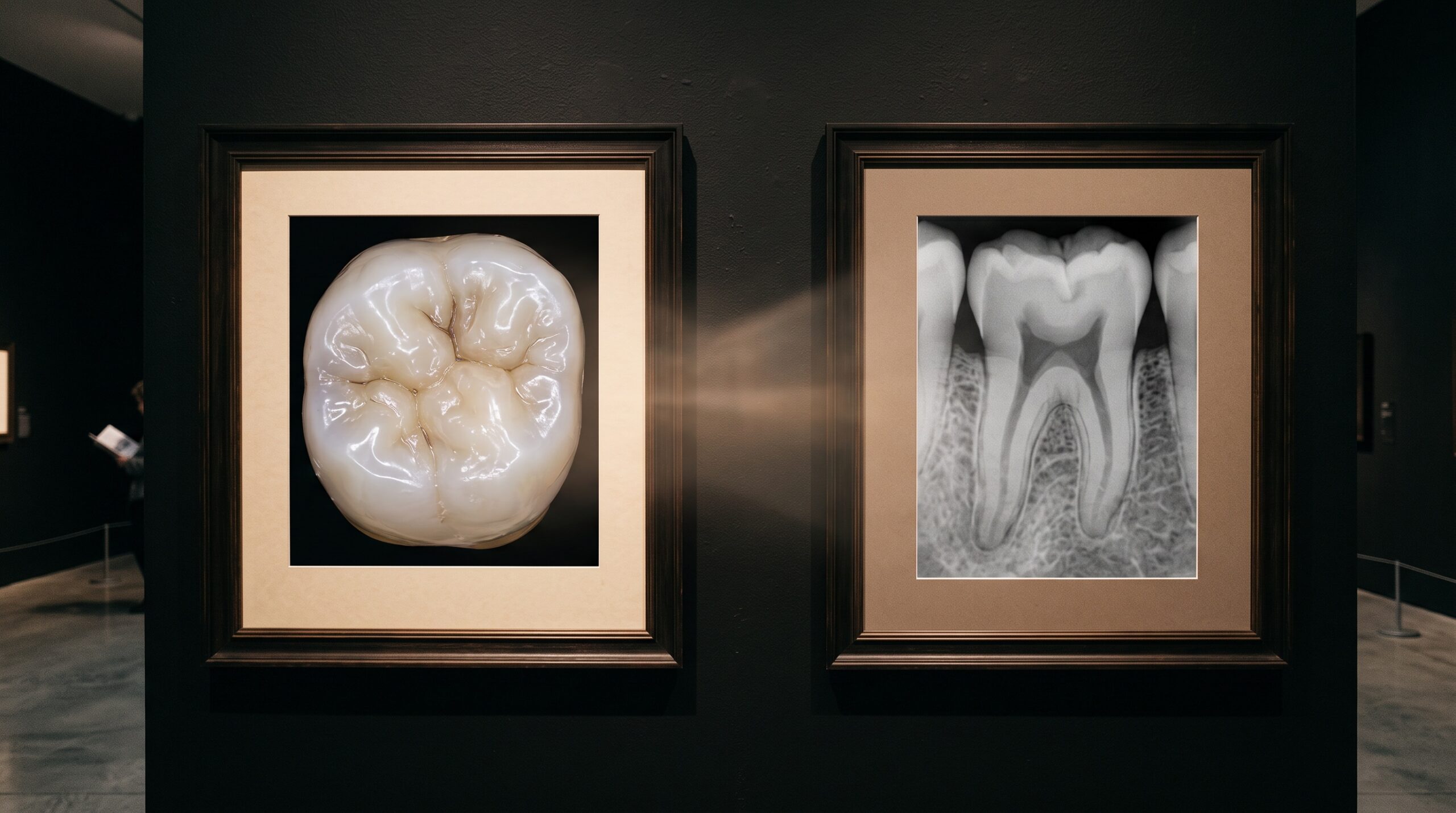
Every tooth has two truths, and until very recently no single picture could hold them both. There is the truth of the surface – the enamel’s exact topography, the wear facet, the margin of a restoration, the color that shifts from cervical amber to incisal grey – and there is the truth of the interior, the root that dives into bone, the pulp chamber, the shadow of decay creeping under a contact. One is the domain of visible light; the other belongs to radiation. For a century the clinician has carried both in the mind at once, glancing from a photograph to a radiograph on a separate screen and quietly performing, without ever naming it, an act of fusion. New unified viewers now perform that fusion in software, and in doing so they change what it means to look at a tooth.
Two Instruments, Two Blindnesses
An intraoral optical scanner is a device of the surface. It sweeps structured or laser light across the arch and reconstructs a dense three-dimensional mesh of everything that light can touch – crowns, gingival contour, the crisp edge of a preparation – with a fidelity measured in tens of microns and, increasingly, in true color. It is exquisite. It is also, in the most literal sense, superficial. The scan is a flawless skin stretched over a void; enamel is opaque to its light, so the moment the surface ends, the scanner’s knowledge ends with it.
The radiograph has the opposite gift and the opposite blindness. X-rays pass through the tooth and render its interior as a map of densities – roots, bone, caries, the fine line of the periodontal ligament – but they flatten that interior into greyscale and discard the surface almost entirely. The color is gone, the true three-dimensional contour of the crown is gone, and, in a projection radiograph, so is depth itself. Each instrument, in other words, is precisely blind where the other sees best. That complementarity is not a defect; it is the whole reason both exist. The problem was never the images. It was that they lived in separate software, in separate coordinate systems, and the join between them existed only in a clinician’s head.
The Registration Problem
To place the two datasets into one frame is to solve a problem that medical imaging has wrestled with for decades: registration. A surface mesh and a radiographic dataset are born in different coordinate systems, at different scales, from different physics, and often at different moments in time. Fusing them means finding the single spatial transformation – a rotation, a translation, sometimes a scaling – that lays one perfectly over the other, so that a point on the scanned crown sits exactly above the same point in the radiographic root.
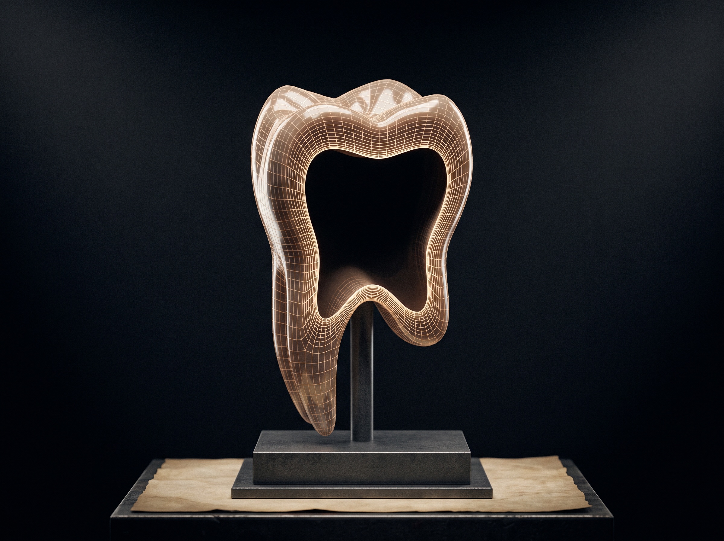
When the radiographic side is a three-dimensional volume, the richest version of this becomes possible: aligning the optical surface to the outer surface of the same structures extracted from a CBCT volume. Algorithms in the iterative-closest-point family nudge the two surfaces together, repeatedly pairing each point on one with its nearest neighbor on the other and shrinking the total distance until the fit can improve no further. Fiducial landmarks – a cusp tip, a shared restoration margin – can seed and constrain the search. The mathematics is elegant, but the practice is unforgiving: metal restorations bloom into streak artifact in the volume and corrupt the very surfaces the algorithm is trying to match, patient movement smears the reference, and a registration that is off by a fraction of a millimeter can quietly place a virtual implant into a nerve. Fusion is only ever as trustworthy as its alignment, and a beautiful composite can be beautifully wrong.
What the Fused Image Makes Visible
When the alignment holds, something genuinely new appears on the screen: a picture that carries both truths at once. The colored, high-resolution surface of the optical scan sits in exact register over the radiographic interior, and the clinician can move between them – fading from the enamel down into the root, seeing the depth of a carious lesion beneath the specific fissure that shows on the surface, understanding a restoration margin in relation to the bone crest below it. The mental fusion that experience once demanded is now made explicit, sharable, and measurable.
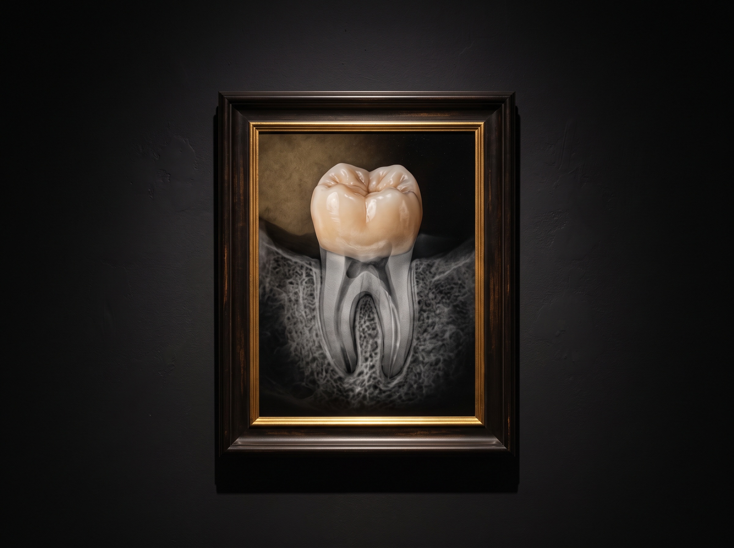
This is, in a sense, the same feat that a panoramic radiograph performs when it assembles one coherent image from a curtain of overlapping blur – the constructed, synthesized picture that reveals what no single raw capture could. Multimodal fusion simply raises the ambition: not one modality resolved into clarity, but two distinct modalities, optical and radiographic, woven into a single navigable object. It is worth remembering that this composite is still an interpretation, a rendering choice – which is exactly why the surface on which it is read, the calibrated diagnostic display, matters as much for a fused image as it does for a plain radiograph.
The Craft of the Blend
A fused viewer introduces a new instrument to the clinician’s hand, and it is not the scanner or the X-ray head – it is the slider between them. The skill is no longer only in acquiring a clean surface or a well-exposed radiograph; it is in the composition: deciding how much surface and how much interior to reveal for a given question, and, crucially, in learning to distrust a seam. A composite that looks seamless can seduce, because the eye reads a smooth join as a true one. The discipline of this new imaging is to interrogate the registration itself – to look for the tell-tale double edge, the surface that floats a hair above its own root – before believing what the blend appears to show.
Future Developments
The near horizon is faster and more automatic alignment, with machine-learning models trained to register surface to volume in seconds and to flag their own uncertainty – to say not merely “here is the fused image” but “this region is confidently aligned and this one is not.” Color, so long the exclusive property of the optical scan, will increasingly be draped over the radiographic and volumetric interior to make anatomy more legible without pretending to measure what greyscale cannot. Further out, fusion becomes temporal as well as spatial: the same tooth registered to itself across months and years, so that the surface wear and the interior change are watched together, as one evolving portrait rather than two disconnected snapshots.
What unifies all of it is a single, quietly radical idea – that the surface and the interior of a tooth were never meant to be seen apart. Two instruments, each brilliant and each half-blind, are finally being asked to speak in one frame. The art of dental imaging has always been the art of making the invisible legible. Fusing the scan and the radiograph is that ambition brought to its natural conclusion: not two ways of seeing held in tension, but one picture, at last, that holds them both.
Sources & further reading:
Sorry, the comment form is closed at this time.