


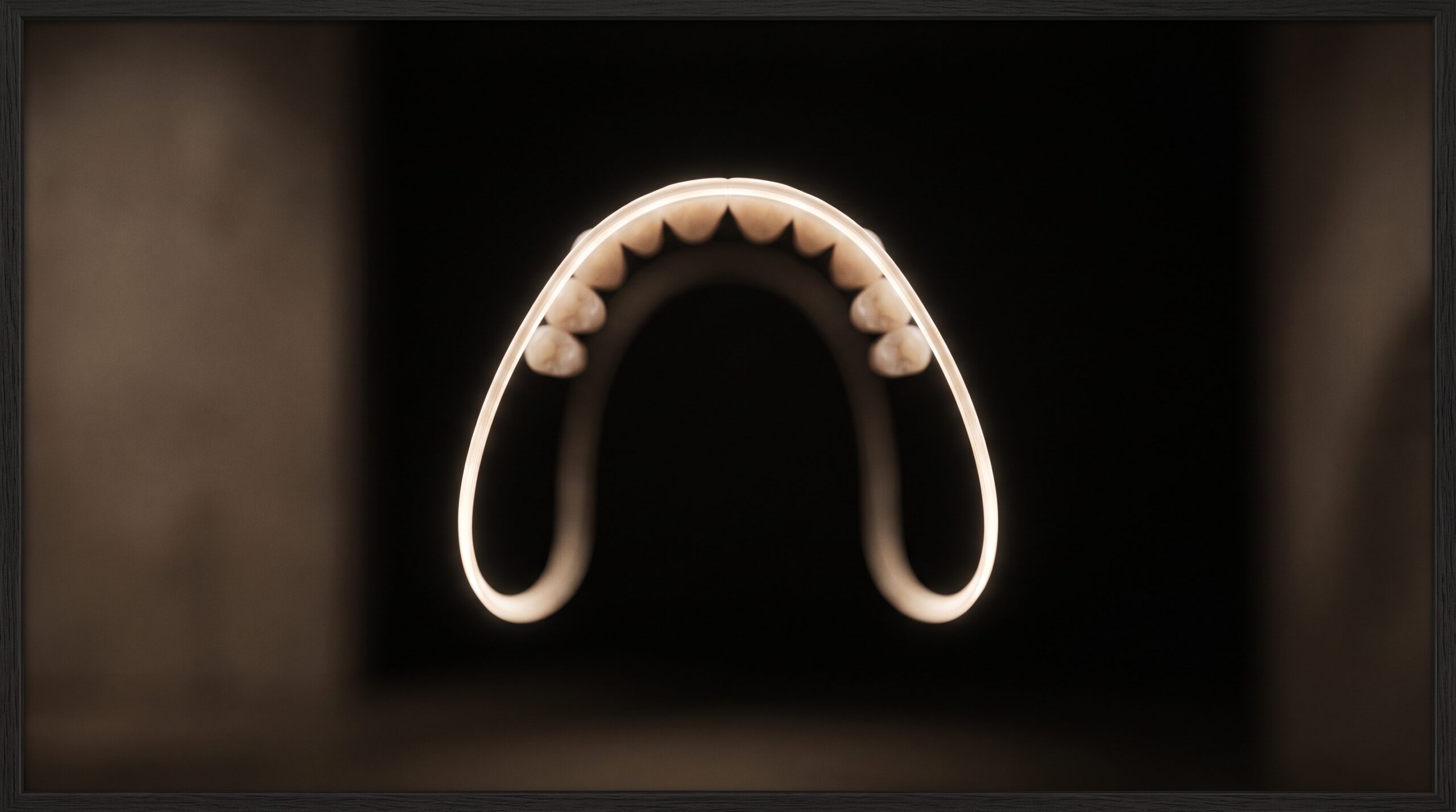
August 12, 2026 The Focal Trough: How a Panoramic X-Ray Builds One Sharp Image From a Curtain of Blur
There is a small quiet marvel in the most routine of dental images. A panoramic radiograph lays the entire dentition — both jaws, every tooth, the condyles, the sinuses, the ramus on each side — flat upon a single curved strip, and it does so in one slow sweep of perhaps fifteen or twenty seconds, the machine gliding once around the patient’s head. Set that beside the geometry it has to defeat and the achievement sharpens. The jaw is not flat; it is a horseshoe, a three-dimensional curve. No single flat detector held to one side could ever bring the whole of that curve into focus at once, any more than a photograph taken from across a room can hold both the near and far walls crisp together. And yet the panoramic image does exactly this. The trick that makes it possible is not a better lens or a brighter source. It is motion, and an invisible curved zone of sharpness the machine carries with it as it turns — the focal trough.
The Problem the Machine Sets Itself
Consider what is being asked. The dental arch curves away from the midline on both sides, so the distance from any fixed point to the front teeth, to the premolars, and to the molars is different at every step. A conventional projection casts a shadow of everything in the beam’s path onto the receptor at once, near and far superimposed, magnified unequally, hopelessly overlapped. To render the arch as a legible ribbon, the machine must do something a still exposure cannot: it must photograph only a thin slice of depth at a time, keep that slice sharp, and let everything nearer or further than it dissolve away. It must, in other words, perform a kind of tomography — imaging by layer — not by holding still, but by moving in a way that privileges one curved plane above all the rest.
Tomography by Motion
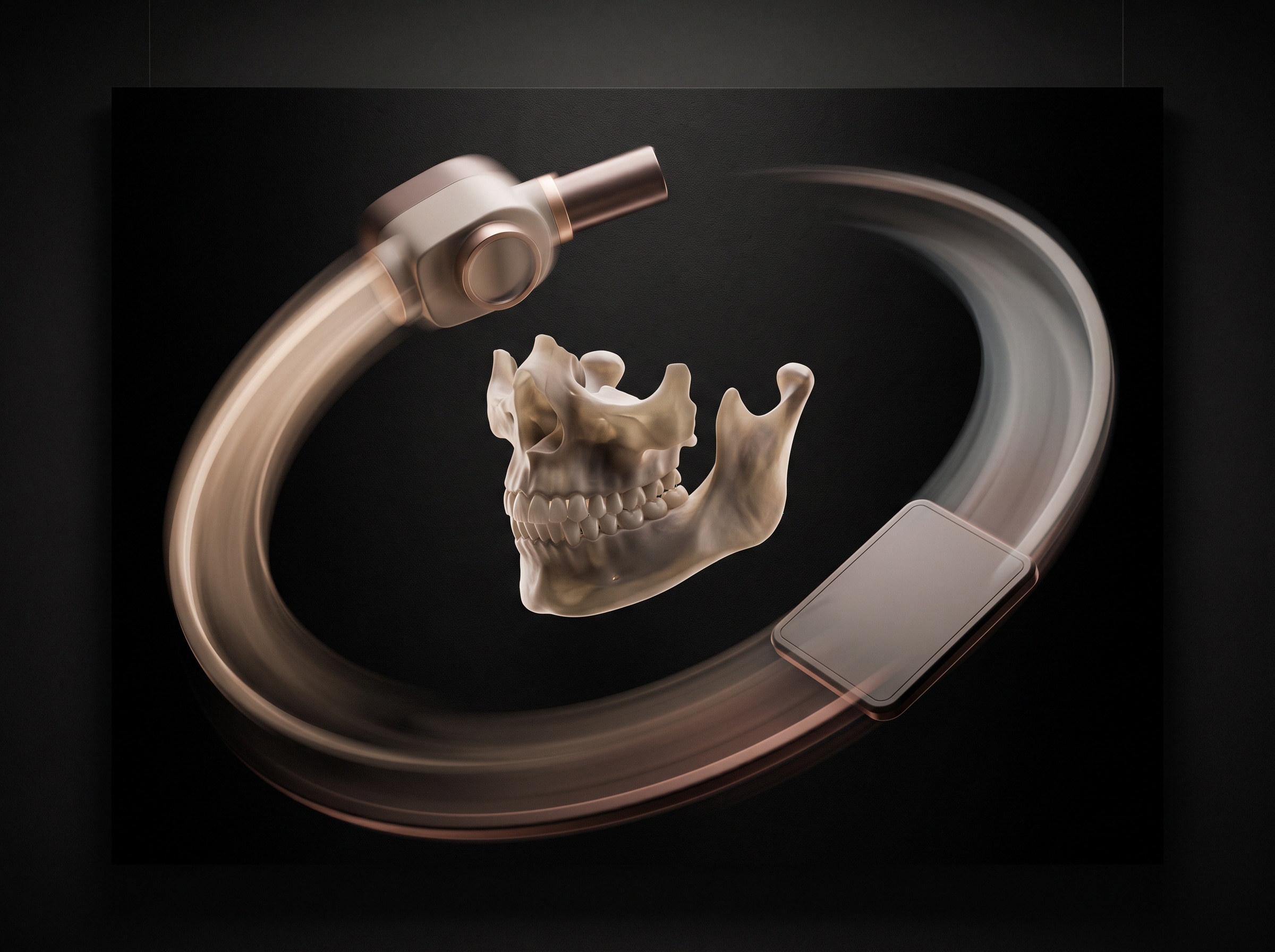
The elegance lies in how the movement is arranged. The X-ray tube sits on one side of the head; the receptor — film once, now a digital sensor — sits directly opposite. The two are rigidly linked and swing together, the source tracing an arc on one side while the receptor sweeps past on the other, and crucially the receptor also translates behind its narrow slit so that fresh detector is continually exposed as the beam moves. The whole assembly rotates not about a single fixed pivot but about a centre that shifts along the way — an instantaneous centre of rotation that migrates to follow the changing curvature of the arch, sitting further back for the flatter anterior region and swinging inward for the sharply curved molar zones. Only structures lying at just the right distance from this moving centre are projected onto the same patch of receptor throughout the sweep. They accumulate as a coherent, stationary image. Everything else is smeared across the moving sensor and blurred into the background — present, but softened past legibility. It is the same principle of motion-defined focus that governs the older art of body-section tomography, refined here into a single graceful pass.
The Focal Trough Defined
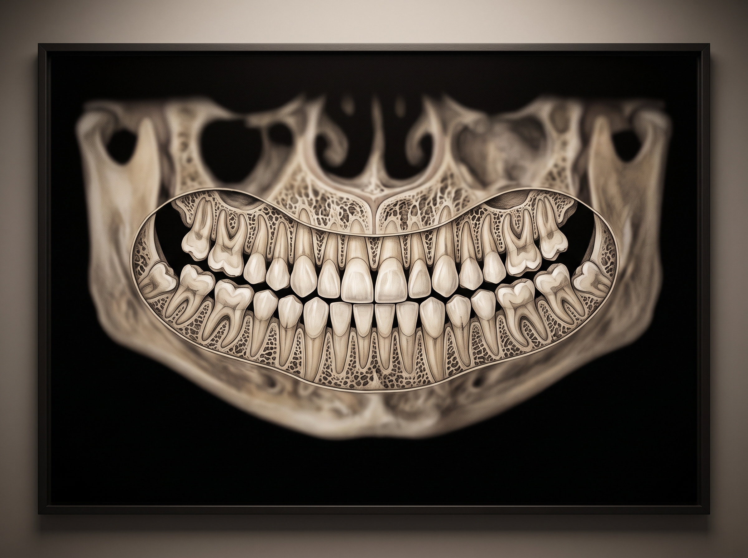
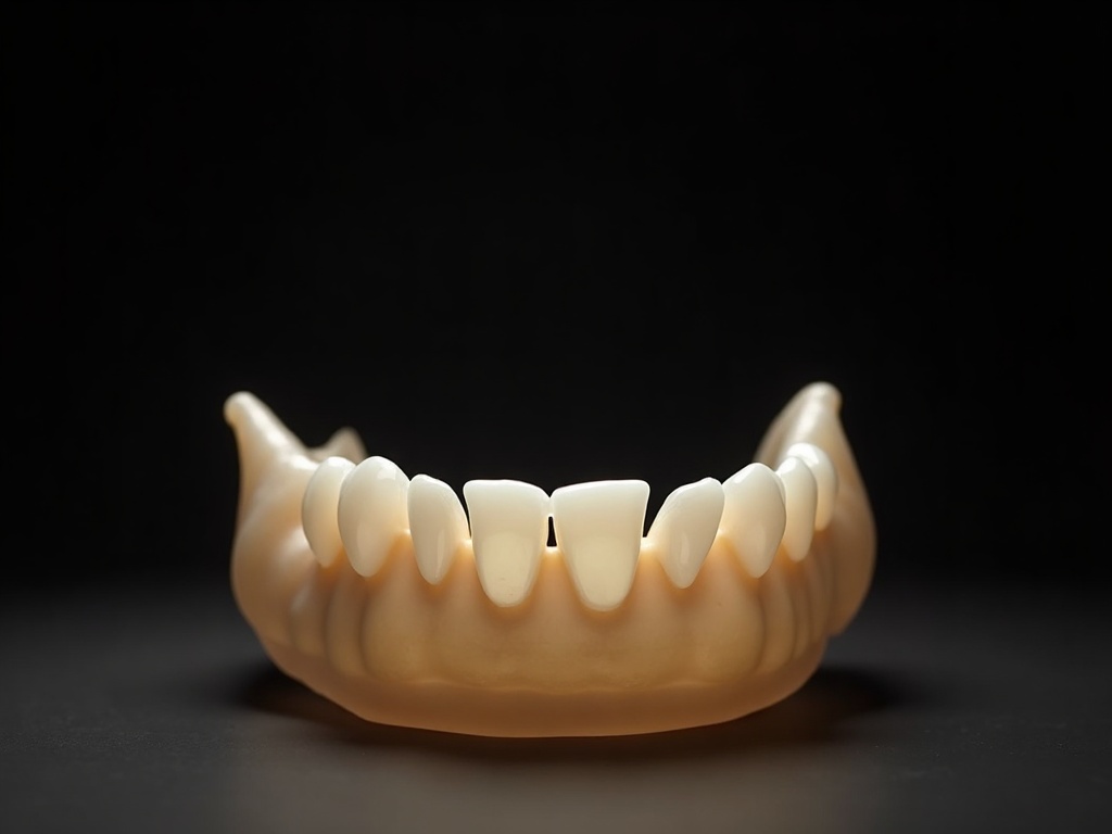
The set of points that stay sharp throughout that sweep is not a plane but a curved, three-dimensional shell shaped to the arch itself — horseshoe-like, following the ideal dental curve. This is the focal trough, sometimes called the image layer or focal plane, and it is the true subject of every panoramic exposure. Anatomy that falls inside it is imaged crisply and at close to its real proportions. Anatomy that lies outside it — buccal or lingual to the shell, in front of it or behind it — loses focus in direct proportion to how far it strays, and is also distorted in size, because objects nearer the source are magnified while those nearer the receptor are compressed. The trough is not uniformly wide, either. It is broadest in the molar regions, where a generous margin forgives small positioning errors, and narrowest at the anterior teeth, where it may be only a few millimetres deep. That narrowing is why the front teeth are the least forgiving part of the whole examination, and why a small error there costs the most. Understanding this shell is the key to reading the resulting image honestly, in the same way that appreciating the physical limits of sharpness in a dental image explains why detail behaves as it does.
Why Positioning Is Everything
Because the trough is fixed in the machine and the patient is not, the entire burden of a good panoramic image falls on placing the arch precisely inside that curved shell. This is the quiet reason for every positioning aid on the unit: the chin rest that sets vertical height, the grooved bite block that fixes the anterior teeth at exactly the right anteroposterior position, the crossing alignment lights that align the midline and the occlusal plane. Each exists to seat the arch in the trough. When positioning drifts, the errors are systematic and readable. A patient positioned too far forward, with the incisors ahead of the trough, yields narrow, blurred anterior teeth; too far back, and they appear widened and unsharp. A slumped or tilted head warps the occlusal plane into an exaggerated smile-shaped curve or flattens it. A tongue not pressed to the palate leaves a dark radiolucent shadow over the maxillary roots. None of these are faults of the X-rays themselves; every one is anatomy wandering out of the zone the machine can hold in focus. Reading a panoramic film well means recognising these signatures for what they are — positioning, not pathology — a discipline that complements the close diagnostic reading of an intraoral image such as what a bitewing reveals and quietly hides.
The Ghosts the Method Leaves Behind
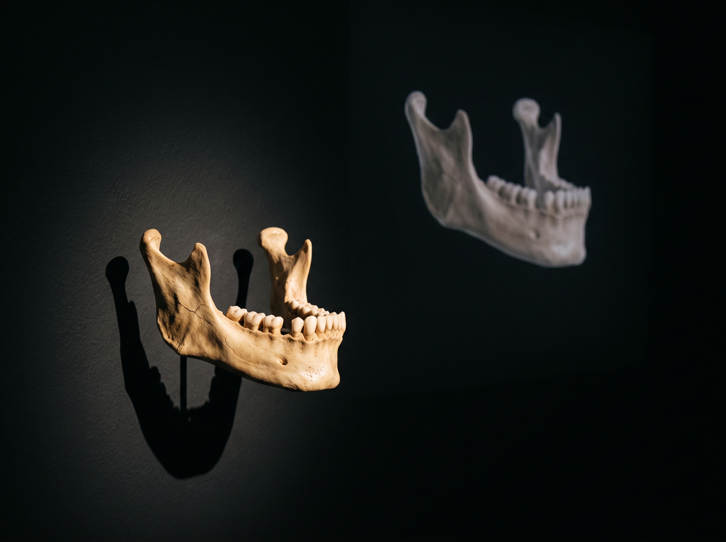
One artefact deserves its own mention, because it puzzles anyone meeting it for the first time. The ghost image is a second, spectral copy of a dense structure — commonly the mandible’s ramus, the hard palate, or a piece of metal jewellery — that appears on the opposite side of the film from the real object, projected higher, magnified, and blurred. It arises because a dense object positioned between the source and the centre of rotation is struck by the beam twice during the sweep: once directly, and once as the beam passes through it again on its way to imaging the far side. The second pass throws a distorted shadow across the receptor. Ghosts are not malfunctions but an inevitable consequence of the rotational geometry, and knowing their rules — always contralateral, always higher, always blurred and magnified — lets the reader dismiss them without alarm. They are cousins to the projection and reconstruction artefacts that trouble three-dimensional imaging too, of the kind explored in the artefacts that distort a CBCT scan, where dense material likewise misleads the machine.
The Trade the Trough Demands
For all its ingenuity, the focal trough is a bargain, and it is worth naming the price. The panoramic image buys its magnificent breadth — the whole dentition and its supporting structures in one legible sweep — by surrendering the thin-layer sharpness and the true three-dimensional depth that other methods provide. It is a survey instrument, superb for a broad overview: impacted third molars, gross bone levels, cysts and fractures, the symmetry of the joints. It is a poor instrument for fine detail confined to a single tooth, where the overlap and the modest resolution of a projected layer cannot compete with a well-angled intraoral film, nor for genuine three-dimensional relationships, where only volumetric imaging suffices. The focal trough is a clever answer to the question how do you flatten a curve without losing it entirely, and like every clever answer it is a compromise, choosing coverage over acuity with full knowledge of the trade.
Future Developments
The most interesting horizon is the dissolving of the fixed trough altogether. Because a modern panoramic sensor captures the raw geometry of the sweep digitally, the newest units no longer commit to a single image layer at exposure time. Instead they record enough information to reconstruct the panorama at a range of depths afterward, letting the clinician shift the effective trough forward or back to bring a wayward anterior tooth into focus without re-exposing the patient — an adaptive, software-defined focal layer in place of a mechanically fixed one. Beyond that, panoramic views are increasingly synthesised directly from cone-beam volumes, where the true three-dimensional data means any curved slice through the arch can be flattened on demand, the trough chosen rather than imposed. Machine-learning tools, meanwhile, are learning to recognise the tell-tale distortions of misplacement and flag a retake, or quietly correct for them, before a blurred study is ever accepted. The curved shell of sharpness that a technician once had to seat a patient into by hand is becoming a thing the machine reasons about after the fact. It is a fitting evolution for an instrument whose whole existence has always rested on a single beautiful idea: that if you cannot hold a curve still, you can move around it until it holds itself sharp.
Sources & further reading:
Sorry, the comment form is closed at this time.