


August 25, 2026 Pearl on the Radiograph: FDA-Cleared Detection and How the Image Gets There
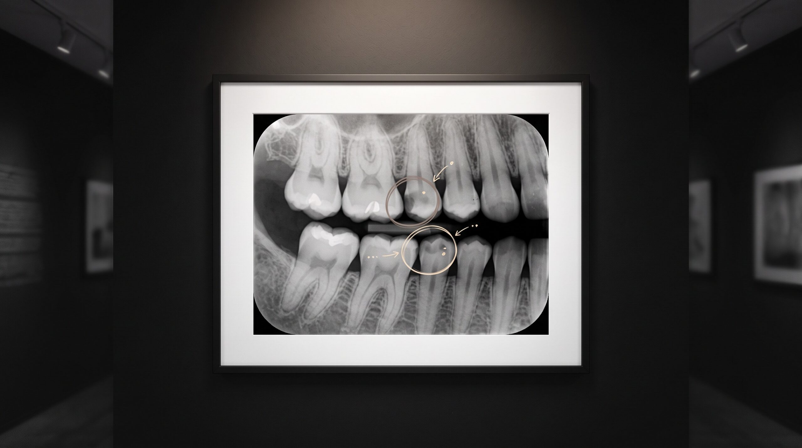
A radiograph is a made thing before it is a diagnosed one. Someone positioned the sensor, chose the exposure, and coaxed the anatomy into the sharp part of the field – and only then did the image become something worth reading. Pearl’s Second Opinion enters at that second moment, laying an automated reading over the picture the clinician has already composed. It is, by a wide margin, the most FDA-cleared name in dental artificial intelligence. But the detections are the spectacle; the quieter and more consequential story is how the image travels from the operatory out to Pearl’s cloud and back again. That pathway – DICOM, TWAIN, or a vendor-built bridge – is what actually decides who gets to use this at all.
The Most-Cleared Eye in Dentistry
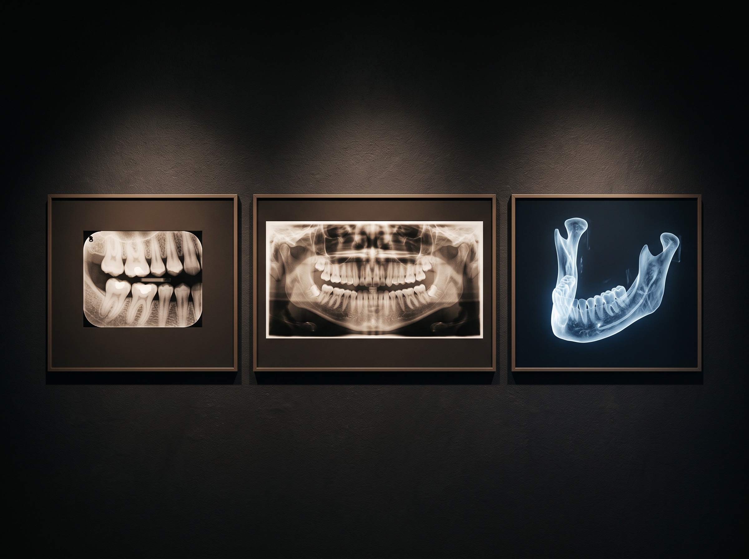
Pearl’s regulatory record is genuinely distinctive, and it is worth stating precisely rather than by vibe. In March 2022, the FDA granted 510(k) clearance to Second Opinion for detecting a broad range of conditions on intraoral radiographs – the bitewings and periapicals that make up most of a practice’s daily film. That clearance was notable because it covered many findings at once rather than a single pathology, making it the first of its kind cleared in the United States. Pearl has since extended the cleared surface twice: Second Opinion 3D received FDA 510(k) clearance for cone-beam CT in 2025, making Pearl the first dental AI company cleared for both 2D and 3D imaging, and a further clearance extended detection onto panoramic radiographs around the turn of 2026.
The honest way to summarize it: Pearl is cleared to assist detection across intraoral, panoramic, and CBCT imaging. It is a clearance to assist – the software surfaces suspected findings for a clinician to accept or dismiss, not to diagnose autonomously. That distinction is not a legal footnote; it is the entire posture of the tool. (We will not quote specific 510(k) numbers here, because clearance identifiers are precisely the sort of detail that should be verified against the FDA database rather than trusted to a press summary.)
What Second Opinion Actually Reads
Across those modalities, the system marks the familiar suspects: interproximal and other caries, calculus, periapical radiolucencies, and signs of bone loss, among others, with the findings drawn directly onto the image as color overlays and outlines. On a bitewing this looks like a set of gentle rings and shaded regions floating over the enamel; on a panoramic it becomes a wider sweep of flagged zones; on a CBCT volume it moves into three dimensions. The clinical value proposition is consistency – the model does not tire on the fortieth radiograph of the day, and it does not skip the corner of the film the eye slid past. What it offers is a reliable second look, and the emphasis belongs firmly on second.
How the Image Gets There
Here is the part that imaging people should care about most, because it is where the marketing goes quiet. Second Opinion is a cloud service. The radiograph does not stay inside the operatory PC to be analyzed; it is transmitted to Pearl’s infrastructure, scored, and the annotations are returned for display. Nothing about the AI happens until the image successfully leaves the building, which means the acquisition-and-transport layer is not a detail – it is the whole precondition.
Pearl reaches practices through three broad kinds of connection. The first is a direct integration with the practice’s imaging or practice-management software, where Pearl builds and configures a bridge so that any image captured or stored in the imaging module is passed automatically – or on demand – to Second Opinion, with detections rendered back inside that same viewer. Open Dental’s imaging module works exactly this way: once enrolled, Pearl supplies the details to stand up the bridge, and images flow to the AI as they are taken. Oryx and other partners embed the same capability natively in their own imaging utilities.

The second route is the DICOM layer – the medical-imaging lingua franca. In practices running a PACS such as MiPACS, integrators confirm the AI at the DICOM layer rather than assuming a generic bridge will do, precisely because an enterprise imaging environment routes and stores images as DICOM objects and expects any consumer of those images to speak the same protocol. The third route is TWAIN, the older acquisition standard by which a piece of software asks a sensor or scanner for an image; a TWAIN bridge can hand the freshly acquired frame to the AI at the moment of capture. Which of the three a given practice uses depends less on Pearl and more on the imaging stack already sitting in the operatory.
Partner-Gated, Not Self-Serve
The strategic reality underneath all this is that Pearl is largely partner-gated. You do not, as a rule, point arbitrary imaging software at an open public Pearl endpoint and start sending radiographs. Integrations are configured by Pearl, over secure connections, typically through relationships with named imaging and practice-management vendors – Open Dental, Oryx, MiPACS, and distribution partners such as Patterson among them. This is a deliberate posture, and a defensible one for a regulated medical device handling protected health information: a curated, tested pathway is easier to validate and to keep compliant than an open door.
But it has a direct consequence for anyone building or choosing imaging software. The question “can my system use Pearl?” is not answered by Pearl’s capability; it is answered by whether your imaging platform is on the partner list, or whether it can present its images in a standard – DICOM most cleanly – that Pearl’s bridge is prepared to consume. An AI that reads brilliantly but will only accept images through a handful of sanctioned doors is, for an unlisted platform, effectively unreachable until that door is built.
Why the Pathway Matters More Than the Model
For imaging software specifically, this reframes the whole conversation about “adding AI.” The differentiator among detection engines is narrowing – several vendors now detect a similar roster of findings with broadly comparable, and separately cleared, competence. What varies enormously is the send-out: whether a platform can get its acquired radiographs and CBCT volumes to a given AI at all, and get the reading back in a form the clinician sees without leaving their workflow. The image must travel cleanly, carry its patient context, and return its annotations to the right study. A platform that already stores and exports clean DICOM is halfway to any DICOM-friendly AI; one locked into a proprietary format faces a bridge-building project for each vendor it wants to reach.
This is the same infrastructural truth that governs so much of modern imaging: the picture is only as useful as its ability to move. Pearl’s consumer-facing story – the reassuring second opinion at the chairside, covered in plain language in our companion piece, Pearl Second Opinion: Chairside AI That Reads Your X-Rays – sits atop an integration story that is far less glamorous and far more decisive. The radiograph is craft; so, increasingly, is the conduit that carries it.
Future Developments
The trajectory of detection is clear and, at this point, almost expected: broader condition coverage, tighter localization, and steady expansion from flat film into the volumetric depth of CBCT, where a finding can be traced through slices rather than guessed at through superimposition. The more interesting frontier is access. As the cleared reading matures into a commodity, the live question stops being can the machine read the picture and becomes which practices are permitted to hand it over. The winners in that next phase may not be the sharpest detectors but the most open pathways – imaging platforms fluent in DICOM, unafraid to let a patient’s radiographs travel out to whichever validated AI serves them best, and disciplined enough to bring the reading home to the same frame the clinician was already studying. The image was always meant to be seen. The work ahead is making sure it can also be sent.
Sources & further reading:
- Pearl – Press Release: Pearl Becomes First Dental AI Company Cleared by FDA for Both 2D and 3D Imaging (Second Opinion 3D)
- Pearl – Press Release: Pearl Expands Dental AI Capabilities with FDA Clearance for Panoramic X-Rays
- Open Dental: Pearl integration (imaging module bridge)
Sorry, the comment form is closed at this time.