


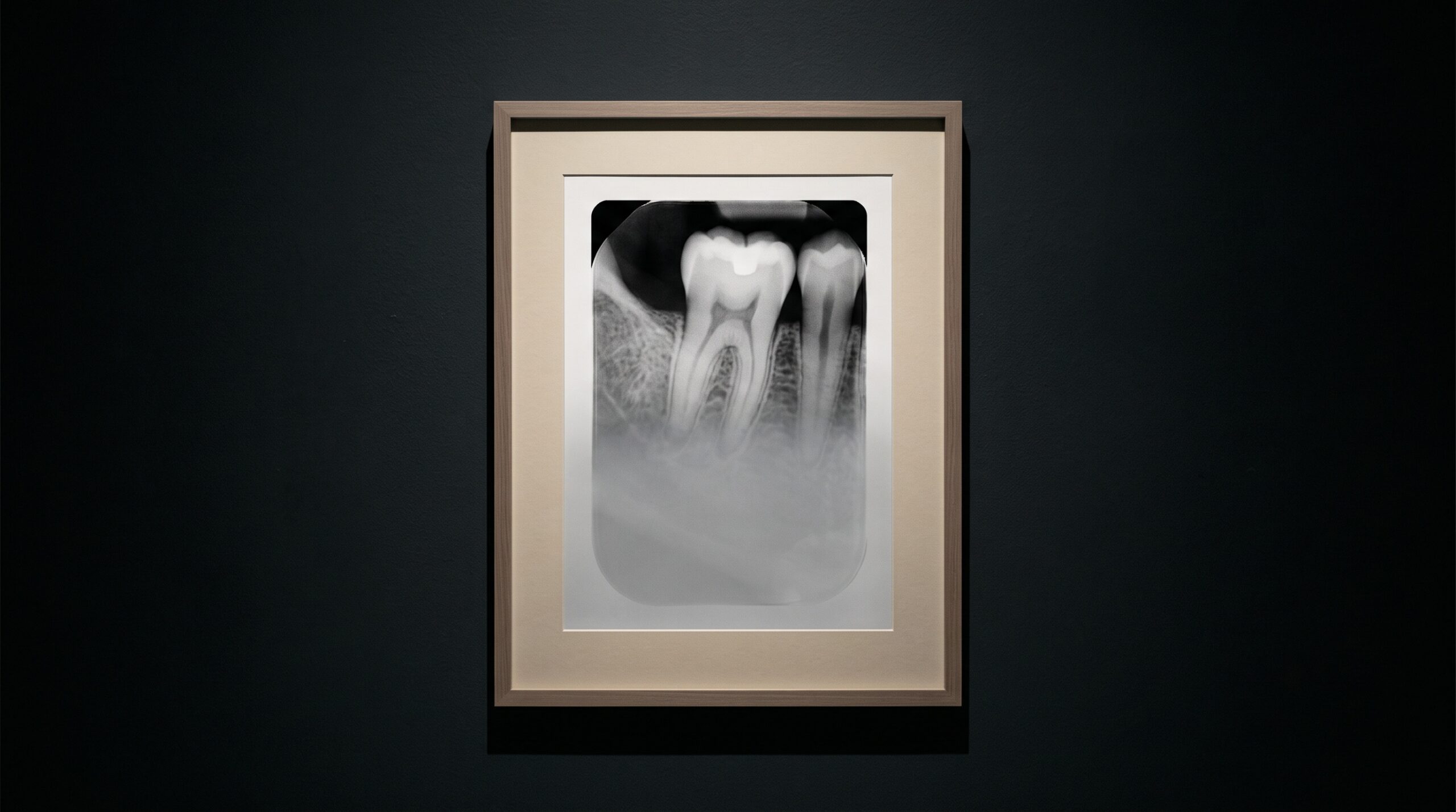
August 19, 2026 The Scattered Photon: How Compton Scatter, Collimation, and the Missing Grid Shape Contrast in Intraoral Radiography
Contrast is the thing a radiograph is really made of. Resolution decides how fine a detail can be drawn, but contrast decides whether the detail can be seen at all – the difference in brightness between a lesion and the sound bone beside it, between enamel and the caries eating into it. That contrast is built by photons behaving honestly: some absorbed by dense tissue and cast as shadow, others passing cleanly through and registering as light. But a third population of photons behaves dishonestly. They do not pass through and they are not cleanly absorbed; they are knocked sideways, and they drift onward to land somewhere they were never aimed, laying a pale veil over the whole picture. This is scatter, and understanding it is understanding why a dental radiograph is collimated the way it is, why it carries no grid, and why the exposure dial is quietly also a contrast dial.
The Two Honest Fates, and the Dishonest One
When an X-ray photon enters tissue, only a few things can happen to it. It can be absorbed outright – the photoelectric effect, in which the photon surrenders all its energy to an atom and vanishes. This is the interaction that builds the image, because dense, high-atomic-number structures like enamel and bone absorb far more readily than soft tissue, and that differential absorption is precisely what casts the shadow we read. The photon can also pass straight through untouched, reaching the sensor to expose the bright background. Both of these are honest: each photon either becomes shadow at the point it was aimed, or becomes light at the point it was aimed.
The third fate is the Compton interaction. Here the photon strikes a loosely bound outer electron, gives up only part of its energy, and ricochets off in a new direction with the rest. It is not absorbed and it does not stop – it wanders, still carrying enough energy to reach the detector, but arriving at a location that has nothing to do with the anatomy above it. A photon that should have reported on the tooth’s apex instead lands over the crown, or the bone, or the empty film margin. It is information detached from its source, and once detached it can only mislead.
A Fog With No Source
Individually a scattered photon is a single misplaced speck. The trouble is that they arrive in enormous numbers, from every direction, with no pattern – and randomness at scale becomes a uniform wash. Scatter does not draw a false structure onto the image; it does something subtler and more corrosive. It adds a roughly even layer of exposure across the entire receptor, lifting the darkest shadows toward grey and pushing the whole image toward the muddy middle. The blacks are no longer black, the whites are no longer clean, and every difference in between is compressed. This is veiling glare, and it is the direct enemy of the differential absorption that makes contrast possible in the first place – the same signal that a well-built sensor works so hard to capture faithfully, described in our look at the true measure of what a sensor can resolve. A useful way to think about image quality is the ratio of scattered photons to primary ones reaching the detector: the higher that scatter fraction climbs, the more the fog thickens and the more the picture’s honest contrast is drowned.
What generates all this scatter is simply irradiated tissue. Every cubic centimetre of tissue in the beam is a workshop manufacturing scattered photons, and the more tissue the beam illuminates, the more fog it produces. That single fact points directly at the most elegant defence dentistry has against it.

Collimation: Refusing the Photons You Never Needed
The cleanest way to deal with scatter is not to clean it up afterward but to avoid manufacturing it. A dental sensor is a small rectangle, yet an uncollimated beam emerges as a wide circle far larger than the receptor, flooding the cheek, tongue, and jaw with radiation that can never form part of the image – and every bit of that surplus tissue is busy scattering. Rectangular collimation trims the beam to the shape and size of the sensor itself, so that almost the only tissue irradiated is the tissue directly in the image path. The gain is twofold and worth stating plainly: the patient receives substantially less radiation dose because far less of them is in the beam, and the image gains contrast because far fewer scattered photons are ever created to fog it. It is one of the rare interventions in imaging where the safer choice and the better-looking choice are the same choice – a discipline of the beam’s edge that governs the picture much as the beam’s own physics governs the subtle intensity gradient we described in the anode heel effect.
The Grid That Isn’t There
Anyone who has looked at medical radiography knows there is a second, more famous weapon against scatter: the anti-scatter grid, a fine lattice of lead strips laid between patient and detector that absorbs photons arriving at oblique angles – the scattered ones – while letting the straight, primary photons through. It is standard in chest and abdominal imaging. It is conspicuously absent from the intraoral setup, and that absence is a deliberate piece of physics rather than an oversight.
A grid earns its place only where scatter is overwhelming, and scatter grows with the volume of tissue irradiated and the thickness of the part. A chest is thick and broad; an intraoral field is small and thin, and once rectangular collimation has already suppressed the irradiated volume, the residual scatter is modest enough that a grid would remove little worth removing. Worse, a grid extracts a heavy toll: because it absorbs a fraction of the useful primary beam along with the scatter, every grid demands a compensating increase in exposure – more dose to the patient to push enough photons through the lead slats. In a discipline built around keeping dose as low as reasonably achievable, spending extra radiation to fight a scatter problem you have already collimated into insignificance is a bad bargain. The short distance between beam, thin tissue, and sensor also lets a modest air gap do quietly what a grid would do expensively: many scattered photons, travelling at an angle, simply miss the small receptor. The grid is left out because the situation was engineered so it is not needed.
The Kilovoltage Dial Is Also a Scatter Dial
There is one more lever, and it cuts both ways. The kilovoltage of the beam – its peak energy – sets how penetrating the photons are. Raise it, and more photons pass through the patient, letting you lower the dose and producing what is called a long grey-scale image: many gentle shades of grey, forgiving and detailed. But higher energy also shifts the balance of interactions toward Compton scattering and away from the photoelectric absorption that builds contrast. So the very setting that grants penetration and a softer tonal range simultaneously raises the scatter fraction and lowers the inherent subject contrast, while a lower kilovoltage yields a short grey-scale image of bold blacks and whites, less scatter, and more contrast – at a higher dose and with less latitude for error. There is no free setting; there is only the trade, chosen deliberately for the task. It is the same seduction we cautioned about when software, rather than physics, is used to manufacture the appearance of contrast after the fact in the ways sharpening and contrast enhancement can deceive – except here the trade is made in photons, honestly, at the moment of capture.
Future Developments
For a century the answer to scatter has been mechanical – collimate the beam, occasionally grid the receptor, choose the kilovoltage with care. The frontier now is computational and spectral. Scatter-correction algorithms already estimate the low-frequency fog mathematically and subtract it, recovering contrast that physics allowed to be lost, a technique that matters most in cone-beam volumes where scatter is far heavier than in a periapical view. Further out lies a more fundamental fix: because a scattered Compton photon has given up part of its energy, it arrives at the detector carrying slightly less than a primary photon. A detector that can measure each photon’s energy rather than merely counting its arrival can therefore recognise many scattered photons by their diminished energy and simply refuse to record them – discarding the fog at the door instead of scrubbing it from the finished picture. As energy-discriminating and photon-counting sensors mature, scatter rejection stops being a matter of lead and geometry and becomes a property of the detector’s intelligence. The scattered photon has always been the image’s quiet vandal; the coming generation of imaging may finally be able to see it coming and turn it away.

Sources & further reading:
No Comments