


October 13, 2026 One Exposure, Many Readings: Window and Level and the Craft of Seeing Inside a Radiograph’s Full Range
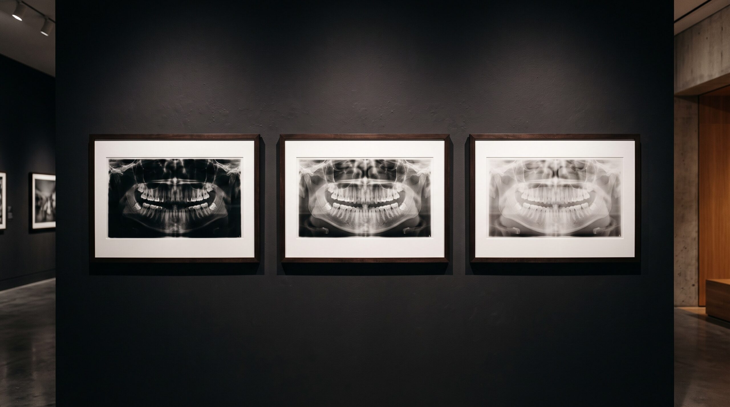
Hang a digital radiograph on the wall and you are looking at a compromise. Not a flaw — a compromise. The sensor that captured it recorded the tissue in thousands of distinct steps of gray, a tonal range far finer than the screen in front of you can display or the eye reading it can resolve. What you see framed and lit is one interpretation of that buried depth: a single mapping of an enormous captured range onto the few hundred grays a monitor can actually paint. The remarkable thing is that the mapping is not fixed. With two controls — window and level — the same exposure can be read a dozen different ways, each one surfacing something the others let sink into black or wash into white. This is windowing, and it is one of the quietest, most underused acts of craft in all of dental imaging.
More gray than the eye can hold
Start with the raw material. A modern intraoral sensor or CBCT detector captures each pixel not as a shade you can name but as a number — typically across a 12- to 14-bit range, which is to say four thousand to sixteen thousand possible levels of gray. A diagnostic display, by contrast, shows on the order of 256 grays per pixel, and the human visual system, even trained and in ideal conditions, can distinguish only a few dozen simultaneous shades in a single scene. The captured image is therefore vastly richer than any one view of it. Most of that richness is invisible at any given moment, not because it was lost, but because it has been compressed to fit what the screen and the eye can take in. The data is all there; you are simply being shown a slice.
This is the fact that makes windowing both possible and necessary. If a radiograph held only as much information as a screen could display, there would be nothing to adjust — what you saw would be all there was. Because it holds far more, the viewer’s first and most consequential decision is which part of the range to look at. That decision is not cosmetic. A subtle periapical change, a faint widening of a membrane space, an early loss of trabecular definition — these can live entirely within a band of grays that the default mapping crushes into a single flat tone. Open that band, and the finding appears.
Two dials, two meanings
Window and level are easy to confuse and worth keeping distinct. The level — sometimes called the window center — chooses the midpoint: which captured gray value sits in the middle of your visible scale, and therefore whether the picture reads bright or dark overall. Slide the level and the whole image lightens or darkens as the window of attention travels up or down the captured range. The window width chooses how wide a band of captured values you spread across the full black-to-white display. A narrow width takes a thin slice and stretches it hard, so small differences in density become bold differences in brightness — high contrast, a short gray scale, dramatic but unforgiving at the edges of the band, which clip to pure black or pure white. A wide width maps a large span gently, so the image carries many soft intermediate tones — low contrast, a long gray scale, forgiving and tonally complete but subtle.
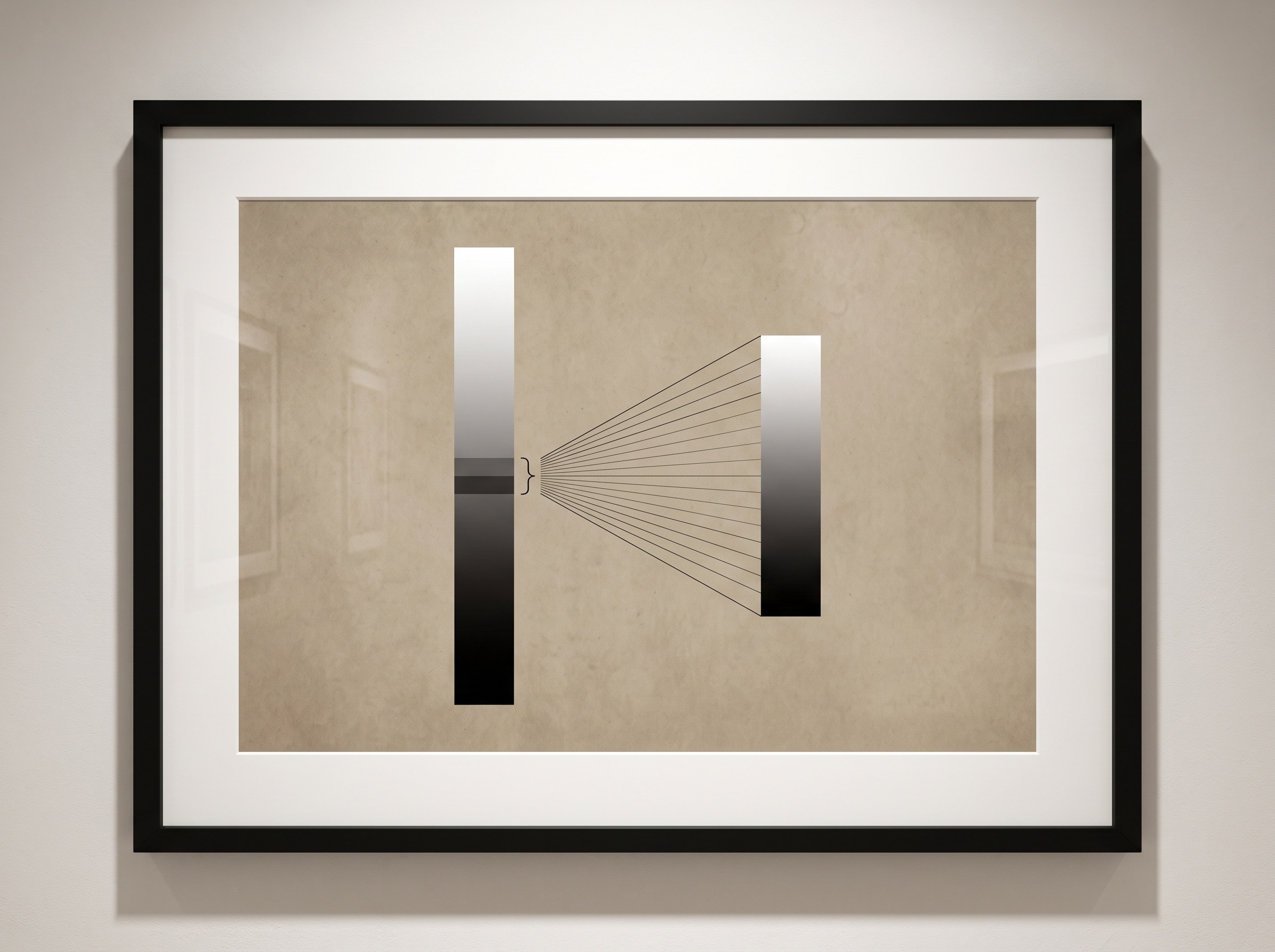
Those two words, width and level, are simply the operator’s hands on the mapping between captured depth and visible range. Everything else in windowing follows from them. And crucially, neither one touches the stored pixel values. Windowing is a viewing transform: reversible, repeatable, lossless. You can tour an image through a hundred settings and the file on disk is exactly as it was. That is what separates it in kind from the irreversible edits covered elsewhere in this gallery — the sharpening, contrast curves, and noise reduction that bake a judgment permanently into the pixels. Windowing changes what you see; processing changes what exists. The discipline is to lean on the former before reaching for the latter.
Reading one image many times
Put the two dials together and a single radiograph becomes a thing to be interrogated rather than merely accepted. The same exposure, viewed with a narrow window, throws trabecular bone into stark relief — ideal for scrutinizing the fine architecture around a root or an implant. Widened, the identical image softens into a long scale that keeps enamel, dentin, pulp, and the faint soft-tissue shadow legible together, so nothing at the extremes is lost. Shift the level up to chase detail buried in the densest cortical bone; drop it down to pull a faint finding out of the darker, less attenuating regions. None of this is retaking the image. It is reading it again, and again, each pass asking a different question of the same buried range.
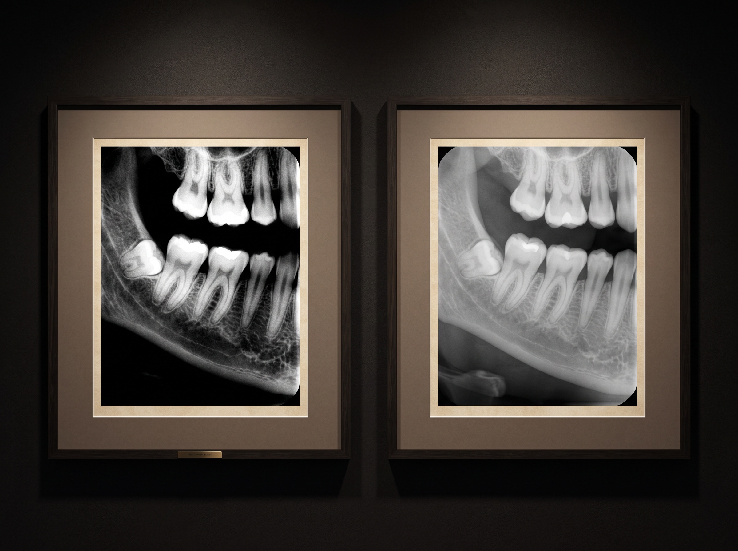
This is the deep continuity between windowing and the inherent contrast and density that the exposure itself establishes. The capture fixes how much tonal information exists; windowing governs how much of it you choose to see at once. A well-exposed radiograph with a generous captured range rewards an active reader who moves the window; a poorly exposed one, its information already collapsed at the time of capture, gives the window little to work with. The two are partners — good technique at the chair creates the latitude that good windowing at the screen then exploits.
Where the habit becomes doctrine: cross-sectional imaging
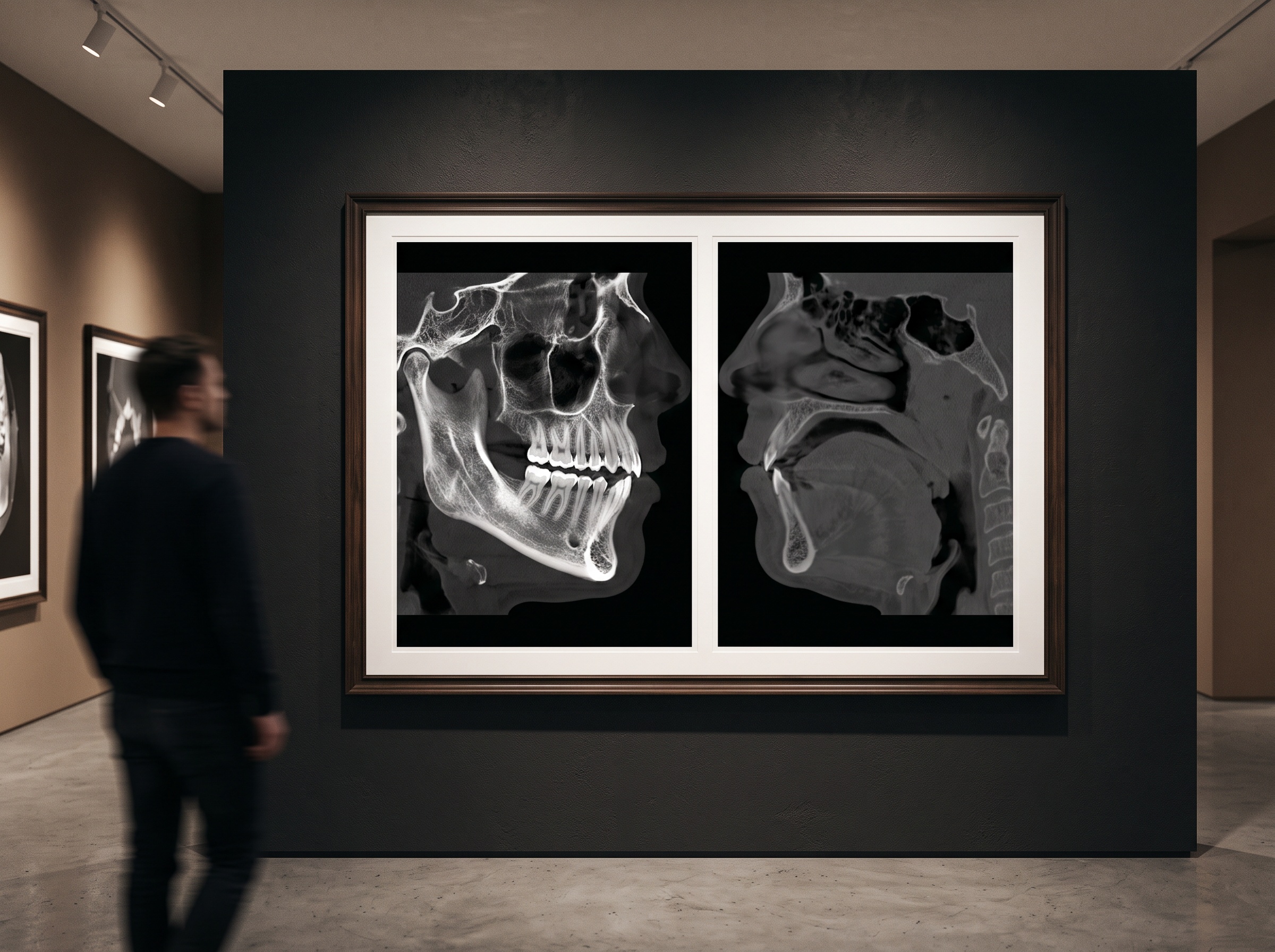
Nowhere is windowing more formalized than in CBCT. A cone-beam volume spans such an enormous range of attenuation — from air to enamel to dense cortical bone — that no single mapping can serve every purpose, so readers adopt named presets: a bone window, narrow and leveled high, to study the hard architecture; a soft-tissue window, wider and leveled lower, to coax out the faint structures beside the bone. Reading a volume means cycling deliberately through these windows, knowing that a finding invisible in one may be obvious in another. It is worth remembering, though, that a CBCT’s gray values are not absolute the way a medical CT’s Hounsfield units are — a caution explored in why a cone-beam scan’s numbers cannot be trusted as true densities — so windowing a CBCT is a tool for revealing relationships and morphology, not for reading off calibrated numbers.
The quiet prerequisite: a display worth trusting
All of this assumes the screen is telling the truth. Window and level decide which grays to send to the monitor; the monitor decides whether those grays arrive as intended. An uncalibrated or washed-out display silently undoes the finest windowing, compressing the very mid-tones the reader just worked to open. That is why windowing and diagnostic display calibration to the DICOM grayscale standard are two halves of one competence: one chooses the slice of information, the other guarantees the slice is faithfully shown. Skilled windowing on a careless monitor is a cartographer drawing a precise map onto warped paper.
Future Developments
The near future of windowing is about removing friction without removing judgment. AI-assisted viewers increasingly propose an optimal window and level for a given image and a given clinical question, learning from how experienced readers settle their dials — a starting point offered, not a verdict imposed. High-dynamic-range displays, carrying well beyond the traditional eight bits per channel, promise to narrow the gap between what a sensor captures and what a screen can show at once, so that less of the range stays hidden and the window has to travel less far. And perceptually tuned mappings, which allot display grays according to where the eye is most sensitive rather than linearly across the range, aim to make a single view carry more genuine diagnostic information. Yet the underlying craft will not be automated away. A radiograph will always hold more than a glance can take in, and the reader who knows to move the window — to ask the same image a second and a third question — will always see what the passive viewer, content with the first rendering, never knew was there. The image was never a single picture. It was always a range, waiting to be read.
Sources & further reading:
- Windowing (CT) — windowing, window width and window level explained (Radiopaedia)
- Grey-scale and bit depth in digital radiography (an overview)
No Comments