


July 28, 2026 Contrast and Density: The Grayscale Craft Behind a Diagnostic Radiograph
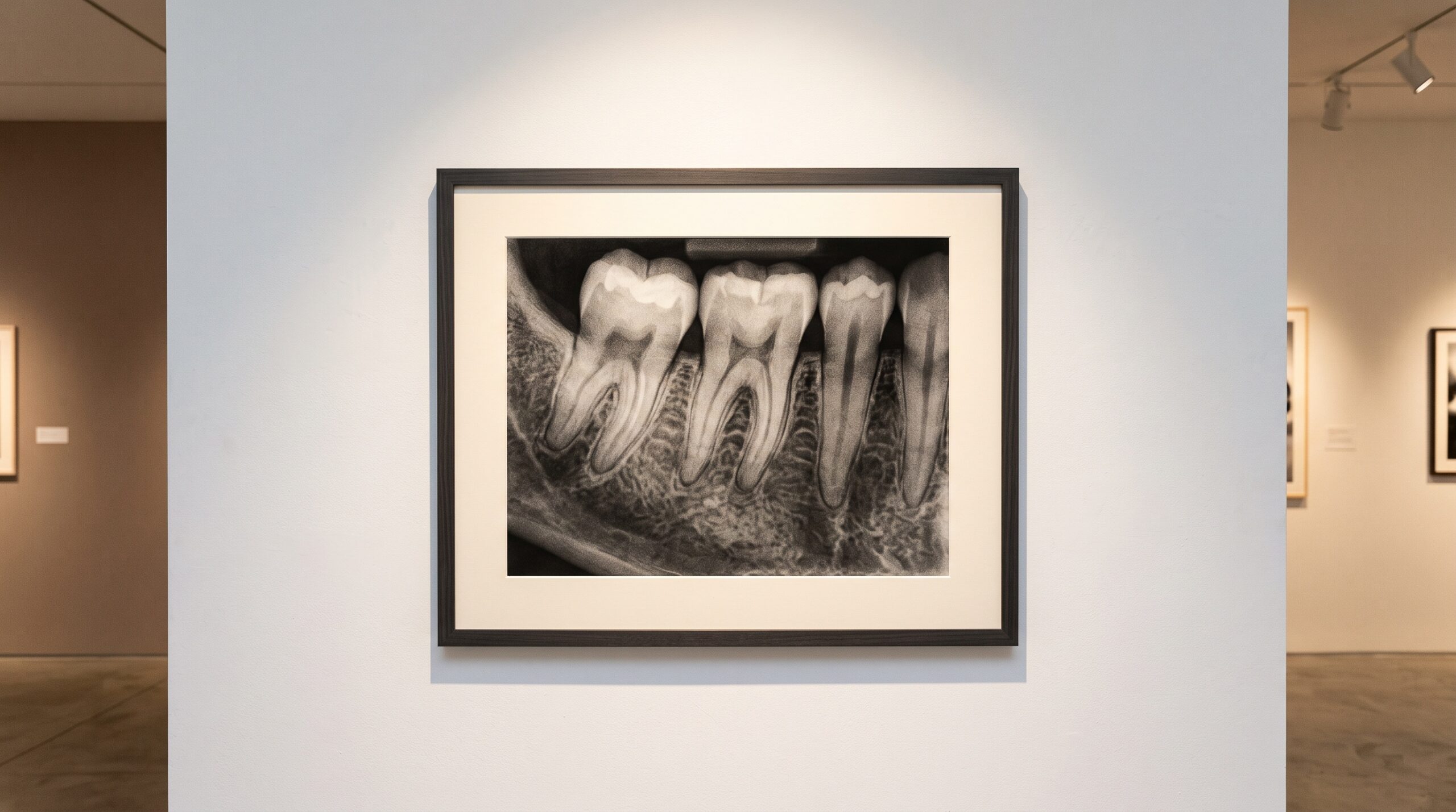
Hang a dental radiograph on a lightbox and, for a moment, forget the diagnosis. Look instead at the image the way you would look at a charcoal study: a composition built entirely from grays. Enamel reads as luminous near-white, the pulp chamber falls away into shadow, bone sits somewhere in the quiet middle. Every clinical judgment you are about to make rests on the relationships between those tones. Radiographic contrast and density are not technical afterthoughts. They are the medium itself.
At PatientGallery we treat the radiograph as an image first and a record second, because the two are inseparable. A picture that cannot be read cannot inform care. So it is worth slowing down to understand exactly how that grayscale is made, and how much of it sits within your control.
Density: how bright, how dark
Density is the overall lightness or darkness of the image — the degree to which the receptor was exposed. In film terms it was literal silver deposited on the emulsion; in a digital sensor it is the signal accumulated in each photosite. Too little exposure and the image is pale and mottled, its subtle structures lost in noise. Too much and the picture goes leaden, thin structures burned into a uniform dark. Density is governed chiefly by milliamperage and exposure time — the sheer quantity of photons delivered — modulated by the distance and the tissue they pass through.
The digital era changed the stakes. Software can rescale a poorly exposed capture after the fact, which tempts operators to treat exposure as forgiving. It is not. Rescaling a thin, underexposed signal simply amplifies its noise along with its detail. The tonal foundation has to be laid correctly at the moment of capture; everything downstream is refinement, not rescue.
Contrast: the difference that reveals
If density is the average tone, contrast is the spread — the difference between the lightest and darkest values, and how the shades in between are distributed. Contrast is what makes a hairline periapical lesion separate from the bone around it, what lets you follow the faint margin of a recurrent carious lesion beneath a restoration. It is governed principally by kilovoltage. Lower kV produces a short-scale, high-contrast image of dramatic blacks and whites and few grays between; higher kV yields a long-scale, low-contrast image rich in intermediate tones.
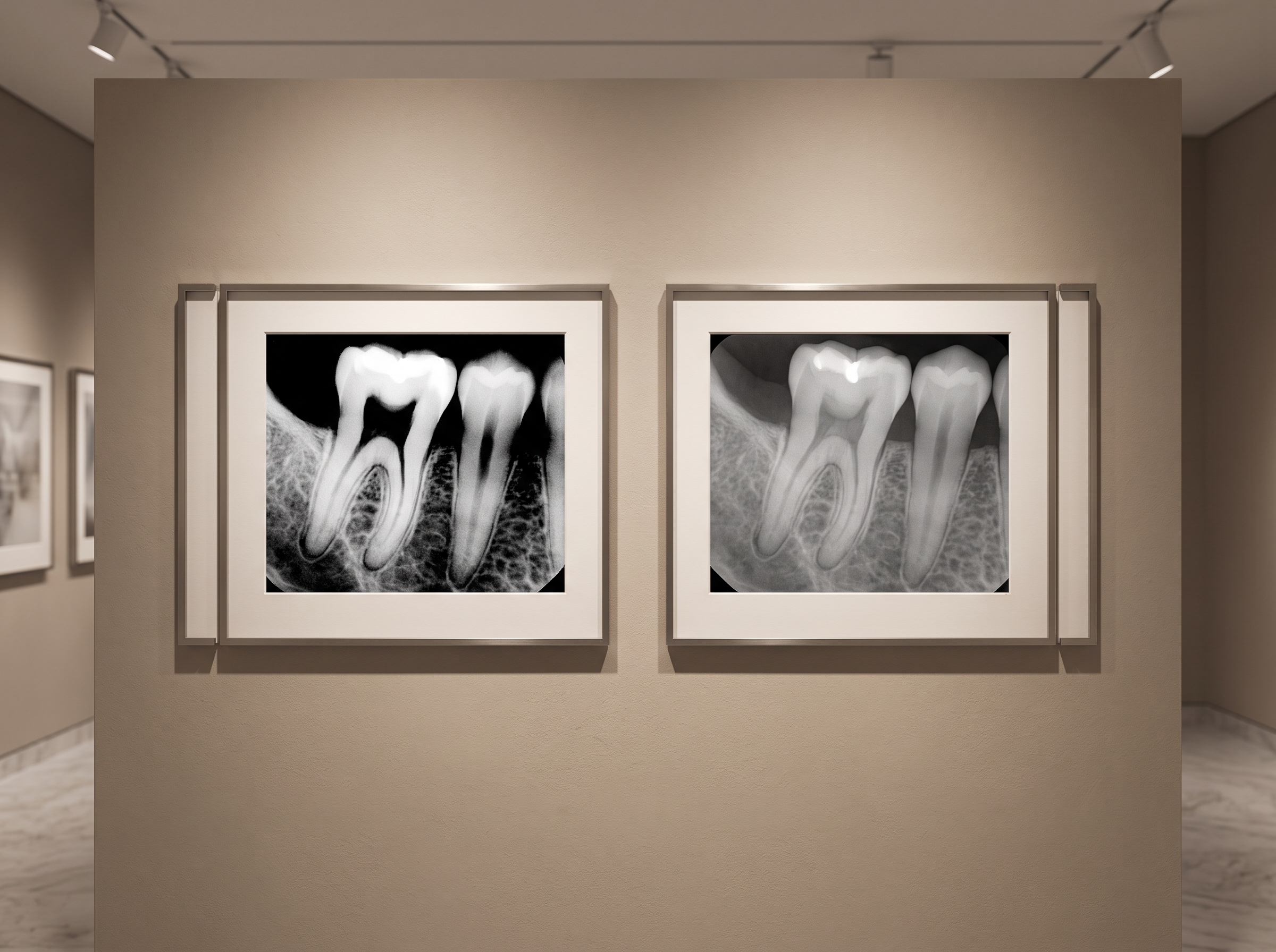
Neither is universally better. A short tonal scale can make a crack leap off the image but crush the subtle gradations of trabecular bone into a single shade. A long scale preserves those gradations but can make a low-contrast lesion whisper where you need it to speak. The craft lies in matching the tonal scale to the diagnostic question, exactly as a photographer chooses a hard or soft light to serve the subject.
Bit depth and dynamic range: the palette behind the picture
Here material science quietly asserts itself. A modern intraoral sensor does not capture eight shades of gray, or even 256. Working at 12, 14 or 16 bits, it records thousands of discrete tonal steps — a dynamic range far wider than any monitor can display or any eye can resolve at once. That surplus is the reason a single exposure can be re-windowed to interrogate enamel, then dentin, then bone, without a second dose to the patient.
Think of bit depth as the size of the palette from which the visible image is painted. The wider the captured range, the more gracefully software can compress it into the eight bits your screen actually shows, and the more headroom you retain for adjustment. It is invisible on the surface and decisive underneath — the difference between an image that survives manipulation and one that falls apart under it.
Windowing: developing the image with light
Because the sensor captures more tones than a display can render, someone has to choose which slice of that range becomes visible. That is windowing — setting the window level (the central brightness) and window width (the range of values mapped from black to white). It is the digital darkroom, and it is where much diagnostic yield is won or lost.
Narrow the window and you amplify contrast within a chosen band, coaxing a faint lesion out of near-uniform bone. Widen it and you recover the full sweep of anatomy in a single view. The radiograph is not one image but a family of images latent in the captured data, and windowing is how you develop the one the diagnosis requires. Treating it as a craft — deliberate, repeatable, matched to the task — is what separates confident reading from squinting hopefully at a default preset.
Noise, the uninvited tone
No discussion of grayscale is honest without noise. Every capture carries a floor of random signal — quantum mottle from the finite number of photons, electronic noise from the sensor itself. Noise is the grain in the photograph, and like grain it competes with the faintest detail. Push exposure too low to spare dose and noise rises to swallow the very structures you were trying to see. The elegant image is not the one with the least noise at any cost, but the one that finds the honest balance: enough signal to reveal, no more dose than the question demands.
Future Developments
The tonal scale is where imaging is quietly being reinvented. Deep-learning reconstruction now denoises low-dose captures without the smearing that once accompanied aggressive filtering, letting clinicians hold the line on radiation while preserving contrast. Perceptual tone-mapping — borrowed from computational photography — promises windowing that adapts to the anatomy rather than to a fixed preset, presenting the reader with an image already optimized for the structure in view. And as sensors push toward ever-wider dynamic range, the single capture becomes an ever-deeper well of latent images, each one waiting to be developed by light.
What will not change is the fundamental truth on the lightbox: a radiograph is a picture made of gray, and the clinician who understands how that gray is built sees more in it. Contrast and density are the grammar of the medium. To master them is to read the image as both science and art — which, in the end, is the only way to read it well.
Sources & further reading:
- CBCT: radiation risk or diagnosis risk? (Nature)
- Deep learning-based assessment of periapical radiographic image quality (Nature)
Related Reading
- One Exposure, Many Readings: Window and Level and the Craft of Seeing Inside a Radiograph’s Full Range
- The Last Instrument: Diagnostic Display Calibration and the DICOM Grayscale Standard for Reading a Radiograph
- Radiopaque Contrast Agents in Dental Imaging: Advanced Material Science for Enhanced Diagnostic Visibility
Sorry, the comment form is closed at this time.