


May 6, 2026 Radiopaque Contrast Agents in Dental Imaging: Advanced Material Science for Enhanced Diagnostic Visibility
Radiopaque contrast agents represent a transformative advancement in dental imaging technology, offering clinicians enhanced diagnostic capabilities through improved radiographic visibility. These specialized materials fundamentally alter how dental structures, restorative materials, and pathological conditions appear on radiographic images, providing unprecedented clarity in complex clinical scenarios.
Understanding Radiopaque Materials
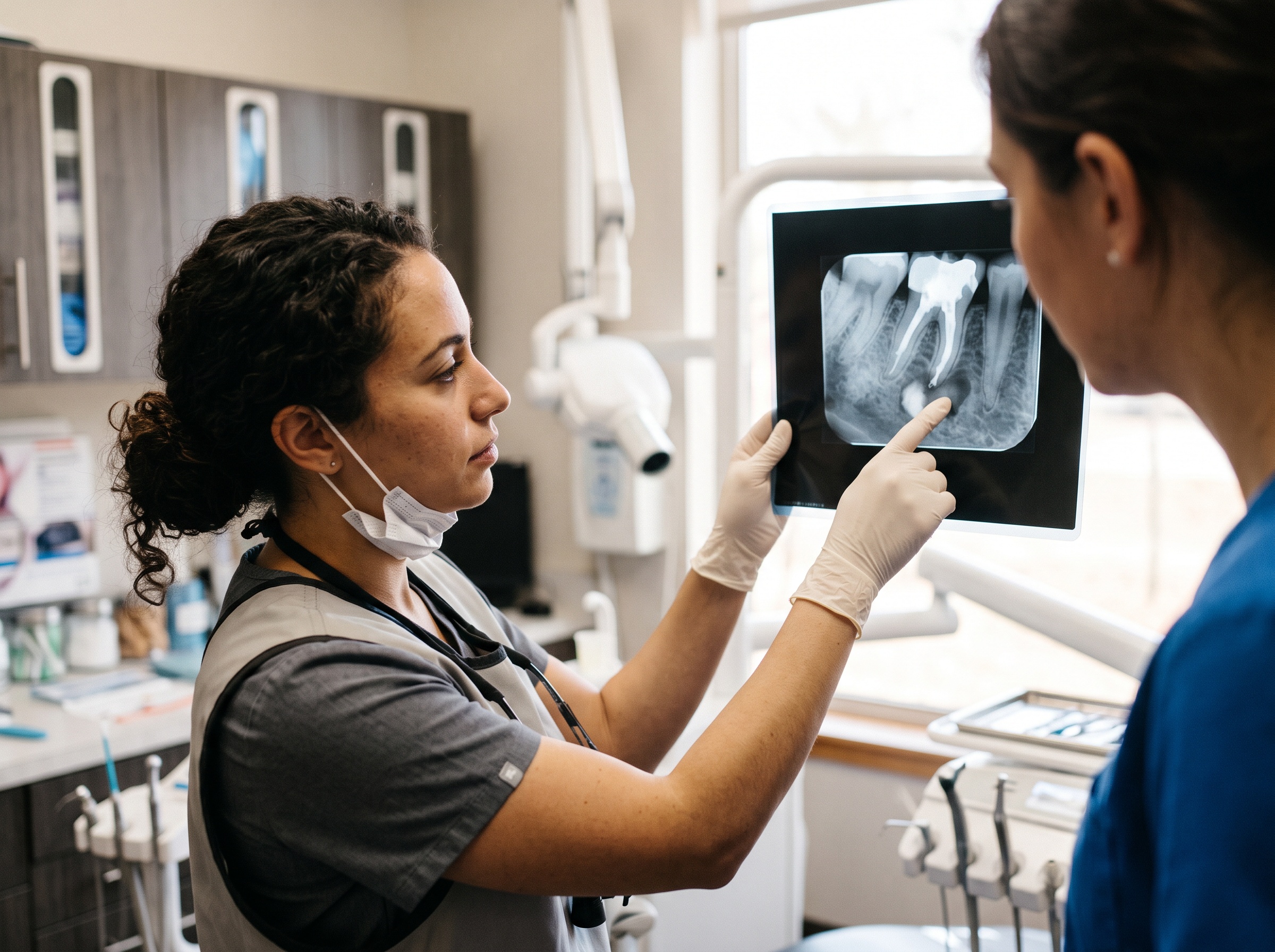
Radiopaque contrast agents work by absorbing X-ray radiation more effectively than surrounding tissues, creating distinct contrast differences on radiographic images. The effectiveness of these materials depends on their atomic number and density, with higher atomic number elements providing superior radiopacity. Modern dental imaging relies on precise contrast differentiation to identify subtle pathological changes and evaluate treatment outcomes.
Common Radiopaque Agents in Dentistry
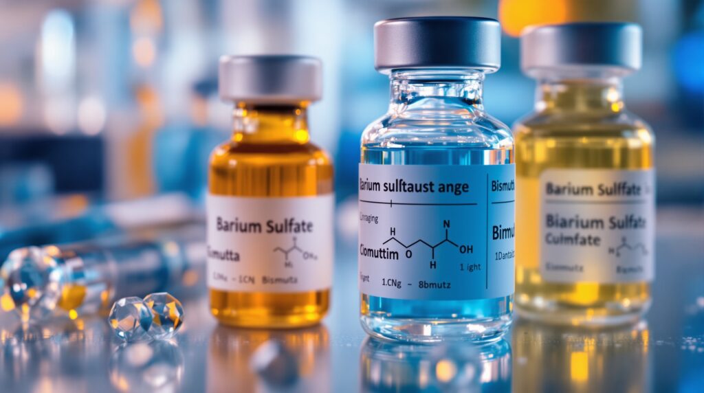
Barium sulfate (BaSO4) stands as one of the most widely utilized radiopaque agents in dental materials. Its exceptional radiopacity, biocompatibility, and chemical stability make it ideal for incorporation into composite resins, glass ionomer cements, and endodontic sealers. Bismuth compounds, including bismuth trioxide and bismuth subcarbonate, offer similar radiopaque properties with additional antimicrobial benefits, particularly valuable in endodontic applications.
Zinc oxide serves as both a therapeutic and radiopaque component in dental materials, providing antimicrobial properties while enhancing radiographic visibility. Zirconium dioxide (ZrO2) represents an advanced option, offering excellent biocompatibility and mechanical properties alongside superior radiopacity, making it particularly suitable for high-stress applications.
Clinical Applications and Benefits
The incorporation of radiopaque agents in dental materials enables precise monitoring of restorative procedures, detection of secondary caries, and evaluation of marginal integrity. In endodontic therapy, radiopaque sealers allow clinicians to assess the quality of root canal obturation and identify voids or inadequate filling. Orthodontic applications benefit from radiopaque markers that facilitate treatment monitoring and appliance positioning verification.

Digital Imaging Considerations
Digital radiography systems respond differently to radiopaque agents compared to conventional film-based systems. Materials incorporating iodoform (CHI3) or barium sulfate demonstrate significantly enhanced radiopacity on digital sensors, requiring adjusted exposure parameters and interpretation protocols. This enhanced sensitivity allows for reduced radiation exposure while maintaining diagnostic quality, aligning with the ALARA principle (As Low As Reasonably Achievable).
Material Selection Criteria
Selecting appropriate radiopaque agents requires consideration of multiple factors including biocompatibility, mechanical properties, chemical stability, and diagnostic requirements. The radiopacity level should exceed that of dentin (typically 1-2mm aluminum equivalent) to ensure adequate contrast. Particle size distribution affects both radiopacity and material properties, with optimal ranges balancing visibility and mechanical performance.
Future Developments
Nanotechnology advances are revolutionizing radiopaque agent development, with nanostructured ceramics and hybrid materials offering enhanced properties. Smart contrast agents that respond to specific biological conditions represent an emerging frontier, potentially enabling real-time monitoring of healing processes and treatment outcomes. These innovations promise to further enhance diagnostic capabilities while maintaining the biocompatibility and performance characteristics essential for clinical success.
The strategic incorporation of radiopaque contrast agents continues to advance dental imaging capabilities, providing clinicians with essential tools for accurate diagnosis, treatment planning, and quality assessment in modern dental practice.
Sorry, the comment form is closed at this time.