


September 5, 2026 The Whole Picture: What a Dental X-Ray Reveals Beyond the Teeth
You come in for your teeth. The radiograph is framed for your teeth, catalogued under your teeth, and discussed – if it is discussed at all – in terms of your teeth. Yet the X-ray beam that made the picture was never so disciplined. It passed through the jaw, the sinuses, the airway, the joints of the jaw, and the soft tissues of the neck, and it recorded every one of them in the same field of grey. A dental radiograph is a portrait with a wide background, and now and then something in that background turns out to matter more than the reason you sat down. Radiologists have a plain name for it: the incidental finding – the clinically important thing an image reveals that no one was looking for.
This is imaging at its quietly most powerful. A caries lesion answers the question the film was taken to ask. An incidental finding answers a question nobody asked, sometimes one the patient did not know to ask, and occasionally one that reaches well beyond the mouth. Reading the whole picture – not just the arch of teeth at its center – is one of the least visible and most consequential parts of the craft.
The Field of View Is Wider Than the Appointment
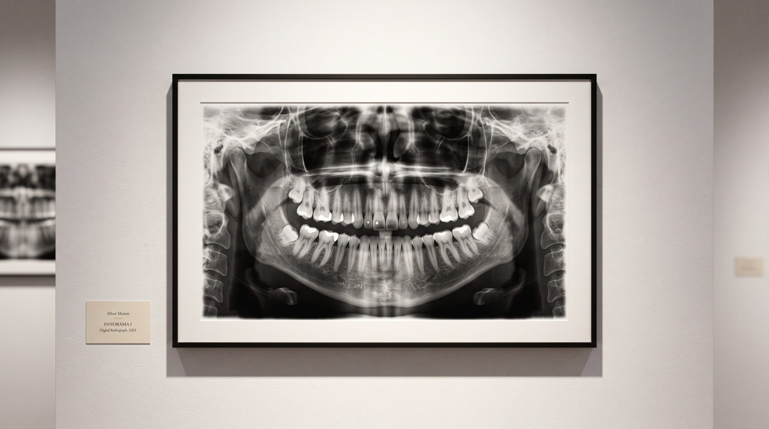
Every imaging modality has a field of view, and the dental ones are generous. A panoramic radiograph sweeps in a single arc from one condyle to the other, capturing both jaws, the maxillary sinuses, the nasal floor, the hard palate, portions of the airway, and the soft-tissue shadows of the neck. A cone-beam CT goes further still, resolving all of that as a navigable three-dimensional volume. Even a humble bitewing includes crestal bone and the edges of structures that are not teeth. The tooth is simply the brightest, most central subject in a much larger composition.
That geometry creates a responsibility. If the anatomy is in the frame, it has been imaged, and if it has been imaged, someone competent is expected to have looked at it. The standard of care in radiology is not to interpret the region of interest and ignore the rest; it is to read the entire captured field. The whole picture was taken. The whole picture must be seen.
The Marquee Example: A Calcification in the Neck
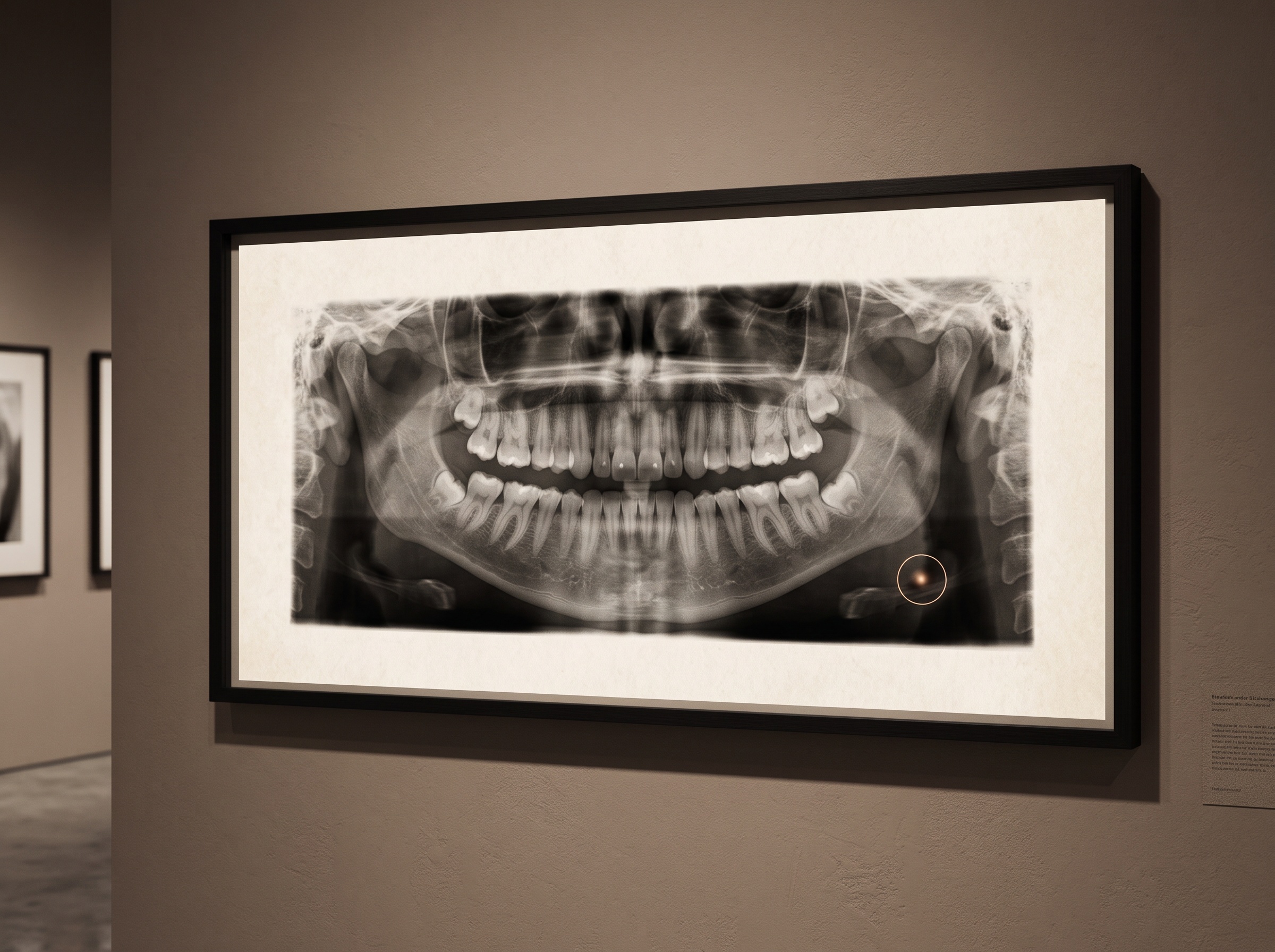
The most striking incidental finding in dentistry sits low in the panoramic image, near the angle of the jaw, at roughly the level of a particular neck vertebra – a small, often nodular radiopacity that has nothing to do with any tooth. It can be a calcified carotid artery atheroma: a deposit of calcified plaque in the wall of the carotid artery, the vessel that feeds the brain. On an ordinary dental panoramic film taken for entirely routine reasons, it can be the first visible hint of atherosclerotic disease in a patient who feels perfectly well.
The literature has taken this seriously for decades. Reviews in the dental and radiology press have long described panoramic radiography as an auxiliary route to detecting carotid atheromas, and more recent work frames the incidental finding of calcified carotid artery atheroma on standard dental radiographs as a genuine public-health opportunity for cardiovascular risk reduction – a chance to flag people who might benefit from medical evaluation before an event, not after. The dentist who notices it and refers is doing something an eye trained only on enamel would never do.
The honest caveat matters just as much as the finding. A calcification on a two-dimensional film is not a diagnosis of stroke risk, and the current consensus is explicit that risk should never be estimated from the image alone. A radiopacity in that region can be other things – a calcified triticeal cartilage, a calcified lymph node, other benign structures – and even a true atheroma must be weighed alongside age, smoking history, cholesterol, blood pressure, and diabetes. The correct move is not alarm in the chair; it is a calm note and a referral to a physician who can evaluate the whole person. The radiograph raises the question. Medicine answers it.
The Everyday Findings: Sinuses, Stones, and Extra Teeth
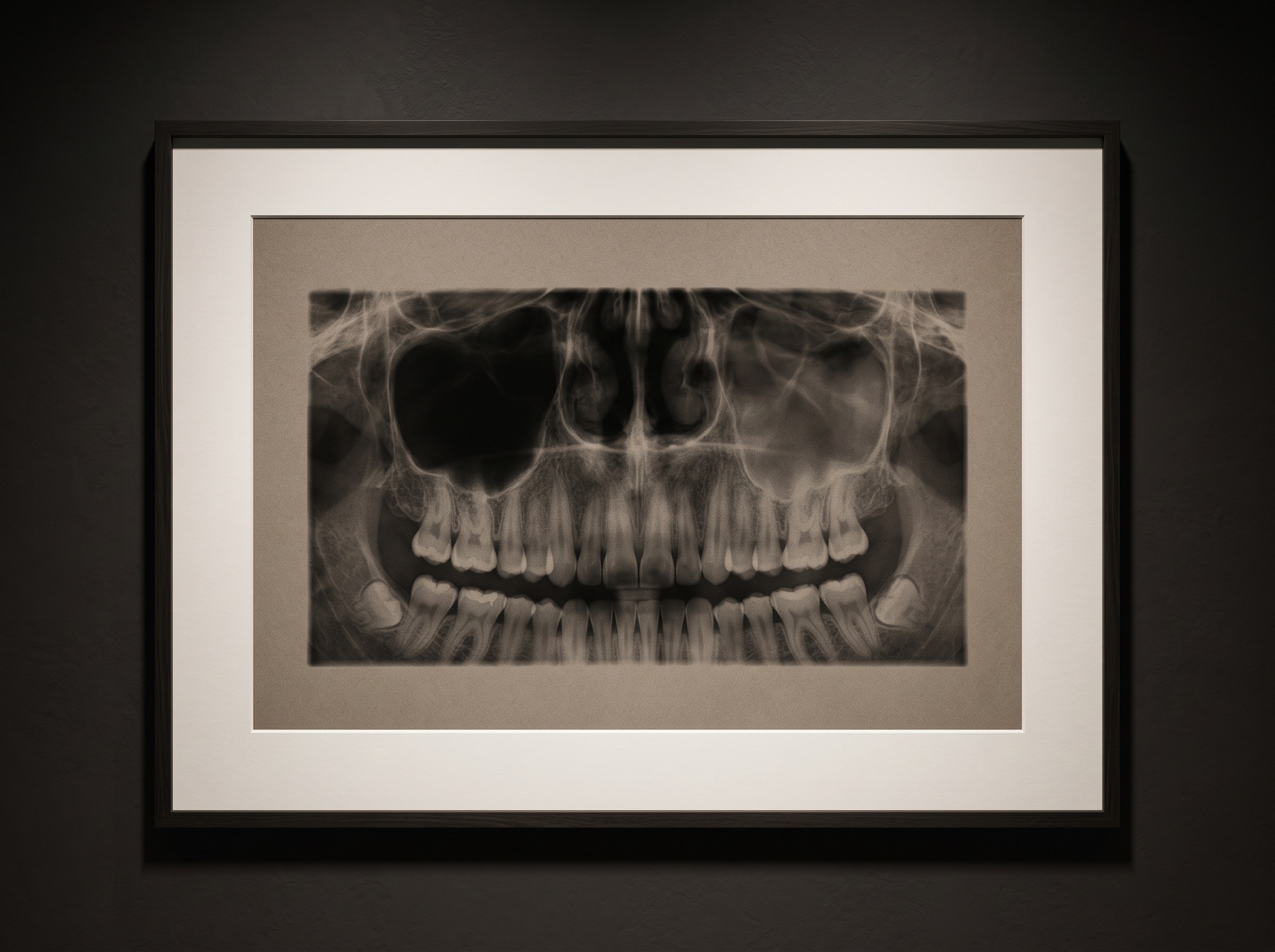
Most incidental findings are less dramatic and more common. The maxillary sinuses sit squarely inside the upper part of a panoramic or CBCT field, and a sinus clouded on one side where its partner is clear can point to sinusitis, a retention cyst, or disease that explains a vague ache the patient had blamed on an upper tooth. Small radiopaque flecks in the soft tissue near the throat can be tonsilloliths – calcified debris in the tonsils – sometimes the quiet cause of chronic bad breath and throat discomfort, and a subject of recent study for their association with other findings on the same films.
Then there are the findings within the bone itself. Impacted teeth that never erupted, supernumerary teeth the patient never knew they carried, retained roots from an old extraction, and the occasional radiolucent shadow in the jaw that warrants a closer look – a cyst, a benign lesion, rarely something more serious. Bone patterns can hint at systemic conditions; a thinned, altered trabecular structure has been studied as a possible signal worth correlating with bone-density concerns elsewhere in the body. And every so often the film simply shows something that should not be there at all: a fragment of a broken instrument, a foreign body, a surgical remnant. None of these were the reason for the appointment. All of them are the radiograph doing more than its narrow job.
How AI Widens – and Sharpens – the Reading
The move from film to three-dimensional imaging enlarged both the opportunity and the duty. A CBCT volume contains vastly more anatomy than a flat film, which means more can be found and, correspondingly, more can be missed by an interpreter who scrolls only to the tooth in question. This is exactly where automated reading is starting to help. Systems that segment a volume structure by structure do not tire of the periphery the way a human eye does; as we described in AI on CBCT, they inventory the whole field rather than fixating on the obvious. The same detection instinct that lets a model read caries and bone loss on a bitewing can, in principle, be pointed at the sinus, the airway, or a suspicious calcification, and surface it for a human to judge.
The judgment remains the point. An algorithm that flags a radiopacity in the neck has not diagnosed anything; it has done what good imaging always does – drawn a trained eye to a place worth looking. Turning that flag into a finding, and a finding into a considered referral, is human work, and it belongs in the structured account of the study rather than in a raw list of pixels, much as we argued about the seam between detection and reporting in From Detection to Diagnosis.
Future Developments
The trajectory is toward the dental radiograph understood, openly, as a broader health record than its filename suggests. As automated screening of the full field of view matures, the incidental finding should become less a matter of whether a particular clinician happened to glance at the corner of the image, and more a reliable, systematic pass over everything the beam captured – the arteries, the sinuses, the airway, the bone – with anything unexpected surfaced for human interpretation and, where warranted, referral. That will sharpen an old ethical question the technology only makes louder: what is captured must be read, and what is read carries a duty to act. For the patient, the reassurance is simple and worth stating plainly. The picture taken for your teeth is looking after more than your teeth – and the craft of dental imaging has always been, at its best, the discipline of seeing the whole frame.
Sources & further reading:
Sorry, the comment form is closed at this time.