


September 7, 2026 Can a Handheld Radiograph Match the Wall-Mount? The Physics of Image Quality in Portable Dental X-Ray
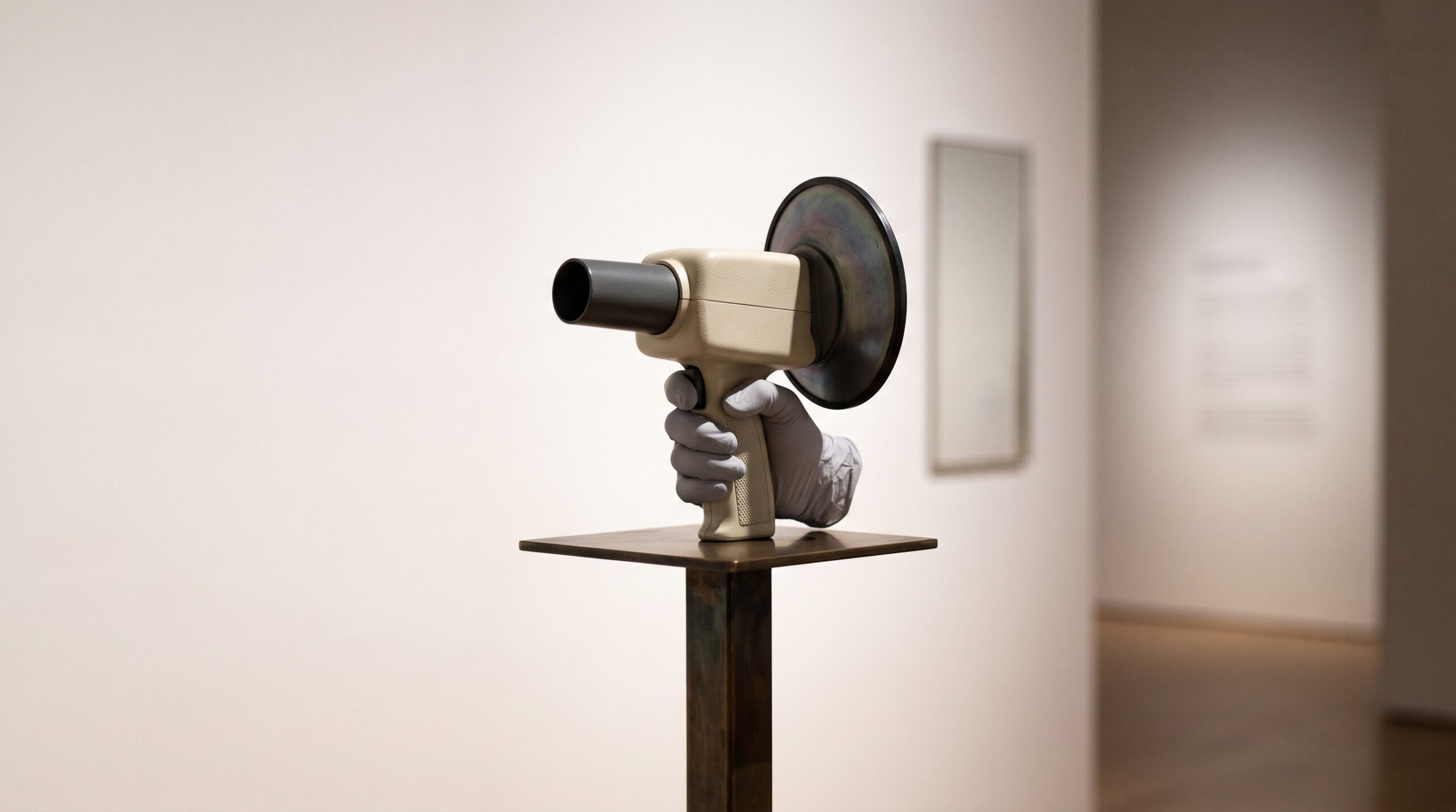
A wall-mounted dental X-ray head is a fixed instrument, and its fixedness is part of its authority. It hangs from a stable scissor arm, swings to the tooth in question, holds its aim without a tremor, and is fired from behind a wall or a leaded screen. Everything about it says permanence. The handheld unit says something else entirely. It puts the same X-ray tube into a single gloved hand, no larger than a hair dryer, and asks the clinician to be aimer, stabiliser, and operator all at once, standing in the room with the patient as the exposure is made. To an eye trained on the wall-mount, it can look like a compromise – convenience bought at the cost of the image. The interesting question, and the one this piece is about, is whether that intuition is actually true. The answer is not a matter of opinion. It is a matter of physics, geometry, and a few numbers a good radiograph must hit.
Those numbers matter because the reason handheld imaging exists is not laziness. It is reach. A patient in a nursing-home bed, in a wheelchair that will not recline into a fixed unit, or in an operatory where moving them is unsafe, still needs a radiograph. Mobile and geriatric dentistry depend on being able to bring the source to the person. The only honest way to justify that is to prove the image does not suffer for it.
The Standard a Radiograph Must Meet
Before asking whether a handheld unit is good enough, one has to define good enough. A 2026 study in the journal Gerodontology by Geibel and colleagues did exactly that, evaluating handheld mobile radiography against an explicit quality bar. It anchored to the specifications of the German Dental Association, which for routine intraoral diagnostics call for an imaging voltage of at least 60 kilovolts, a spatial resolution of at least five line pairs per millimetre, and a distance from the focal spot to the end of the tube of at least 200 millimetres. Each of those three numbers guards a different property of the image. The voltage governs how the beam penetrates and how contrast is rendered; the resolution is the fineness of detail the system can actually resolve; and the source distance controls geometric distortion and magnification. That resolution figure is the one clinicians feel most directly – as we explored in How Sharp Can a Dental Image Truly Be?, five line pairs per millimetre is a demanding threshold, the point at which the finest structures either hold their edges or dissolve into an undifferentiated grey. A device is not diagnostic simply because it makes an image. It is diagnostic when the image clears bars like these.
The Tube That Fits in a Hand
What makes it possible to meet that standard from the palm of a hand is a generation of compact, high-frequency, constant-potential generators. Older portable sources pulsed and sagged, delivering a ragged beam that wasted dose on low-energy photons and struggled for consistent contrast. A modern handheld runs a stable direct-current potential, holding a clean sixty to seventy kilovolts throughout the fraction of a second the exposure lasts, from a small focal spot that keeps the geometry crisp. Bolted to the front is not a cosmetic snout but a working optical element: a collimating cone that enforces the mandated source-to-skin distance and trims the beam to a rectangle no larger than the sensor. That cone is doing quiet double duty on image quality – the fixed distance tames magnification and keeps the beam’s intensity even across the field, the same evenness we examined in The Anode Heel Effect, where the geometry of the tube itself subtly shades one edge of every radiograph. The handheld did not shrink the physics. It repackaged it.
The Operator Becomes the Tripod
Here is where the portable device faces a threat the wall-mount never does. On a fixed arm, the source cannot move during the exposure; the geometry that existed when the button was pressed is the geometry that persists until the beam stops. In the hand, the operator is the tripod, and a living hand is never perfectly still. A tremor of a fraction of a millimetre during a short exposure blurs the finest detail – the radiographic equivalent of a slightly shaken photograph, softening exactly the sharp edges the five-line-pair standard demands. The physics offers a defence: the same high-frequency generator that stabilises the voltage also permits very short exposure times, and a shorter exposure freezes motion the way a fast shutter does. Technique supplies the rest. The device is braced against the patient’s face or a bite-block holder rather than floated in free space, and the sensor is fixed by an aiming ring so that source, tooth, and detector share one honest line. Handled this way, the geometry becomes as reproducible as a fixed unit’s – and reproducibility, not any single heroic image, is what a diagnostic workflow actually needs.
Dose, Scatter, and the Shield That Makes It Defensible
The obvious objection to standing in the room, X-ray source in hand, is radiation to the operator. The answer is engineered into the front of the device: a leaded backscatter shield, the round disc mounted at the mouth of the cone. Its job is to intercept the radiation that scatters back off the patient toward the person holding the unit – the Compton-scattered photons we examined in The Scattered Photon, which fly in every direction and are the dominant occupational exposure in intraoral work. Combined with tight rectangular collimation, which keeps the primary beam confined to the sensor and generates less scatter to begin with, the shield brings operator dose to levels that measured studies find acceptable for routine hand-held use. And because the collimator restricts the field so precisely, patient dose is well controlled too – which places a real burden on the clinician not to over-expose out of caution. As we cautioned in The Dose You Cannot See, a forgiving digital sensor will still produce a usable image from too much radiation, letting exposure quietly creep upward unseen. Portability does not suspend that discipline; it demands it.
What the Evidence Actually Shows
Set against these standards, the handheld acquits itself well. The Gerodontology evaluation concluded that radiographs produced with a mobile handheld system met the quality requirements for diagnosis in the mobile setting – that the images were, for their clinical purpose, the equal of what a fixed unit would have produced. This is the crucial and slightly counterintuitive point. The limiting factor in handheld imaging is not the device’s inherent capability, which now clears the physical bar comfortably. It is the operator’s technique: the steadiness of the hold, the fidelity of the sensor placement, the honesty of the geometry. A wall-mount forgives a mediocre operator because it removes several ways to go wrong. A handheld returns those variables to human hands, which means it can produce an image indistinguishable from the fixed unit’s – or a poor one – depending entirely on the care of the person wielding it. The instrument is capable. The craft is the variable.
Future Developments
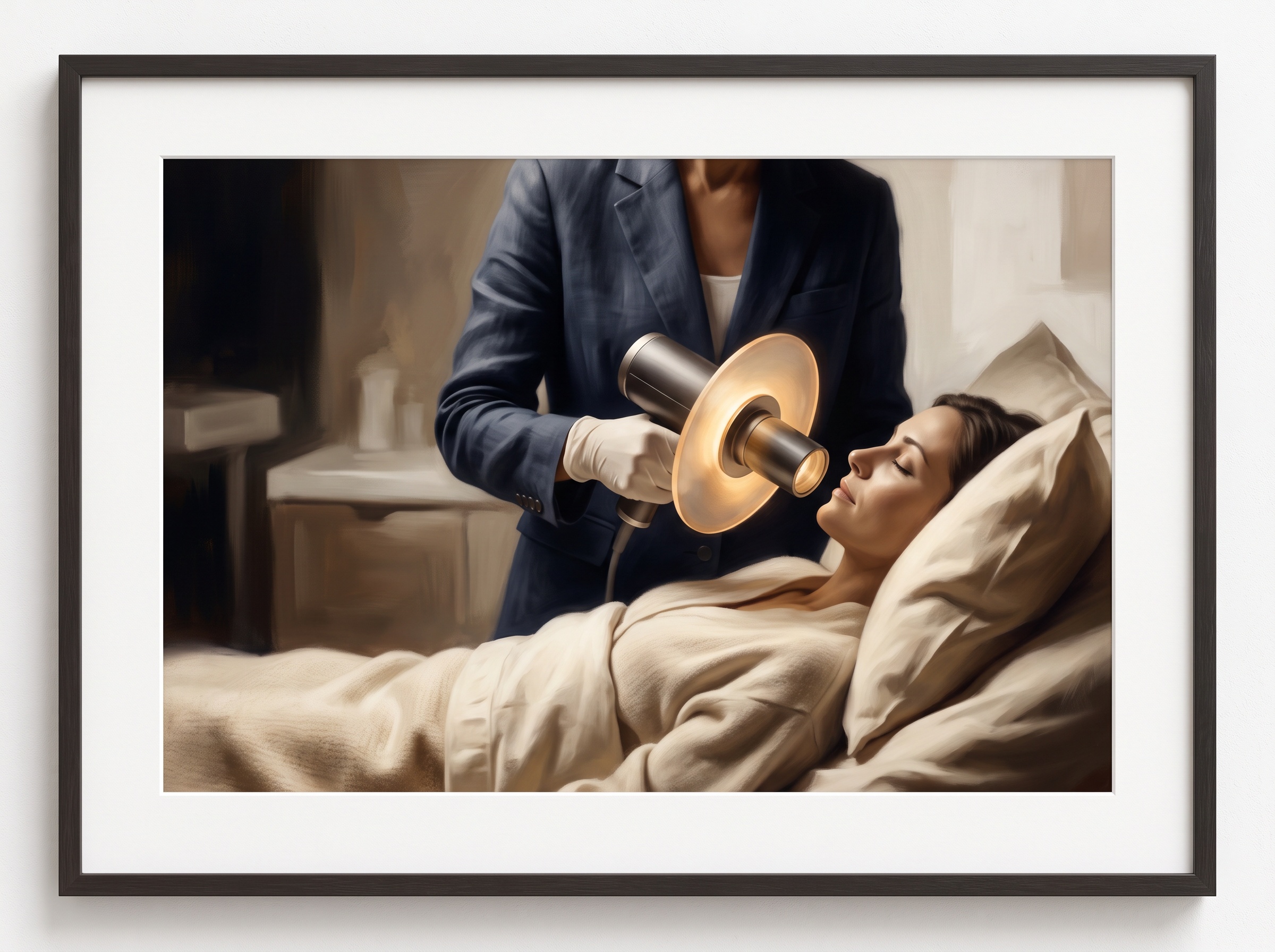
The trajectory points toward closing the last of that gap without taking the device out of the hand. Ever-more-sensitive CMOS sensors need less radiation for the same signal, shrinking exposure times further and making residual motion blur harder to induce. Acquisition-time quality feedback – the same kind of image-scoring intelligence beginning to grade radiographs the instant they are captured – could soon warn an operator that a shot was soft or the geometry off before the patient is dismissed, turning the retake from an afterthought into a caught error. Stabilisation borrowed from handheld cameras, and lighter shielding materials, will make the units steadier and less fatiguing to hold. The deeper significance is not technical but human: the handheld radiograph dissolves the old assumption that diagnostic-quality imaging requires the patient to come to a fixed studio. It lets the studio travel – to the bedside, the wheelchair, the home – and arrive with its standards intact. That is imaging as craft in its most generous form: not the pursuit of the most impressive machine, but the discipline of carrying a real standard into the places a fixed machine can never reach, and refusing to lower it on the way.
Sources & further reading:
Related Reading
- The X-Ray That Fits in a Bag: NASA’s Portable Imaging Trials and the Craft of Radiography Far From Any Hospital
- The Discipline of Restraint: The New Imaging Guidance and the Art of the Radiograph Not Taken
- The Blur Built Into Every X-Ray: Focal Spot Size, Penumbra, and the Geometry of a Sharp Radiograph
Sorry, the comment form is closed at this time.