


August 11, 2026 The Color a Screen Cannot Show: Gamut, Metamerism, and Why a Tooth Refuses to Be Reproduced
Of all the things a dental image is asked to carry faithfully — dimension, sharpness, contrast, texture — color is the one that most stubbornly resists. A radiograph never pretends to be in color, and we forgive it. But a clinical photograph of an anterior tooth promises the truth of a shade, and on that promise a great deal rides: the ceramist who will match a crown to it, the patient who will judge the result in a bathroom mirror, the record that must still mean something a year later. And yet between the light that leaves the enamel and the color that finally glows on a screen, something is always lost. Not through carelessness, but through the nature of the machinery itself. A tooth is a spectrum; a photograph is three numbers. Understanding exactly what falls into the gap between them is the beginning of reproducing color well.
What a Color Space Actually Fences In
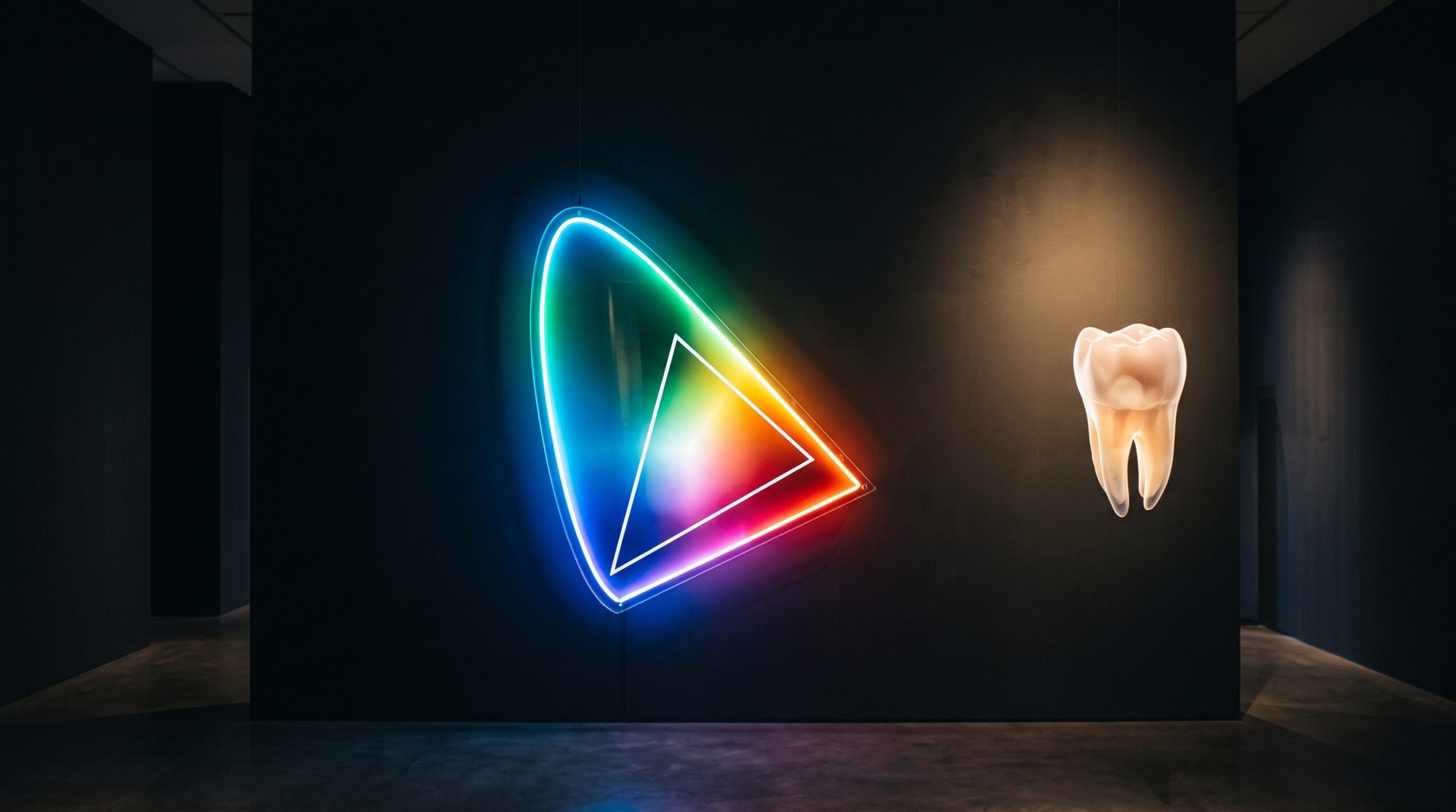
Every camera, every screen, every printer can reproduce only a finite region of all the colors the human eye can see. That region is its gamut, and the honest way to picture it is the horseshoe of the CIE chromaticity diagram: a map of every visible hue, its curved edge tracing the pure spectral colors of the rainbow. No device fills the horseshoe. Instead, each carves out a triangle inside it, anchored on its particular red, green, and blue primaries. Everything within the triangle can be shown; everything outside it simply cannot, no matter how the numbers are adjusted. The familiar sRGB space — the default of the web, of most monitors, of countless cameras set to their factory state — describes a notably modest triangle. Wider spaces such as Adobe RGB and ProPhoto RGB stretch the triangle outward, reclaiming saturated greens and cyans that sRGB clips flat. The choice of working space is therefore not a technicality. It quietly decides, before a single shade is judged, which colors your image is even permitted to contain.
The Metamerism Trap
The deeper difficulty is not the size of the fence but the crudeness of the instrument reading the color in the first place. Human vision does not measure a spectrum. It samples the incoming light with three types of cone, collapses that light into three signals, and hands the brain a summary. A profound consequence follows: two physically different spectra can produce the same three signals and therefore look identical. These are metamers, and they are the quiet saboteur of every shade match. A ceramic crown and the natural tooth beside it may agree perfectly under the surgery’s daylight-balanced lamp and then part ways at the window, or under the warm bulbs of a living room, because their underlying spectra were never the same — only their appearance under one particular light. The camera inherits this frailty and adds its own, for its three color filters do not see quite as our cones do. A pairing the sensor calls a match, the eye may not; a difference the eye dismisses, the sensor may exaggerate. This is why a faithful dental shade match captured through photography depends so heavily on controlling the light, and why a shade judged under one illuminant is never quite a promise under another.
Three Numbers for an Infinite Spectrum
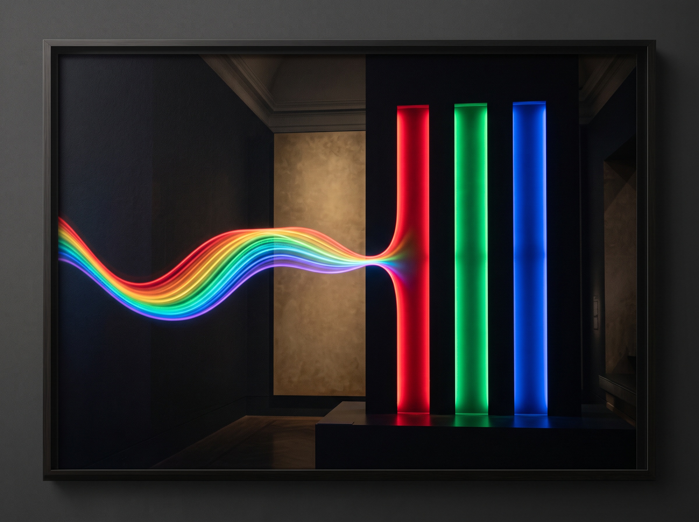
Step back and the whole pipeline reveals itself as a series of ruthless compressions. A continuous spectrum of light — an unbroken curve across the visible band — strikes the sensor and is reduced, at every pixel, to three tristimulus values: a red, a green, a blue. Whatever spectral subtlety distinguished the light is gone the instant it is recorded; only the triad survives. Those three numbers are then quantized into discrete steps according to the file’s bit depth. An 8-bit channel allows 256 gradations; the smooth transition from the incisal translucency to the cervical warmth of a tooth must be described in those 256 rungs, and where the rungs are too coarse, the gradient fractures into visible banding — steps of flat color where nature laid down a continuous blend. Capturing in a higher bit depth, and in a raw format that preserves the sensor’s full range before it is squeezed into a delivery space, keeps those gradations fine enough that the compression stays invisible. It cannot restore the lost spectrum, but it can keep the surviving numbers honest.
Where the Tooth Falls Outside the Fence
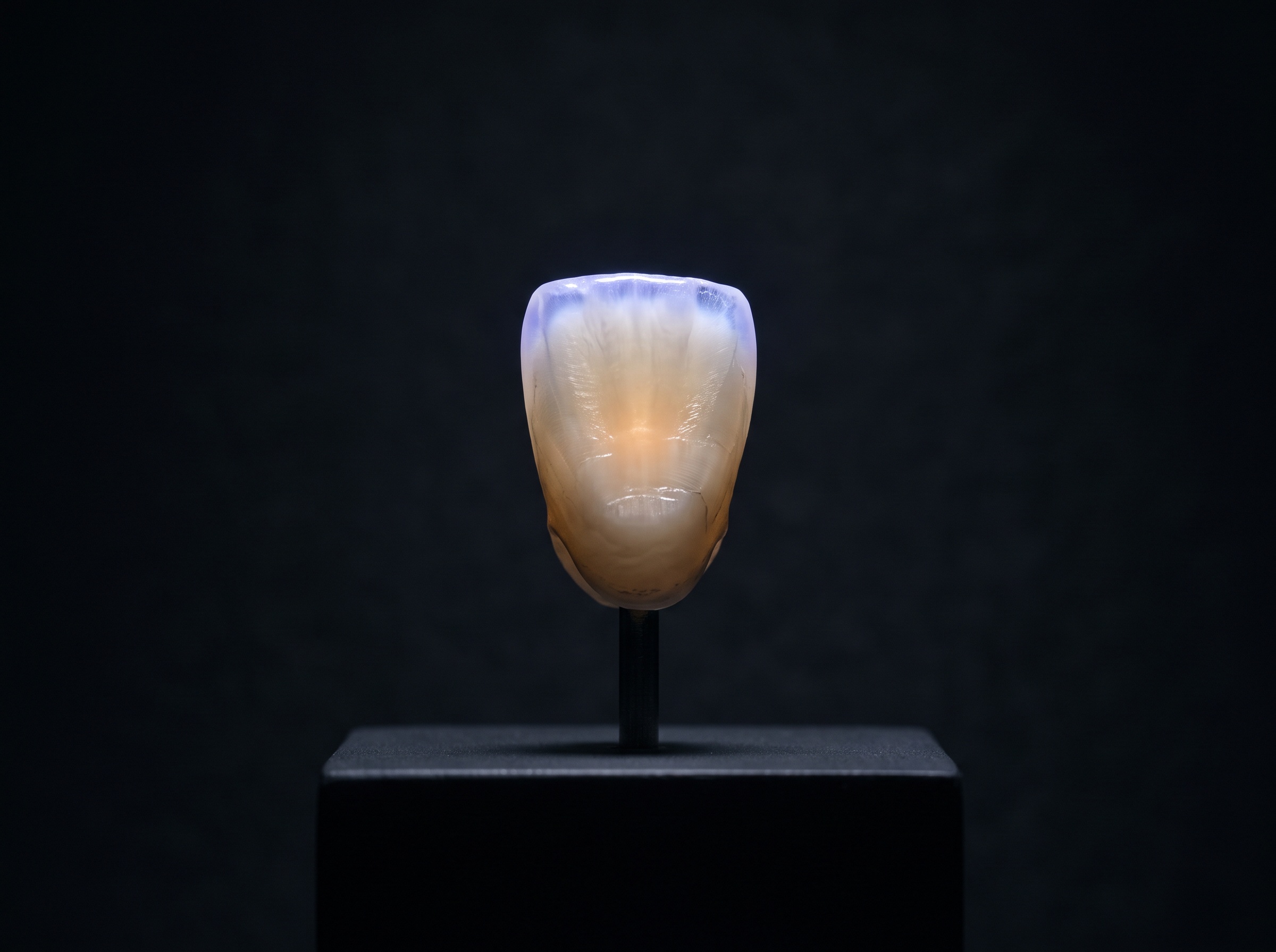
Even a generous gamut and a deep bit depth meet their match in enamel, because a tooth is one of the most optically complicated surfaces a camera is ever asked to render. It is not a flat swatch of color but a layered, semi-transparent event. Light does not merely bounce off it; it enters, scatters through the translucent enamel, reflects from the dentin beneath, and returns altered, carrying the depth we read as vitality. The incisal edge glows with a faint blue opalescence, scattering short wavelengths the way the sky does. Under ultraviolet, the tooth fluoresces, emitting a soft light of its own that no reflective color model anticipates. These are not colors a triangle on a chromaticity chart handles gracefully; the most vivid opalescent blues and the living luminance of fluorescence sit at or beyond the edge of what an ordinary display can show. Glare compounds the problem, laying a veil of the lamp’s own color across the surface and hiding the very structure we are trying to record — which is why techniques such as cross-polarized photography that suppresses surface glare matter so much, stripping away the reflection to let the true internal color speak. The tooth, in short, keeps asking for colors that live outside the fence.
Managing Color Honestly
If perfect reproduction is impossible, disciplined reproduction is not, and the craft lies in refusing to leave any link in the chain to chance. It begins in the frame: a neutral gray card or a known color reference photographed in the same light gives every downstream step a fixed truth to calibrate against, converting a subjective impression into a measurable one. It continues with ICC profiles — the small translation tables that tell each device precisely which real colors its numbers stand for — so that a value captured by the camera means the same color when it reaches the screen and, later, the ceramist’s monitor. It depends, finally, on the last device in the chain being trustworthy, because none of this discipline survives an uncalibrated display that quietly distorts every clinical color it is asked to show. Consistent, diffuse, daylight-balanced lighting; a reference in the frame; a wide-enough working space; a calibrated screen at the end — none of it defeats metamerism or conjures colors past the gamut boundary, but together they ensure that what is lost is lost predictably, the same way every time. Predictable loss can be corrected for. Random loss cannot.
Future Developments
The horizon belongs to instruments that refuse the three-number compromise at its source. Multispectral and hyperspectral imaging capture not three broad channels but many narrow ones, sampling the reflectance curve densely enough to record the spectrum itself rather than a metameric summary of it — and a device that knows the spectrum can predict a tooth’s appearance under any light, dissolving the metamerism trap that has haunted shade matching since its beginning. Spectrophotometric shade devices already bring a version of this rigor to the single-point measurement; the coming work is to extend it across the whole surface of an image. Displays, meanwhile, keep widening their triangles, with wide-gamut and high-dynamic-range panels reclaiming the saturated, luminous colors that sRGB has long clipped away, so that more of the tooth’s true character can survive to the screen. The dream is a pipeline that carries a tooth’s full optical signature intact from capture to display to fabrication, with nothing collapsed and nothing invented. We are not there. But each advance narrows the gap between the spectrum that leaves the enamel and the color we finally see — and in that narrowing, the tooth comes a little closer to being reproduced as faithfully as it deserves.
Sources & further reading:
Sorry, the comment form is closed at this time.