


July 19, 2026 The Art of the Shade Match: How Dental Photography Captures a Tooth’s True Color
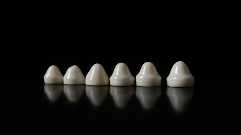
Ask a ceramist about the hardest object to reproduce in all of dentistry and the answer is often disarmingly small: a single front tooth, sitting beside its natural neighbor. Match it perfectly and no one will ever notice your work — which is precisely the point. Miss by a fraction of a shade and the eye catches it instantly. This is where dental photography stops being documentation and becomes an art of perception.
Why the Human Eye Cannot Be Trusted
Chairside shade selection has always fought against biology. The eye adapts to surrounding color in seconds, tires quickly, and is thrown off by the color temperature of operatory lighting, the color of the walls, even a patient’s lipstick. Two clinicians can look at the same tooth under the same light and record different shades — and the same clinician can disagree with themselves an hour later. The tooth has a fixed color; our perception of it does not.
The Camera as an Objective Witness
A calibrated camera changes the equation. Unlike the eye, a photograph can be captured under controlled, repeatable conditions and examined at leisure, side by side with a shade tab in the same frame. The image becomes a permanent, shareable record that travels to the laboratory exactly as the clinician saw it — no memory, no translation, no “a little more yellow at the cervical.” The ceramist receives evidence rather than description.
Cross-Polarization: Seeing Through the Shine
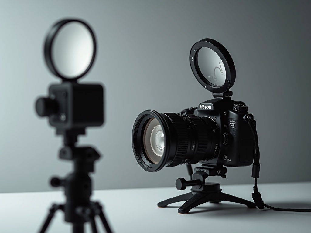
The single most transformative technique is cross-polarization. By placing a polarizing filter over the light source and another over the lens, oriented at ninety degrees, the specular glare that bounces off wet enamel is eliminated. What remains is the light that has actually entered the tooth and returned — the true internal color. Suddenly the white spots, the translucent incisal edge, the subtle warmth near the gum, and the internal craze lines all appear with clarity that the naked eye simply cannot achieve.
Reading a Tooth in Three Dimensions of Color
Great shade matching means abandoning the idea of a single “color.” Ceramists think in three axes. Value — the lightness or darkness — is the most important and the easiest to get wrong; a crown that is too high in value looks like a bright artificial chip no matter how perfect its hue. Chroma is the intensity or saturation of the color, typically strongest near the neck of the tooth. Hue, the actual pigment, varies least between teeth but still matters. A well-exposed, color-managed photograph lets all three be measured region by region, rather than averaged into a single guess.
Calibration Is Everything
A photograph is only trustworthy if the color pipeline is disciplined. That means a consistent light source with a known color temperature, a custom white balance set from a neutral gray card in the frame, a fixed exposure, and a color reference tab photographed alongside the tooth. With a gray or color reference in every image, the laboratory can correct for any residual shift and read absolute color values. Without it, even a beautiful image is just an opinion in higher resolution.
Where Science Meets the Gallery
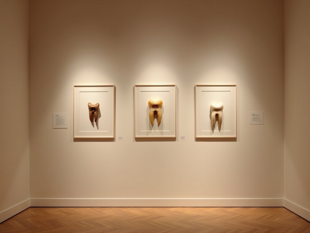
There is a reason this work rewards an artistic eye. Once glare is removed and color is trustworthy, a tooth reveals itself as something closer to a small painting — layered, luminous, and individual. The finest restorative results come from clinicians and ceramists who treat each image not as a clinical formality but as a portrait worth studying. That is the philosophy at the heart of organizing patient images as a gallery: when every photograph is captured with intention, the archive becomes both a diagnostic tool and a record of craft.
Future Developments
The next chapter is already arriving. Spectrophotometric analysis built directly into imaging software promises to translate a calibrated photograph into objective color coordinates automatically, while machine-learning models trained on paired photographs and finished restorations are beginning to suggest layering recipes to the ceramist. As these tools mature, the camera will not replace the artist’s eye — it will give it a more faithful canvas to work from, closing the gap between the tooth the patient owns and the one the laboratory builds.
Sorry, the comment form is closed at this time.