


August 3, 2026 Cross-Polarization and the Craft of Clinical Dental Photography
Ask any ceramist about the hardest part of their work and the answer rarely concerns hands or heat—it concerns information. A shade tab held to a tooth in operatory light captures a fleeting impression, but the photograph that follows too often lies. Bright specular highlights bloom across the enamel, washing out the very gradients the lab needs to read. Cross-polarization is the quiet correction to that problem: a way of photographing teeth that removes surface glare and lets true color, translucency, and texture speak for themselves.
Why Enamel Defeats the Naked Lens
Light striking a tooth splits into two populations. Some of it penetrates the enamel and dentin, scatters through the internal structure, and returns carrying the tooth’s actual color—its chroma, value, and the layered translucency that makes natural dentition read as alive. The rest bounces straight off the glossy enamel surface as specular reflection: bright, white, and utterly uninformative about color. To the eye and to the camera, these two signals arrive superimposed. The specular component dominates the highlights, flattening contrast and masking the subtle transitions the clinician most needs to see.
This is why conventional flash photography, for all its sharpness, is a poor shade-matching tool. The brightest, most attention-grabbing regions of the image are precisely the regions carrying the least diagnostic information. The craft of clinical shade capture begins with a single goal: separate the internal, color-bearing light from the surface glare—and discard the glare.
The Optics of Crossed Polarizers
Polarization makes that separation possible. A linear polarizing filter placed over the flash constrains the emitted light to a single plane of vibration. When that polarized light reflects specularly off the smooth enamel surface, it largely retains its orientation. But light that penetrates the tooth and scatters internally becomes depolarized—its orientation randomized by the journey through enamel and dentin.
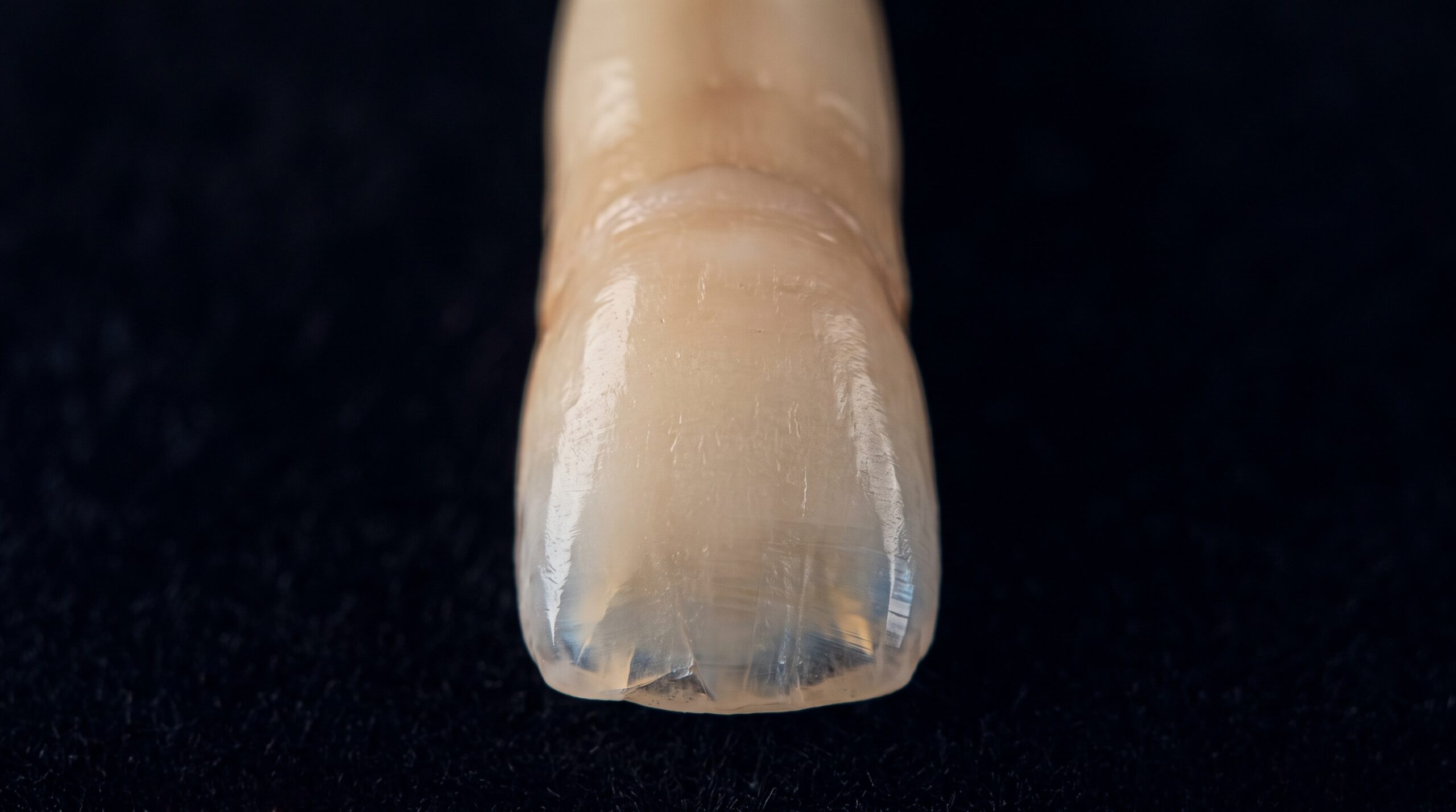
Now place a second polarizer over the lens, rotated ninety degrees to the first. This crossed analyzer blocks light that still carries the flash’s original orientation—the specular glare—while freely passing the depolarized, internally scattered light that encodes true color. The result is a photograph in which the enamel’s mirror-like highlights simply vanish, replaced by an even, matte rendering of the tooth’s genuine internal appearance. Mamelons, translucent incisal halos, hypocalcification spots, hairline cracks, and the fine seams of existing restorations all emerge with a clarity the glossy original could never provide.

Building the Instrument: Gear That Earns Its Place
Cross-polarization asks little of a well-equipped clinician. A macro lens in the 90–105mm range and a flash system are the foundation; the polarizers are the specialization. The essential debate concerns the light source. A ring flash encircles the lens and produces flat, shadowless, highly repeatable illumination—forgiving and consistent, though its frontal light can suppress the sense of surface relief. Twin or dual flashes mounted on adjustable arms sculpt the tooth with directional light, revealing texture and contour more expressively, at the cost of demanding more skill to standardize.
For polarized shade work, many clinicians favor the evenness of a ring flash or a purpose-built polarizing flash bracket, precisely because repeatability matters more than drama. Filters can be commercial cross-polarizing attachments or a matched pair of linear polarizers. Rounding out the kit are black contrastors to eliminate distracting background reflections and shadows behind the incisal edges, cheek retractors to clear the field, and clean mirrors for indirect views. Each accessory exists to remove a variable.
Standardization: The Discipline Behind Repeatability
A cross-polarized image is only as valuable as it is comparable. Shade is relative, and a photograph taken at an unknown exposure tells the lab nothing it can trust. The remedy is a fixed, documented protocol: a locked manual exposure—a representative starting point being ISO 100, f/22, and 1/125s with flash, adjusted and then held constant—a consistent working distance and magnification ratio, and identical framing from patient to patient.
Two additions elevate the result from qualitative to quantitative. First, include a shade tab in the frame, positioned in the same plane as the tooth, so the lab reads the target and the reference under identical light. Second, incorporate a neutral gray or a calibrated color reference card, enabling precise white-balancing in post-processing. With these anchors, a cross-polarized photograph stops being a pretty picture and becomes measurable data.
Communicating Shade to the Laboratory
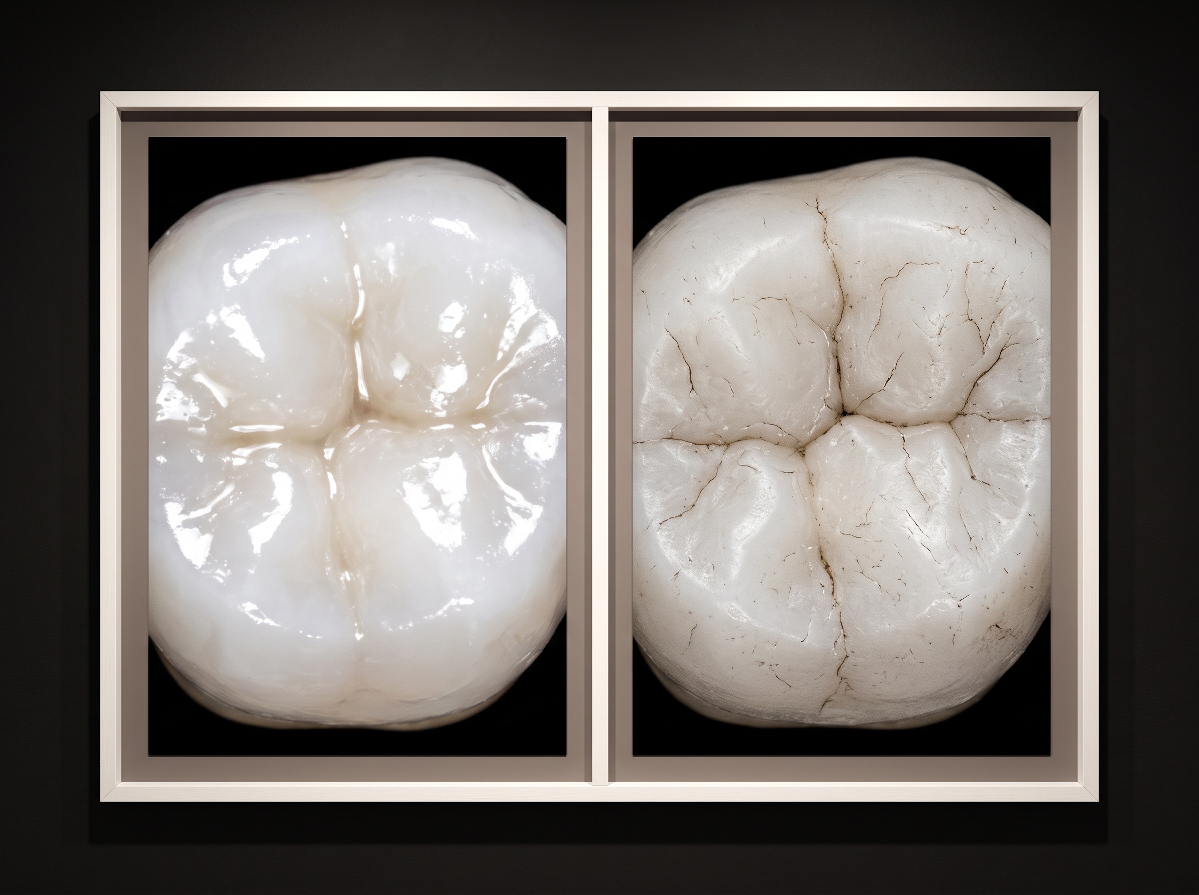
The ceramist cannot see the patient; they can only see what you send. A cross-polarized series translates the chairside reality into a language the lab can act on. The glare-free image reveals the true value and chroma at cervical, middle, and incisal thirds; the translucency map at the incisal edge; the position and intensity of mamelons; and any characterizations—white spots, crack lines, tea-stain gradients—that a monochrome shade designation could never convey.
Best practice is a paired approach: a conventional, non-polarized image to preserve the natural gloss and surface reflectivity the patient will actually see, alongside the cross-polarized frame that exposes the underlying color architecture. Together they give the technician both the destination and the blueprint. Organizing these images within a structured imaging platform—where polarized and standard series sit side by side, tagged to the patient and the case—keeps that visual dialogue coherent from consultation through delivery. This is one place a system such as PatientGallery quietly earns its keep: not by replacing the clinician’s eye, but by preserving and transmitting what that eye has captured.
Future Developments
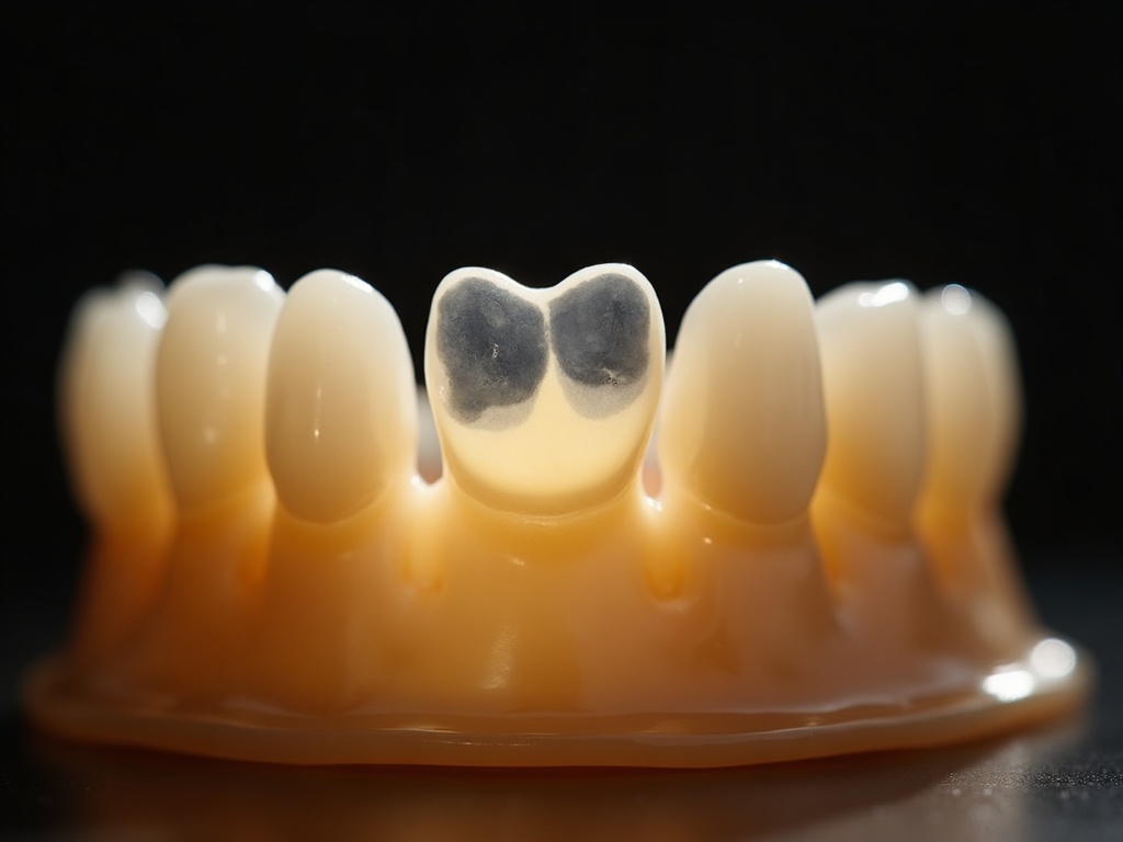
Cross-polarization is a mature technique built on stable optics, yet its future is anything but static. The trajectory points toward calibration and computation. AI-assisted shade capture is beginning to translate cross-polarized images into quantified color values—mapping chroma, value, and translucency across the tooth automatically and flagging characterizations a hurried eye might miss. Chairside spectrophotometry and colorimetry, long the objective gold standard for single-point measurement, are converging with photographic workflows, so that a calibrated image and a numerical reading validate one another. As these tools integrate into imaging platforms like PatientGallery, the cross-polarized photograph becomes not merely a record but a computable object—searchable, comparable across visits, and shareable with the lab as data rather than impression. And yet the craft persists. Behind every calibrated pixel remains a clinician composing light, choosing when the tooth is most honestly seen. Dental imaging has always lived at the intersection of science and art; cross-polarization is where the physics of light and the aesthetics of the smile meet most gracefully—and the next decade promises only to sharpen that meeting, never to dissolve it.
Sources & further reading:
Sorry, the comment form is closed at this time.